Organelles Of Cell Endoplasmic Reticulum Ribosomes Vacuoles Ribosomes

Organelles Of Cell Endoplasmic Reticulum, Ribosomes, Vacuoles

Ribosomes Definition: The ribosomes are small, dense, rounded and granular particles of the ribonucleoprotein. They occur either freely in the matrix of mitochondria, chloroplast and cytoplasm (i. e. , cytoplasmic matrix) or remain attached with the membranes of the endoplasmic reticulum and nucleus.

Structure of Ribosome

Occurrence and Distribution: The ribosomes occur in cells, both prokaryotic and eukaryotic cells. In prokaryotic cells the ribosomes often occur freely in the cytoplasm. In eukaryotic cells the ribosomes either occur freely in the cytoplasm or remain attached to the outer surface of the membrane of endoplasmic reticulum. The yeast cells, reticulocytes or lymphocytes, meristematic plant tissues, embryonic nerve cells and cancerous cells contain large number of ribosomes which often occur freely in the cytoplasmic matrix.
 two")
Types of Ribosomes: Recently according to the size and the sedimentation coefficient (S) two types of ribosomes have been recognized. Ø 70 S Ribosomes Ø 80 S Ribosomes

70 S Ribosomes. The 70 S ribosomes are comparatively smaller in size and have sedimentation coefficient 70 S and the molecular weight 2. 7× 10⁶ Daltons (Dalton is the unit of molecular weight (MW); one Dalton equals the weight of hydrogen atom). They occur in the prokaryotic cells of the blue green algae and bacteria and also in mitochondria and chloroplasts of eukaryotic cells.

80 S Ribosomes: The 80 S ribosomes have the sedimentation coefficient of 80 S and the molecular weight 40 × 106 Daltons. The 80 S ribosomes occur in eukaryotic cells of the plants and animals. The ribosomes of mitochondria and chloroplasts are always smaller than 80 S cytoplasmic ribosomes and are comparable to prokaryotic ribosomes in both size and sensitivity to antibiotics, although their sedimentation values vary in different phyla, e. g. , 77 S in mitochondria of fungi, 60 S in mitochondria of mammals and 60 S in mitochondria of animals in general. The ribosomes of chloroplasts are 70 S type.

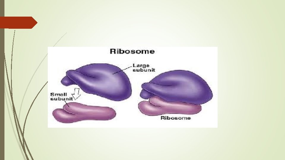
Structure of Ribosomes: The ribosomes are oblate spheroid structures of 150 to 250 Ao in diameter. Each ribosome is porous, hydrated and composed of two subunits. One ribosomal subunit is large in size and has a domelike shape, while the other ribosomal subunit is smaller in size and occurring above the larger subunit and forming a cap-like structure.

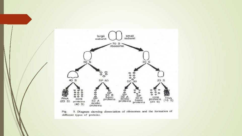
Chemical Composition of Ribosomes: Ribosomes are composed of about equal amounts of r. RNA (ribosomal ribonucleic acid), and proteins, with a little proportion of lipids and certain metallic ions such as Mg, Ca and Mn. Proteins and r. RNA are the major constituents of ribosomes. Their proportion in both types of ribosomes (70 S and 80 S) is variable. Ribosomal proteins in small subunits are prefixed by an alphabet ‘S’ and in large subunits by ‘L’. Most of ribosomal proteins act as enzymes which catalyze protein synthesis at various stages.

Ø Ribosomal RNA: It generally represents more than 80% of the total RNA present in cells. It is represented by a highly folded filament of RNA which may measure up to 7000 A in extended forms. Protein molecules are usually attached to this r-RNA filament. About 60% of total r-RNA (both 28 S and 18 S) presents a helical configuration like DNA.

Continue. . Ribosomal RNA is found in three forms: In eukaryotic cells, the RNAs are larger and 18 S is found in small subunit, and 28 S and 5 S in the larger subunit. Eukaryotic ribosomes also have an extra small RNA, which is called 5. 8 S RNA and it is transcribed in the nucleolus as a single unit along with 18 S and 28 S RNA. 5 S RNA is synthesized outside the nucleolus. Whereas in prokaryotes three RNA molecules are 16 S RNA in the small subunit, and 23 S and 5 S in the large subunit. The 30 S and 50 S ribosomal subunits associate to form 70 S ribosomes only when they are involved in protein synthesis. The 70 S ribosomes frequently form clusters called polysomes

Ø Ribosomal Proteins: Protein contents of the ribosomes are very complex and designated as core proteins. About 50 – 55 proteins have been isolated. In prokaryotic ribosomes, about 21 proteins are found in smaller 30 S subunit and about 34 proteins in the larger 50 S subunit. The proteins are core proteins. All the proteins are different with the exception of one that is present in both subunits. Eukaryotic ribosomes contain a higher content of protein than prokaryotic ribosomes. For instance, Tsoetal, (1958) determined a protein content of 60% for Pea seedling particles and 55% for those from rabbit reticulocytes (compared to 37% for E. coli). Ribosomal proteins are small (7000 to 32, 000 Dalton in molecular weight) and are rich in basic amino acids. Ribosomal proteins can be dissociated from the ribosome and these are called split proteins (SP).

Ø Ribosomal Enzymatic Proteins: Most of the ribosomal proteins act as enzymes thereby catalyzing protein synthesis. Initiation proteins F 1, F 2 and F 3 initiate the process of protein synthesis, whereas transfer proteins (G – factor, Ts-factor) help in translocation of ribosomes over m. RNA and transfer of t-RNA residue from one site of the ribosome to the other site.

Endoplasmic Reticulum Definition: The cytoplasmic matrix is traversed by a complex network of inter-connecting membrane bound vacuoles or cavities. These vacuoles or cavities often remain concentrated in the endoplasmic portion of the cytoplasm; therefore, known as endoplasmic reticulum, a name derived from the fact that in the light microscope it looks like a “net in the cytoplasm” (Eighteenth-century European ladies carried purses of netting called reticules).

Occurrence: The occurrence of the endoplasmic reticulum varies from cell to cell. The erythrocytes (RBC), egg and embryonic cells lack in endoplasmic reticulum. The spermatocytes have poorly developed endoplasmic reticulum. The adipose tissues, brown fat cells and adrenocortical cells, interstitial cells of testes and cells of corpus luteum of ovaries, sebaceous cells and retinal pigment cells contain only smooth endoplasmic reticulum (SER). The cells of those organs which are actively engaged in the synthesis of proteins and cells of some endocrine glands are found to contain rough endoplasmic reticulum (RER) which is highly developed.

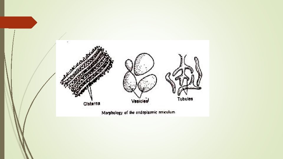
Morphology: Cisternae: The cisternae are long, flattened, sac-like, unbranched tubules having the diameter of 40 to 50 μm. They remain arranged in parallel order in the form of bundles or stakes. RER usually exists as cisternae which occur in those cells which have synthetic roles as the cells of pancreas, notochord and brain.

Ø Vesicles: The vesicles are oval, membrane-bound vacuolar structures having the diameter of 25 to 500 μm. They often remain isolated in the cytoplasm and occur in most cells but especially abundant in the SER. Ø Tubules: The tubules are branched structures forming the reticular system along with the cisternae and vesicles. They usually have the diameter from 50 to 190 μm and occur almost in all the cells.

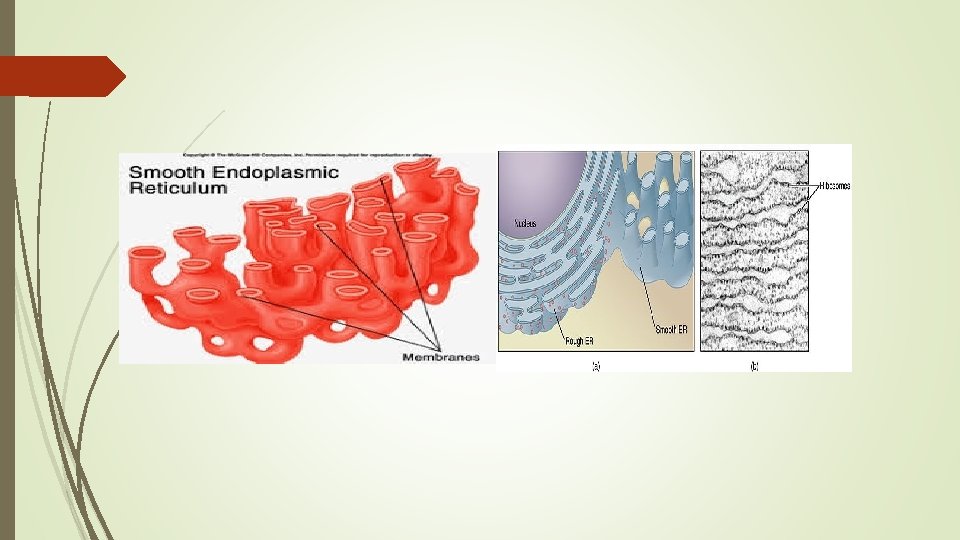
Types of Endoplasmic Reticulum Ø A granular or Smooth Endoplasmic Reticulum: This type of endoplasmic reticulum possesses smooth walls because the ribosomes are not attached with its membranes. The smooth type of endoplasmic reticulum occurs mostly in those cells, which are involved in the metabolism of lipids (including steroids) and glycogen. The smooth endoplasmic reticulum is generally found in adipose cells, interstitial cells, glycogen storing cells of the liver, conduction fibers of heart, spermatocytes and leucocytes. The muscle cells are also rich in smooth type of endoplasmic reticulum and here it is known as sarcoplasmic reticulum.

Ø Granular or Rough Endoplasmic Reticulum: The granular or rough type of endoplasmic reticulum possesses rough walls because the ribosomes remain attached with its membranes. Ribosomes play a vital role in the process of protein synthesis. The granular or rough type of endoplasmic reticulum is found abundantly in those cells which are active in protein synthesis such as pancreatic cells, plasma cells, goblet cells, and liver cells. The granular type of endoplasmic reticulum takes basophilic stain due to its RNA contents of ribosomes.

Role of Endoplasmic Reticulum: Ø Mechanical Support: ER contributes to the mechanical support of the cytoplasm by dividing existence compartmentsØ Intracellular circulation: The endoplasmic reticulum may act as a kind of circulatory system for intracellular circulation of various substances. Membrane flow may be an important mechanism for carrying particles, molecules and ions into and out of the cells by way of vascular system. The “pinocytosis, ” or “cellular drinking” also takes place by endoplasmic reticulum

Protein synthesis: Proteins may be synthesized to be utilized within the cell or these may have to be exported outside the cell to the site of their utility. It is the later kind of proteins in whose synthesis; endoplasmic reticulum plays an important role. For instance, rough endoplasmic reticulum, which has attached ribosomes carries synthesis of secretary proteins on these ribosomes and export them. Cell differentiation: Some specific instances of development have been studies in detail which more or less confirm the contention that the ER is important in the process of cell differentiation. Not only this much, ER also plays role in coordinating the differentiation.

Ø Transportation of message from genetic material: ER provides passage for the genetic material to pass from the nucleus to the various organelles in the cytoplasm, thereby controlling the synthesis of proteins, fats and carbohydrates. Ø ATP synthesis: ER membranes are the sites of ATP synthesis in the cell. The ATP is used as a source of energy for all the intracellular metabolism and transport of materials.

Vacuoles Definition: A vacuole is a membrane-bound organelle that is present in all plant and fungal cells and some protists, animal and bacterial cells. The most conspicuous compartment in most plant cells is a very large, fluid-filled vacuole. There may be several vacuoles in a single cell. Each vacuole is separated from the cytoplasm by a single unit membrane, called the tonoplast. Generally, they occupy more than 30 percent of the cell volume; but this may vary from 5 percent to 90 percent, depending on the cell type.
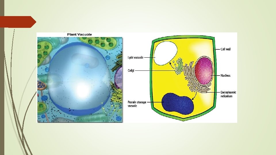

Structure of Vacuoles: They generally have no basic shape or size; its structure varies according to the requirements of the cell. In immature and actively dividing plant cells the vacuoles are quite small. These vacuoles arise initially in young dividing cells, probably by the progressive fusion of vesicles derived from the Golgi apparatus. A vacuole is surrounded by a membrane called the tonoplast or vacuolar membrane and filled with cell sap. The tonoplast is the cytoplasmic membrane surrounding a vacuole, separating the vacuolar contents from the cell’s cytoplasm

Functions: Ø Storage of molecules: Plant vacuoles can store many types of molecules. It can act as a storage organelle for both nutrients and waste products. Some of the products stored by vacuoles have a metabolic function. Ø Homeostatic Activity: The vacuole has an important homeostatic function in plant cells that are subjected to wide variations in their environment. Ø Maintenance of Turgor Pressure: Many plant cells maintain turgor pressure at remarkable constant levels in the face of large changes in the tonicity of the fluids in their immediate environment by changing the somatic pressure of the cytoplasm and vacuole in part by controlled breakdown and re-synthesis of polymers such as polyphosphate in the vacuole, and in part by altering.

Ø In Other Cells In fungal cells, they are involved in many processes including the homeostasis of cell p. H and the concentration of ions, osmoregulation, storing amino acids and polyphosphate and degradative processes. In animal cells, vacuoles perform mostly subordinate roles, assisting in larger processes of exocytosis and endocytosis.
- Slides: 31