Oral surgery with laser LASER OPERCULECTOMY Historical overview


 WAVELENGTH EMISSION AREAS Argon (Ar) 488 Continuous")

 Continuous")


- Slides: 12
Oral surgery with laser LASER - OPERCULECTOMY
Historical overview � The word laser is an acronym for light amplification by stimulated emission of radiation. � Lasers have been is use in the medical community since the 1970 s. � In 1989 the first laser specifically designed for use in dentistry was introduced. � Since then, the number of clinical applications is constantly increasing. � Laser light is different from ordinary light by being: - monochromatic (generates a laser beam of a single color) - coherent (identical in phisical size and shape)
Laser types in dentistry LASER WAVELENGTH (nm) WAVELENGTH EMISSION AREAS Argon (Ar) 488 Continuous wave Soft tissue Nd(YAG) 1 064 Free running pulse emission Soft tissue 800 -830/904/980 Continuous wave Free running pulse emission Soft tissue 2 780 – 2 940 Free running pulse emission Osseous tissue Enamel Dentin Cementum Continuous wave Soft tissue Diode Erbium (Er) CO 2 10 600
Safety � Lasers can cause eye and skin damage. � Protocol: - wavelength-specific protective eyewear - minimizing reflective surfaces presence of a designated safety officer laser mainenance and calibration specialized staff pacient education
Tissue interaction Tissue temperatrure increases Inter- and intracellular water boils away (100 degrees) Continuous exposure to laser energy tissue CARBONISATION Soft tissue vaporisation Hard tissue expansion and disruption
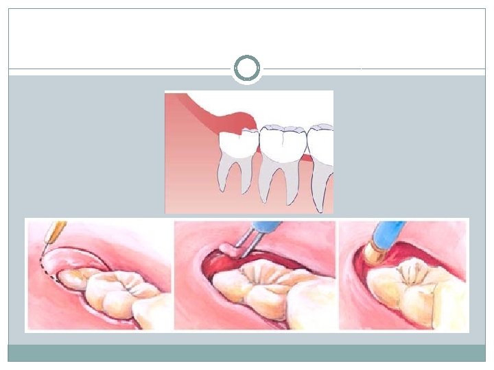
Clinical case �Laser in use : BIOLITEC �Settings : 5 – 7 watts continuous wave
-The laser has been used to make a semilunar incision over the partially erupted tooth. -Excision of the tissue will allow the tooh to completely erupt, with no further treatment needed. Preoperative view – pericoronal tissue around a partially erupted left third molar.
It has been decided that the molar should not be extracted. The surgical field is clean and dry. Immediate postoperative view showing a completely exposed third molar.
The use of lasers accelerates the treatment and decreases the amount of drugs used. Two weeks postoperative view showing a completely healed tissue.
Advantages vs. Disadvantages � High precision � Relatively costly � Reduce the amount of bacteria � Extensive training in the surgical site � Hemostasis and no sutures � Reduce swelling and postoperative pain � Less traumatising for the pacient � Promotes cellular healing, leading to faster recovery times � Lasers are only end cutting; side cutting and shaping cannot be performed with laser
References 1. Gáspár L. : The use of four different lasers in oral soft tissue surgery In: Loh Hong Sai : Lasers in Dentistry, Monduzzi Editore, Bologna, Italy, 1995. 2. Waidelich, W. , Waidelich, R. , Hofstetter, A. : Laser in der Medicin Springer Verlag, Berlin, Heidelberg, 1992. 3. Donald J. Coluzzi, DDS; Robert A. Convissar, DDS: Atlas of laser applications in dentistry, Quintessence Publishing Co, Inc, 2007. Thank You for Your attention