ORAL PIGMENTATION Dept of Oral Medicine And Radiology


 •")

 3")


- Slides: 52
ORAL PIGMENTATION Dept of Oral Medicine And Radiology Yenepoya Dental College Mangalore
Pigment Color / Coloring Endogenous Originate with in the body Exogenous pigments formed as reaction of chemicals of exogenous origin
Different types of pigments • HEMOGLOBIN : Red /Blue – Associated with vascular lesions • HEMOSIDERIN : Brown – Blood extravasation Trauma hemoglobin degraded to hemosiderin • MELANIN : Black – Hyperfunction of melanocyte or neoplastic transformation of melanocytes
Clinical classification of pigment acc to color COLOR FOCAL DIFFUSE MULTIFOCAL BLUE /PURPLE Varix, Hemangioma, Hemangioma Kaposi’s sarcoma, HHT, BROWN Melanotic macules, nevus, melanoma Ecchymosis, Physiologic melanoma, drug pigmentation, induced, hairy neurofibromatosis, tongue, petechiae OLP, addison disease, PJ Syndrome GRAY BLACK Amalgum tattoo, graphite hairy tongue tattoo, nevus, melanoma Metal ingestion
ENDOGENOUS PIGMENTATION PIGMENT COLOR DISEASE PROCESS HEMOGLOBIN Blue, red , purple Varix, hemangioma, kaposi’s sarcoma, angiosarcoma, HHT HEMOSIDERIN Brown Ecchymosis, petechiae, varix, hemorrhage mucocele, hemochromatosis Melanotic macule, nevus melanoma, basilar melanoma MELANIN Brown, black, gray
EXOGENOUS PIGMENTATION I. Accidental pigmentation II. Iatrogenic pigmentation III. Pigmentation due to drugs and metals IV. Localized pigmentation
BLUE / PURPLE VASCULAR LESIONS Hemangioma Varix and Varices Angeosarcoma Kaposi’s sarcoma Hereditary Hemorrhagic Telangiectasia
HEMANGIOMA BENIGN PROLIFERATION OF BLOOD VESSELS CONGENITAL ACQUIRED CAPILLARY CAVERNOUS PORT WINE STAINS BLANCHES on pressure
• SITE: • Tongue • Lips • BM / Palate • Multinodular • COLOR: - Depends on depth • Reddish blue close to epithelium • Blue connective tissue • Flat reddish blue macule PORTWINE STAIN TRAMLINE CALCIFICATION
HEMANGIOMA
• DIFFERENTIAL DIAGNOSIS : • Mucocele • Ranula • TREATMENT : • Sclerosing technique ( Sod Tetradecyl Sulfate intralesionaly) • Cryosurgery
VARIX AND VARICES DISTENDED VEIN RESULT FROM PARTIAL BLOCKAGE OF VEIN • VARICES / VARICOSITY Pathological dilatation of vein or venules • VARIX Focal dilatation of group of venules or vein
VARICES • SITE : - Ventral surface of tongue • AGE : - Progressively prominent with age • COLOR: - Blue / Red / Purple elevations • SYMPTOMS: - Painless • CAVIAR TONGUE Many sublingual veins
VARIX SITE : - Lower lip / areas prone to pinching trauma SIZE : - 2 – 4 mm AGE : - Elderly persons COLOR: - Blue / Red / Purple raised pigmentation, nodular lobulated or MARGINS & SHAPE Sharply delineated borders & smooth,
VARICES & VARIX • DIFFERENTIAL DIAGNOSIS • • Ranula Hemangioma HHT multifocal & hemorrhagic Nevi do not blanch on pressure • MANAGEMENT – Electrosurgery / Cryosurgery – Intralesional injection of Sod Tetradecyl Sulfate
ANGIOSARCOMA • Malignant vascular neoplasm • Rapidly proliferative / Nodular tumors • SUPERFICIAL Red, Blue or Purple nodular tumor
KAPOS’S SARCOMA • Common neoplasm in HIV pt’s • SITE Posterior hard palate / Multifocal • Nodular growth, Protruding below the occlusal plane • COLOR – Cutaneous Red macule becomes blue, purple & brown nodular – Oral Flat red macule • Rx Electrocautery
HEREDITARY HEMORRHAGIC TELANGIECTASIA • GENETICALLY TRANSMITTED • SITE Vermilion border & mucosal surface of lips/ tongue/ BM • SHAPE Multiple round or oval papules. 0. 5 cm • COLOR Red or brown • MANAGEMENT Electrocautery
Melanin associated pigmentationsmme Role of tyrosine Albinism Vitiligo
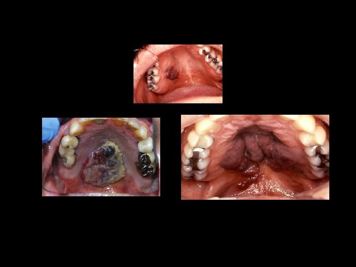
BROWN MELANOTIC LESIONS • Labial & oral melanotic macule • Nevus & nevi • Melanoma • Drug induced melanosis • Physiologic pigmentation • Cyanosis • HIV oral melanosis • Addison’s Disease • Peutz-Jegher’s Syndrome • Neurofibromatosis • Albright’s syndrome • Hyperfunction of pituitary gland • Pregnancy & female sex hormones
LABIAL & ORAL MELANOTIC MACULE EPHELIS/ FOCAL MELANOSIS/ SOLITARY LABIAL LENTIGO • Melanin by basal layer melanocytes without in melanocytes • Common oral pigmentation • AGE 41 – 45 yrs / Both sex • SITE Vermilion border of lower lip/ gingiva/ palate/ BM
• COLOR: – Brown / Brown black – Small flat macule – Single / multiple • DIFERENTIAL DIAGNOSIS: – Amalgam tattoo – Ecchymosis – Nevi
NEVUS • Benign proliferation of melanocytes • Most common Intramucosal (63 -70 %) • Common on skin; rare in oral mucosa • Smooth nodule • Hard palate common location
4 types
• • Women – 2/3 rd cases Age – 32 years (avg) 3 rd & 4 th decades Small - < 6 mm D/D: • • • Oral melanotic macule Retention phenomenon Cyst Hemangioma Early melanoma
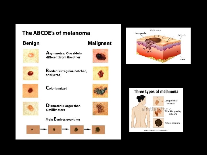
ORAL MELANOMA • Melanoma malignant tumor of nevus cells • Have a nodular component • Slightly more in males • 4 presentations – pigmented macule – pigmented nodule – large pigmented exophytic lesion – Amelanotic variety • Color mucosal pink – brown – blue – black • May ulcerate but no rolled out borders
• Rate of growth slow • Vertical phase rapid • Usually painless • Rapid infiltration fixed to underlying tissues • Spread lymphatic & hematogenous routes • Many develop from nevi
ORAL MELANOMA A – asymmetry C – color variation B – border irregularity D – diameter > 6 mm D/D: • Oral melanotic macule • Amalgam tattoo • Nevus • Focal hemosiderin deposit
DRUG INDUCED MELANOSIS • DRUGS: – Estrogen – Bleomycin, Quinoline/ Hydroxyquinoline/ antimalarial, zidovudine – Minocycline – Rx of acne – Drugs intake more than 4 months • COLOR: – Gray / Blue Black – Localized to hard palate or multifocal throught
PHYSIOLOGIC PIGMENTATION • Common characteristic of darker races • SITE: - Gingiva / Tongue • COLOR: • Brown macules • Multiple / Diffuse • Generalized / Localized
CYANOSIS Proportion of reduced Hb to Oxygenated Hb in blood • Localized / Generalized • Apparent when reduced Hb < 5 g/100 ml • COLOR: • Generalized bluish • Generalized green (congenital heart defect) • Cold weather Peripheral cyanosis of lips
HIV ORAL MELANOSIS • SITE: – Buccal mucosa – Gingiva – Palate – Tongue • COLOR: – Diffuse, multifocal, macular brown pigmentation
ADDISON’S DISEASE CAUSES CLINICAL FEATURES • Chr Adrenal insufficiency • Skin & MM pigmentation • Bilateral adrenocortical • Cheek /Gingiva /Tongue /Lips destruction TB / fungal • Nausea/ Vomiting/ Diarrhea & Anemia • Bilateral tumor metastasis • Bluesh black/ pale brown/ deep • Leukemic infiltration chocolate • Amyloidosis • Skin Bronze color
ADDISON’S DISEASE D/D: - • Hyper pitutarism • Argyria • hemochromatosis MANAGEMENT Adequate corticosteroid maintanance
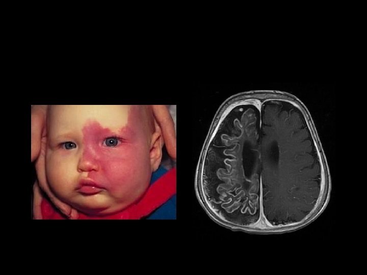
PEUTZ-JEGHER’S SYNDROME HEREDITORY INTESTINAL POLYPOSIS SYNDROME CLINICAL FEATURES: • SEX Equally • SITE Intestinal polyps ORAL MANIFESTATION: • SITE BM, gingiva, tongue, hard palate • SYMPTOMS Abdominal pain • AGE from birth • COLOR Bluish black macules • Multiple melanotic brownish • SIZE 1 -5 cm • Facial pigmentation macules around lips • SIZE 0. 5 cm
PEUTZ-JEGHER’S SYNDROME
CAFÉ AU LAIT SPOTS
NEUROFIBROMATOSIS VON RICKLINGHAUSEN’S DISEASE CLINICAL FEATURES: - • TRIAD Areas of pigmentation, sessile or pedunculated tumors of skin & mucous membrane & nerves • TUMORS Flexiform, soft, smooth, fluctuant • Café-au-lait spots
ORAL MANIFESTATION • Pigmentation -mucosa, lips • JAWS Intrensic bone formation RADIOGRAPHIC: • Central cyst like radiolucency • Irregular area of bone destruction • Pits & superficial cavities
ALBRIGHT’S SYNDROME • • Type of fibrous dysplasia Endocrine disturbances Skin pigmentation Common on lips • D/D: – Adisons disease – PJ Syndrome
HYPER FUNCTION OF PITUITARY GLAND • Increased secreation of ACTH & MSH PREGNANCY • Pregnancy increased ACTH levels • Pituitary function increased • CHLOASMA GRANDARUM Pigmentation of circumoral tissue & nipples
BROWN HEME ASSOCIATED LESIONS Ecchymoses and Petechiae Hemochromatosis Carotenemia Jaundice Hematoma
PETECHIAE & ECCHYMOSIS • Submucous or subcutaneous hemorrhages • Same mechanism • Bluish macules different sized • Petechiae pin point • Ecchymosis > 2 cm • Initally red bluish brown color
HEMOCHROMATOSIS BRONZE DIABETES • TETRAD: – Liver cirrhosis – Diabetes – Cardiac failure – Bronze skin • ORGANS INVOLVED: – Liver – Skin – Pancrease – Adrenal gland
HEMOCHROMATOSIS CAUSES: • Increased dietary iron intake • Excessive blood transfusion q SEX : - 80% in men q Deposition of excess iron (Ferritin & Hemosiderin) in body tissue q COLOR: - Blue Gray Skin (Genital, face, arm
CAROTENEMIA Chronic excess level of carotene pigments food containing carotene such as carrots, sweet potatoes & egg Palms , Soles & areas of Soft palate Orange / Yellow Skin & Oral mucosa
JAUNDICE COLOR: • Yellow / Green • Skin & Mucous membrane
HEMATOMA • Pool of effused blood confined within tissues • Elevated bluish mucosal swelling • Fluctuant, rubbery, well defined • Pressure never blanches stinging sensation • After 24 hours black blue green disappears within 23 weeks
HEMATOMA • Local cause trauma • Systemic cause hypertension blood dyscrasias D/D: • • Mucocele / Ranula varicosity Hemangioma Superficial cyst