Oral Mucous Membrane O Mucous Membrane Moist lining

Oral Mucous Membrane

O Mucous Membrane: Moist lining of the gastrointestinal tract, nasal passages and other body cavities that communicate with the exterior. In the oral cavity the lining is called as oral mucous membrane or oral mucosa.

Functions of the Oral Mucosa O 1. Protection: Barrier for mechanical trauma and O O O microbiological insults 2. Sensation: Temperature (heat and cold), touch, pain, taste buds, thirst; Reflexes such as swallowing, etching, gagging and salivating 3. Secretion: Salivary secretion 4. Thermal regulation: Important in dogs not in humans. 5. Permeability and absorption: generally is not permeable except for the floor of the mouth which is the thinnest area i. e. used to absorb certain drugs.

Organization of the Oral Mucosa 1. Masticatory Mucosa: 25% of total mucosa. Gingiva (free, attached and interdental) and hard palate. Primary mucosa to be in contact with food during mastication. MASTICATORY MUCOSA IS USUALLY KERATINIZED. 2. Lining Mucosa: 60% of total mucosa. Covers the floor of mouth, ventral (underside) tongue, alveolar mucosa, cheeks, lips and soft palate. Does not function in mastication and therefore has minimal attrition. On-keratinized; soft and pliable. 3. Specialized Mucosa: 15% of total mucosa. Covers dorsal tongue and composed of cornified epithelial papillae.
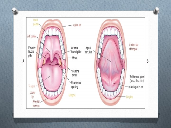


Clinical features: O 1 - color: depend on the concentration and state of dilatation of bl. v. in the underlying c. t. and thickness of the epithelium, degree of epithelization, and amount of melanin pigment in the epith. O 2 -mosit: due to presence of minor salivary glands. O 3 -smooth in general except for dorsum of the tounge, rugae of the palate. Healthy gingiva show stippling. O 4 -firmness: its firm in the palate and immobile while in the lip and cheek is soft and pliable.

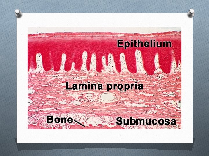
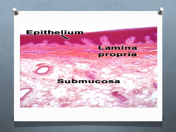
Histological features of oral mucosa: It composed from 2 main parts; oral epithelium and lamina propria. The interface between the epith. And C. T. is usually irregular and upward projections of C. T. called C. T. papillae. (Rete ridges or pegs). The epithelium composed of 2 main types:
 the cells are cuboidal or columnar cells containing bundles of tonofibrills")
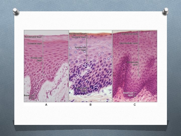
Basal layer: (germinativum) the cells are cuboidal or columnar cells containing bundles of tonofibrills and other cell organelles; site of cell divisions which provide cells for turnover. Prickle layer: Larger ovoid cells containing conspicuous tono fibril bundles; membrane-coating granules appear in upper part of this layer. Granular: flattened cells containing conspicuous keratohylaine granules associated with tonofibrills; membrane-coating granules fuse with cell membrane. Nuclus showed sign of degeneration. Keratinized: extremely flattened and dehydrated cells in which all organelles have been lost; cells filled only with packed febrile material; when pyknotic nuclei are retained, parakeratinization occur.

B-Non-keratinized epithelium: O Basal layer: cuboidal or columnar cells containing O O separate tonofilaments and other cell organelles; site of most cell divisions. Prickle cell: larger ovoid cells containing many dispersed tonofilaments membrane-coating granules appear in upper part of layer; filaments become numerous. Intermediate: Slightly flattened cells containing many dispersed tonofilaments and glycogen. Superficial: Slightly flattened cells with dispressed filaments and glycogen; fewer organelles are present' but nuclei persist.


Non-keratinocytes in oral epithelium O Melanocyte: it is located in basal layer of epith. It's dendritic no desmosomes or tonofilaments; premelanosomes and melanosomes present. Function: synthesis of melanin pigment granules (melanosomes) and transfer to surrounding keratinocytes. O Langerhan's cell: predominantly suprabasal. It's dendritic; no desmosomes or tonofilaments have Langerhan's granule. Function is antigen trapping and processing.

O Merkel cell: located in basal layer. Its non -dendritic has desmosomes or tonofilaments and special vesicles. Function is tactile sensation. O Lymphocyte: variable location. Are large circular nucleus; scant cytoplasm with few organelles; no desmosomes or tonofilaments. Function associated with inflammatory response in oral mucosa.

The interface is a structureless layer about 12 micron thick called basement membrane when seen in light microscope and basal lamina in electron microscope. it composed of 2 lamina: a-lamina lucida: this is clear cell free zone, it's towards epith. b-lamina densa: this is dark zone toward C. T. It's filamentous and granular, contain collagen fibers.

Connective tissue of oral mucosa: O 1 -Lamina propria: is the C. T. that lies immediately beneath the epith. Its function provides metabolic needs for avascular epith. Via bl. v. Present in it. It is divided into: O Papillary layer: forms finger like projections of connective tissue that extend deep into the epith. It's prominent in masticatory mucosa. O Reticular Layer: netlike and refers to the arrangement of collagen fibers. It's prominent in lining mucosa. O Cells of L. P. are:

Fibroblasts: stellate or elongated with abundant R. E. R. its function is secretion of fibers and ground substance. Histiocytes: spindle-shaped or stellate; often darkstaining nucleus, has lysosome vesicles. It is a precursor of macrophage. Mast cells: round or oval with basophilic granules staining. It secret certain inflammatory mediators and vasoactive agents (histamine, heparin and serotonin). Endothelial cells: associated with a basal lamina contains numerous pinocytotic vesicles. It's function lining vascular channels throughout L. P. Inflammatory cells: present in area of inflammation either acute or chronic. Includes macrophages, neutrophil, lymphocyte, and plasma cells.

2 - Submucosa: O Is a C. T. that lies under L. P. and provide attachment with underling bone or muscles. It found in the cheek, lip, and palate. It consists of large bl. v. Lymphatic's and nerves. Function is nutrition and defense. O

- Slides: 21