Oral infections 2 1 Fungal infections Dr Brdy

Oral infections 2. 1. Fungal infections Dr Bródy Andrea Semmelweis Egyetem Orális Diagnosztikai Tanszék

Significant yeasts from a medical point of view Filamentous fungi • Dermatophytons – obligate pathogens • Moulds - opportunists Yeasts - opportunists Dimorf fungi – obligate pathogens

Candida infection Stool transplantation! • Local: stomatitis, fungal vulvovaginitis, etc. • Systemic candidiasis – mostly in immuncompromized person – life-threatening disease Systematic candidiasis diagnosed by naturopath: Does not exist in this form.

Oral cavity candidiasis • Opportunist pathogens, Member of the normal microflora Their colonisation in the oral cavity does not mean candidiasis • The appearance of pseudohyphas implies candidiasis

Fungal infections of the oral mucosa • The prevalence of the fungal infection of the skin and mucosa is growing all over the world. • Its significance lies in the fact that it may significantly worsen the life quality and expectancy of the infected individuals. • Could be the source of an life-threatening infection • The most common human fungal infection. • It is an underdiagnosed disease – need improve the knowledge of the dentists
 As an opportunist pathogen")
Pathogenic candida species • Most wide-spread: Candida albicans (70 -90%) As an opportunist pathogen it may be detected in the mouth of many healthy individuals. • Non-albicans strains als common: Candida glabrata, krusei, tropicalis, parapsilosis, guillermondii • Candida dubliniensis: it belongs to the most recently recognised species that was primarily isolated from HIV infected individuals’ oral cavity. The number of cases when it’s found in oral disorder, e. g. parodontitis is growing.

Predisposing factors • Different immune deficiency conditions (the pseudomembranosus form developes in nearly 90% of HIV infected patients) • Diabetes mellitus • Smoking • Sjögren syndrome • Long term antibiotic treatment • Bad oral hygiene, trauma • Childhood and infancy • Hormonal changes • Radiotherapy, chemotherapy, steroids

Local predisposing factors • Old and wrong dentures, K+B bridges, braceas • At night, during sleeping the number of yeasts increases in the mouth • Altered microflora of the oral cavity Chlorhexidine, contraceptives, oral sex, decrease of the Ph of the saliva • Bad eating habits • Smoking • Bad oral hygiene

Classification of the Candida infections of oral mucosa • Different classification based on origin, clinical picture or localizaion • Primer oral candidiasis: affects the tissue of the mouth and the surrounding area • Secunder oral candidiasis: oral manifestation of generalized candida infection

Primer forms Secunder forms • Acute forms: - pseudomembranosus - erythematosus • Chronic forms - hyperplastic papillar plaqued (Candida leukoplakia) - erythematosus (- chronical multifocal candidiasis) • Lesions related to Candida (multifactorial diseases): - denture stomatitis - angular cheilitis - median rhomboid glossitis - linear gingival erythema • Oral manisfestations of systemic mucocutan candidiasis

Relationship between the Candida and the host body Risk factors Microflora Pathological colonisations Local infection Systemic infection

Acute pseudomembranosus oral candidiasis • Always indicates a severe background disease in healthy individuals (except babies) • In AIDS patients its appearance precisely indicates the stage of the disease

Pseudomembranosus candidiasis Unknown origin

Patient with asthma

Long continued flue

Recurrent vaginal infections

Erythematosus candidiasis • Most common form • Could exist secundarly from the pseudomembranosus form or primarly • Could be the first sign of HIV infection • Most times the patient has removable denture – glossitis and/or angular cheilitis are often related to it

Acute erythematosus • It is often found in HIV positive patients but also appears in immune deficiency conditions and as a consequence of sexual infections. • Usually goes with symptoms, causes a burning, stinging sensation on the mucosa

Acute erythematosus form RA

Chronical erythematosus form Pseudomembranosus plaque Diabetes

Chronical erythematosus form Asthma spray

Hyperplastic candididasis – papillary form

Hyperplastic candididasis - candida leukoplakia • More common in smokers - may recover as a result of quitting • Precancerosis – danger of malignization is high – around 15% • If it does not react to antifungal therapy then biopsy is advised • It is not clear whether in this form the yeast has a pathogenic role
 • Most times it is found on the mucosa of")
Hyperplastic candididasis (candida leukoplakia) • Most times it is found on the mucosa of the bucca and less often on the edge of the tongue • Its surface may be homogenic or papillomatosus • Not possible to wipe off Differential diagnosis: pseudomembranosus form

Lesions related to Candida • • Denture stomatitis Angular cheilitis Median rhomboid glossitis Linearis gingival erythema

Denture stomatitis Could be experienced the chronic inflammation of the lip, mucosa and the angulus oris, possibly the patient complains about chronic mouth burn and a stinging sensation Inflammations appear in 50 -70% of patients with removable denture, but often do not cause complaints

• The erythema follows the outline of the denture • Appears in women more often • Often the papillomatosus form may be detected on the palatum • It is rarely treated properly

Lingua fissurata

Candidiasis developed by a K+B bridge

Newton classification of oral stomatitis • Newton I: local erythematosus – red spots on the mucosa • Newton II: diffuse erythematosus - on denture covered mucosa • Newton III: hyperplastic granulomatosus

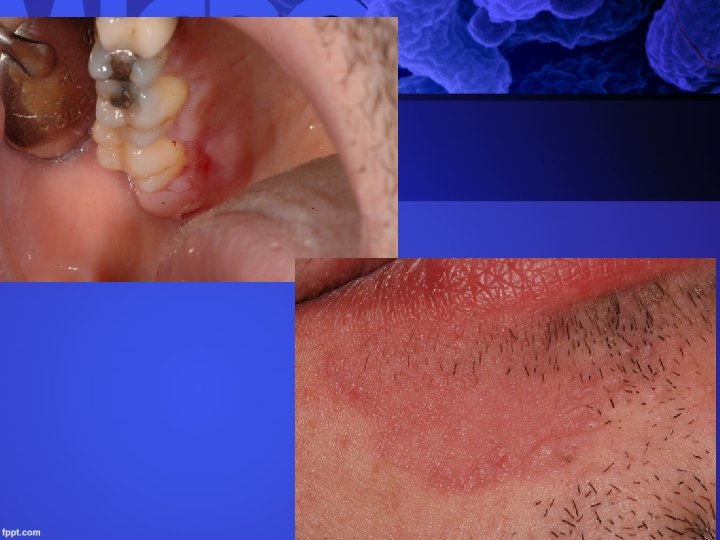
Cheilitis angularis • The erythematosus inflammation of the angulus, with cracks around the contacting areas • Often overinfected with Staphylococcus aureus or other bacteria • Bad denture – low bite height • Maceration by infected saliva

Median rhomboid glossitis • Chronic inflammation that goes with papilla atrophy • Not always clarified origin but often recovers as a result of anti-fungal therapy

Differential diagnosis - denture stomatitis • Glossitis: smoothy and shiney surface also in lack of B 12 vitamins, follic acid and iron • Allergy – acrylic or other • Hyperplastic form: leukoplakia, lichen and lichenoid reactions, pemhigus, carcinoma

Linear gingival erythema • It was first recorded in HIV infected patients • Not caused by plaque, does not imply pockets development • 2 mm-s line around the marginal gingiva • Mixed infection –bacteria and fungals • Could developing in cases with good oral hygine too • In a number of cases Candida dubliniensis was isolated from the deformation

Chronical mucocutan candida infection • Developes in immune deficient patients • Persisting mucocutan infection, that does not react to locally applied drugs • Systematic azol treatment is necessary

Treatment of denture stomatitis • Antifungal therapy + replacement or professional cleaning of the denture • Regular disinfection of the denture is necessary afterwards – e. g. with chlorhexidine (must be fully removed otherwise it may discolour the denture) • Nystatin and chlorhexidine neutralize each otherefore the denture must be washed off and air dried

Polyenes – non toxic used per os • Nystatin The most commonly used local drug Should be a first choice medicine generally when the infection is not too serious or old or the patient hasn’t immundeficiency • Amphotericin B cream, suspension Also effective with the non-albicans species Not absorbsed from the digestive tracts

Azoles Imidazoles: clotrimazole, etoconazole, miconazole, isoconazole only dermatological and gynecological packings are available in Hungary Ketoconazole: Also has wide spectrum but is very hepatotoxic – could be fatal Formulation for local use: cream, tablet, shampoo

Triazoles Flukonazol • The British National Formulary has listed it as suitable for dental use • Non-albicans types are less sensitive or resistant to it • A first choice systemic drug if there is no suspicion of a non-albicans type causing the infection • Suspension is available • Few siginificant drug interaction

Triazoles Itrakonazol • Has a wider spectrum than fluconazol, therefore is well suitable for fluconazol resistant infections of immune deficient patients • Absorvation is not reliable, therefore cannot be applied in systemic infections • May be liver toxic so liver functions must be monitored throughout the treatment • Drug interaction must be paid attention to: cyclosporin, terfenadin, astemizol, digoxin. The level of cyclosporin and digoxin must be monitored.

Other antifungal compounds • terbinafin • voriconazol • echinocandins • posaconazol • 5 -fluorocitozin Not used in oral Candida infections

Certification of fungal infection • Cultured of a fungus doesn’t mean infection • Microscopic investigation is useful pseudohyphae • Quick tests • Cultures

Bacteria caused oral diseases and their symptoms Dr Bródy Andrea

Gingivostomatitis ulcerosa • The ulcerous, painful, acute inflammation of the gingiva • Its symptoms are bad breath and a yellowish coloured ulcer ring around the edge of the gum • In severe cases systematic antibiotic treatment may be necessary (metronidazol, amoxicillin) • If untreated, it may leave an irreversible parodontal and bone damage behind • Mixed infection

Impetigo contagiosa Streptococcuses, Staphylococcus aureus • Causes superficial pyoderma • It appears most often on the face, around the mouth • Spreading yellowish scab developed from the small pustulas • It may appear in the oral cavity in the form of painful and deep ulcers
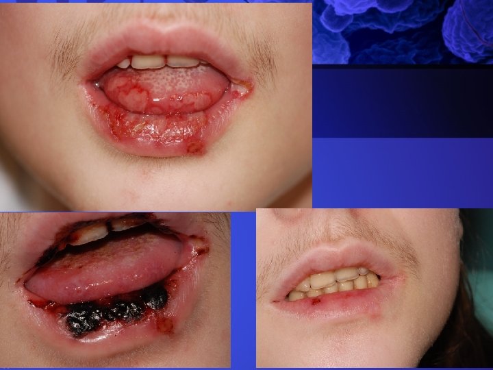

Plaut-Vincent angina Fusobacterium Plauti-Vincenti and Borrelia Vincenti • Affects the oral cavity only • Caused by spirochetas and fusobakteria • May also develop from gingivostomatitis ulcerosa • Necrotic ulcers, bad breath, enlarged lymph nodes, pain • Good outcome

Actinomycosis • Actinomyces israelli – bacteria, not a fungus • Most commonly appears in the angulus of the mandibula • The pathogen gets in through lesions

Diphteria • Rare in Europe due to vaccination • General symptoms – fever, vomiting, faintness, toxic and neurological systematic symptoms • Regional lymp nodes swelling • Ulcers covered with membranes on the palatum, pharinx, larynx, tongue, bucca, in the nose – danger of suffocation • Differential diagnosis: Plaut-Vincent angina, herpangina, mononucleosis, leukaemia
 • Acute disease spread by droplet infection •")
Scarlatina Streptococcus pyogenes (beta haemolytikus ) • Acute disease spread by droplet infection • General symptoms, sore throat • The oral mucosa is inflammed, swollen, red (stomatitis scarlatina) • Tongue is red - strawberry tongue • Differential diagnosis: diphteria, mononucleosis infectiosa, candidiasis

Syphilis – STD

Thank you for your attention
- Slides: 53