
Oral cavity neoplasms Dr Farahnaz Bidari Robbins Pyogenic
Oral cavity neoplasms Dr. Farahnaz Bidari Robbins
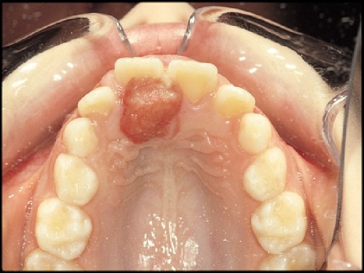
Pyogenic granuloma § § § Usually in gingiva Pregnancy tumor ulcerated
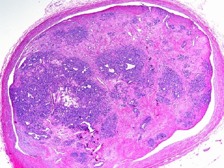
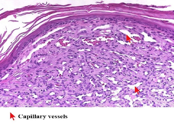
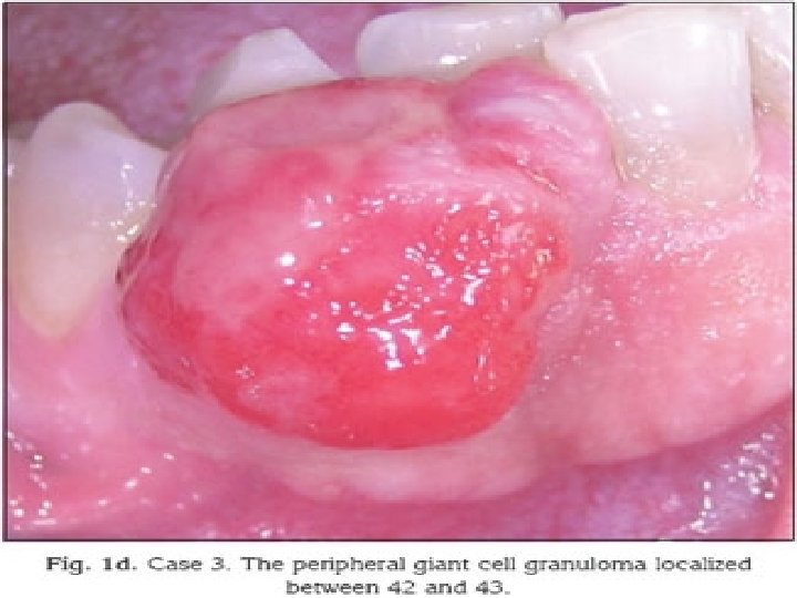
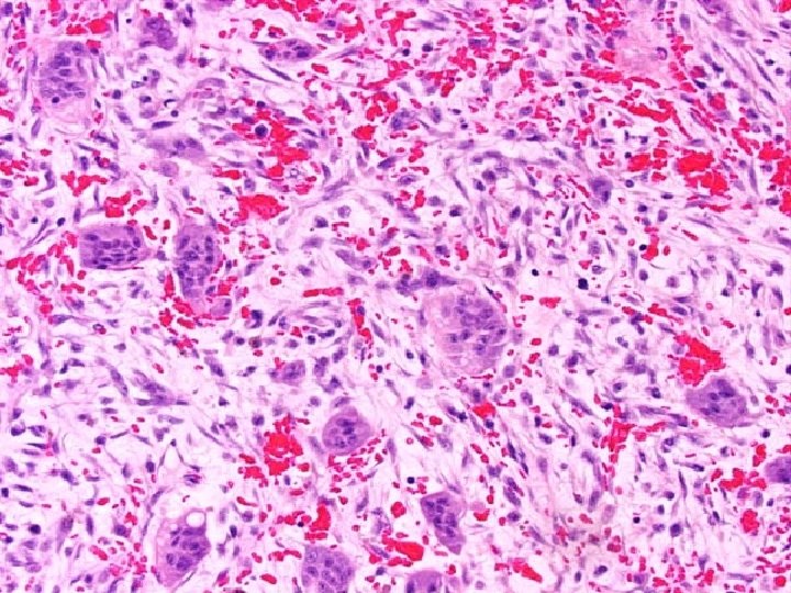
Peripheral giant cell granuloma § § Epulis Clinically similar to pyogenic granuloma Giant cells Fibrovascular stroma
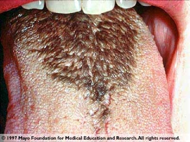
Hairy leukoplakia § § § 80% HIV infected White patches of fluffy thikening Fungal and bacterial overgrowth
Tumors and precancerous lesions § 1) Leukoplakia and erythroplakia § 2)squamous cell Ca.
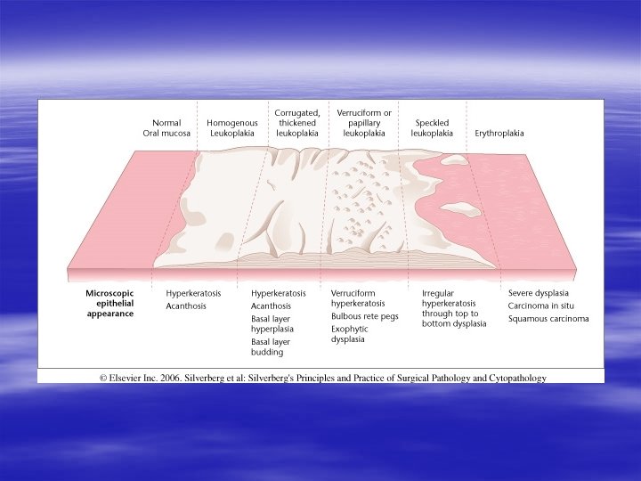
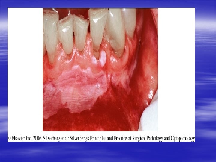
Leukoplakia § White plaque ( a clinical not pathologic term) § Rule out of known causes like lichen planus and candidiasis § Favoured location : buccal , floor of mouth , tongue § Dysplasia (1 -16%)
Erythroplakia § Red , velvety, possibly eroded area § Less common and much higher risk of malignant transformation than leukoplakia § Dysplasia (50%)
Risk factors § § Use of tobacco , most common antecedent Alcohol Persistent irritants ( hot pizza) HPV specially type 16
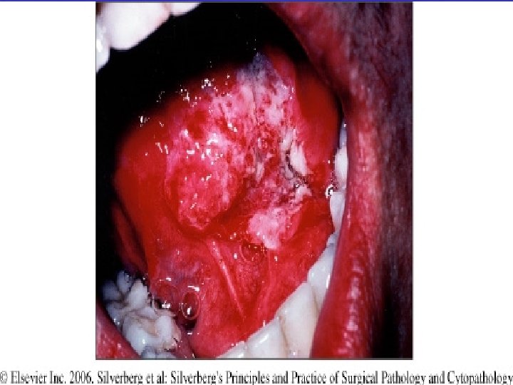
Squamous cell ca. § § § 95% of cancers Many of them detected late Age : 50 -70 y
pathogenesis § § § Tobacco = 2 -4 fold Tobacco + alcohol = 15 fold Marijuana , betel nuts ( india) , HPV( waldeyer ring) , sunlight area and pipe ( lower lip) § Chromosomal abnormality : INT 2, BCL 1 oncogens
Morphology § § Irregular or verrucous mucosal thikening Favoured location : floor of mouth , tongue Grade : 1 -3 Prognosis : beter with lip lesion and poorer with tumors in floor of mouth
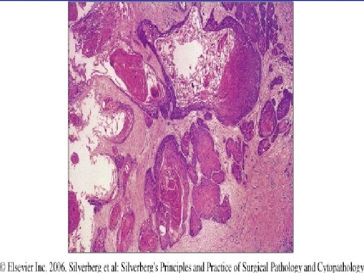
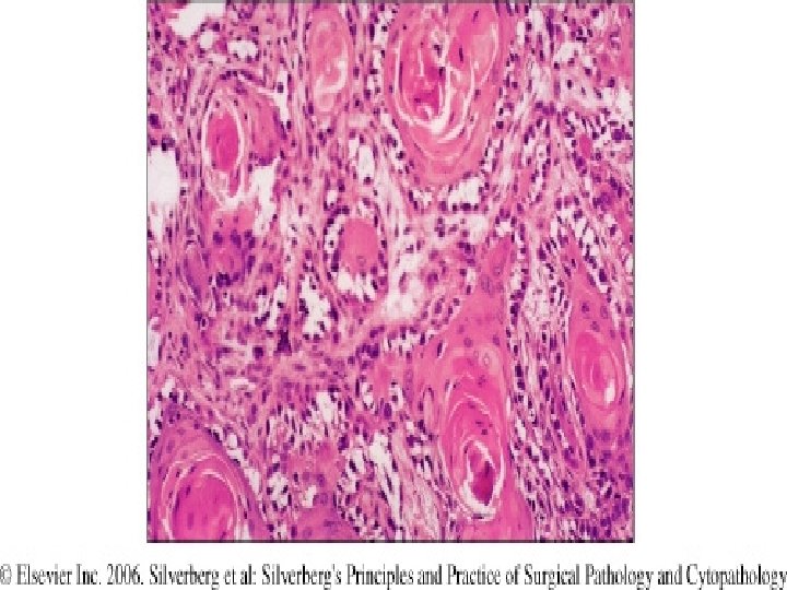
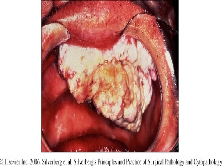
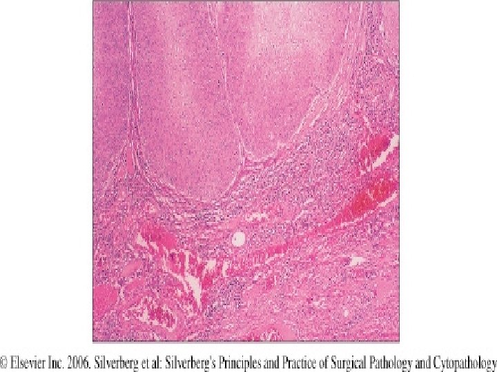
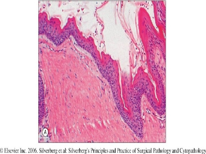
Odontogenic cysts and tumors § Odontogenic cysts : lined with keratinizing squamous epithelium § Multiple form is a component of nevoid basal cell carcinoma syndrome(Gorlin syn)
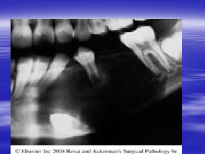
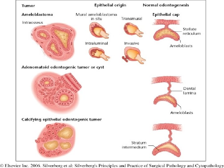
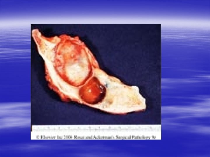
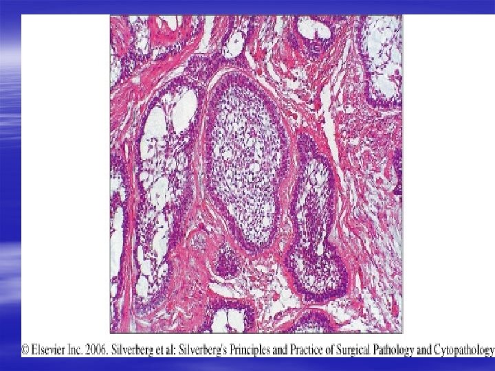
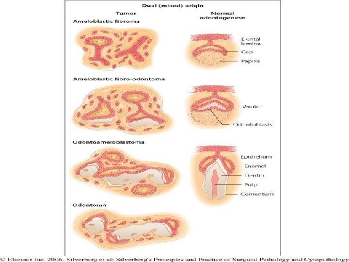
Odontogenic tumors § Neoplasms of the jaws which differentiated toward tooth structures § Ameloblastoma: most common , from inner dental epithelium of developing tooth follicle ( ameloblastic layer) , enamel and dentine are not seen, cystic, slow growing and locally invasive
Odontoma § Odontogenic tumors featuring production of calcified parts of teeth
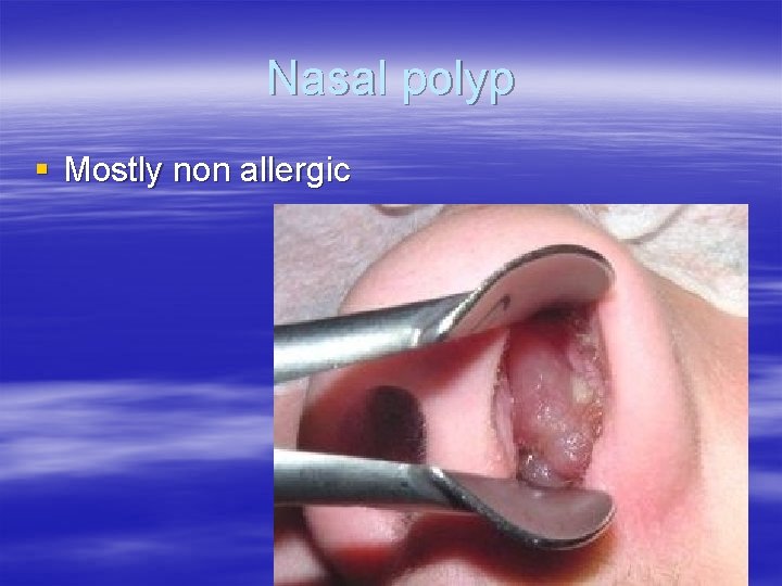
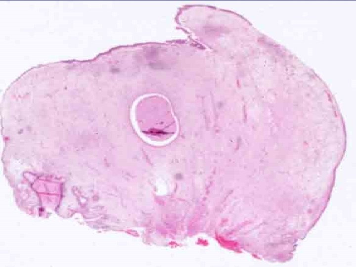
Nasal polyp § Mostly non allergic
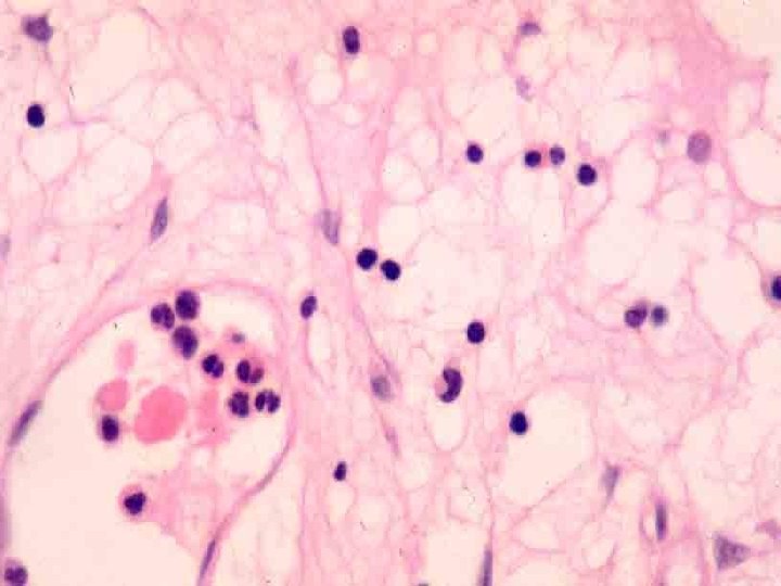
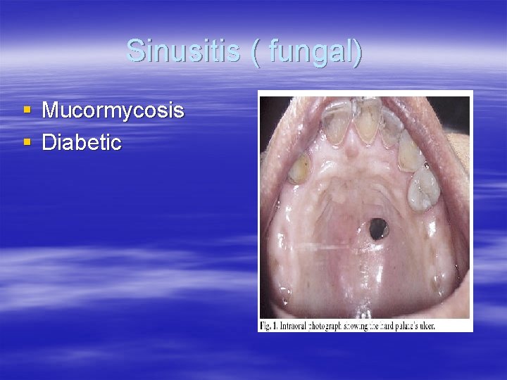
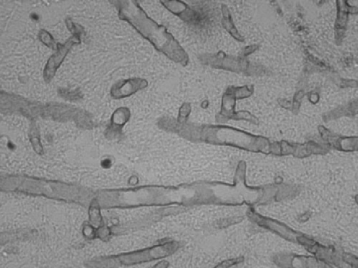
Sinusitis ( fungal) § Mucormycosis § Diabetic
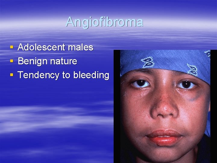
Angiofibroma § § § Adolescent males Benign nature Tendency to bleeding
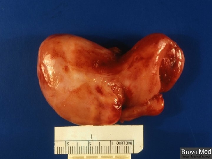
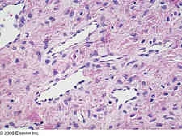
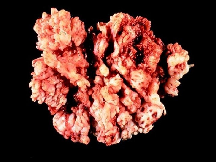
Nasal papilloma § § § Benign neoplasms HPV 6, 11 Inverted papilloma : invasive locally
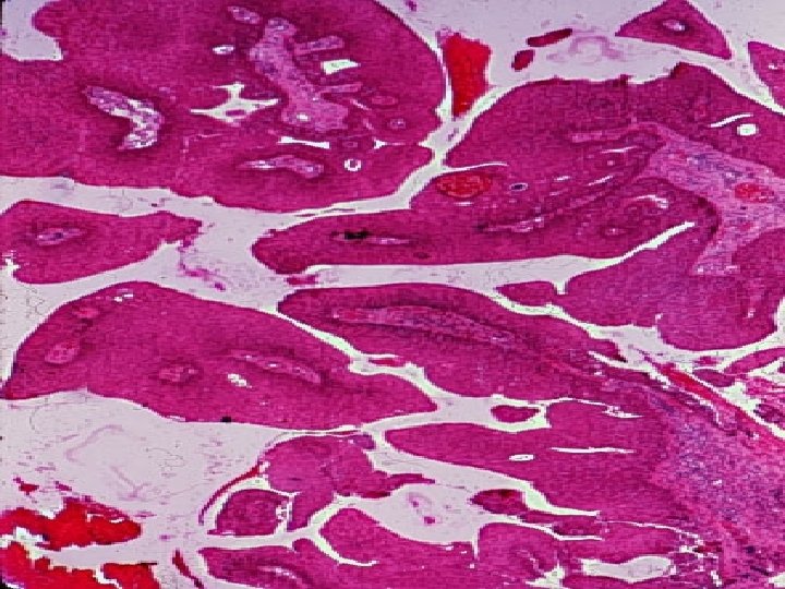
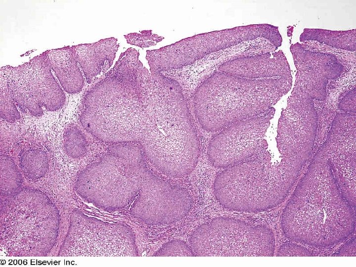
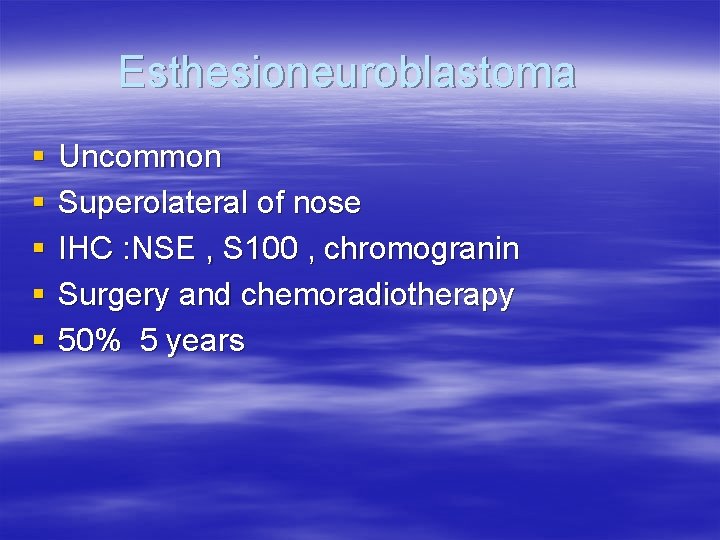
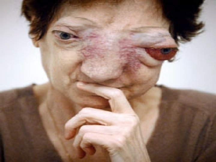
Esthesioneuroblastoma § § § Uncommon Superolateral of nose IHC : NSE , S 100 , chromogranin Surgery and chemoradiotherapy 50% 5 years
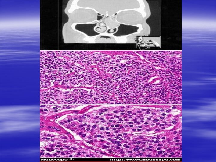
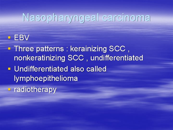
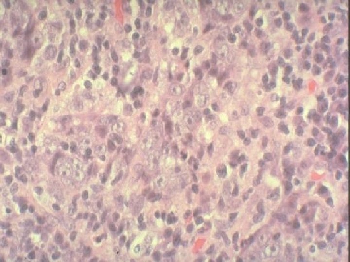
Nasopharyngeal carcinoma § EBV § Three patterns : kerainizing SCC , nonkeratinizing SCC , undifferentiated § Undifferentiated also called lymphoepithelioma § radiotherapy
Vocal cord polyp § Most often in heavy smokers § Singers nodule
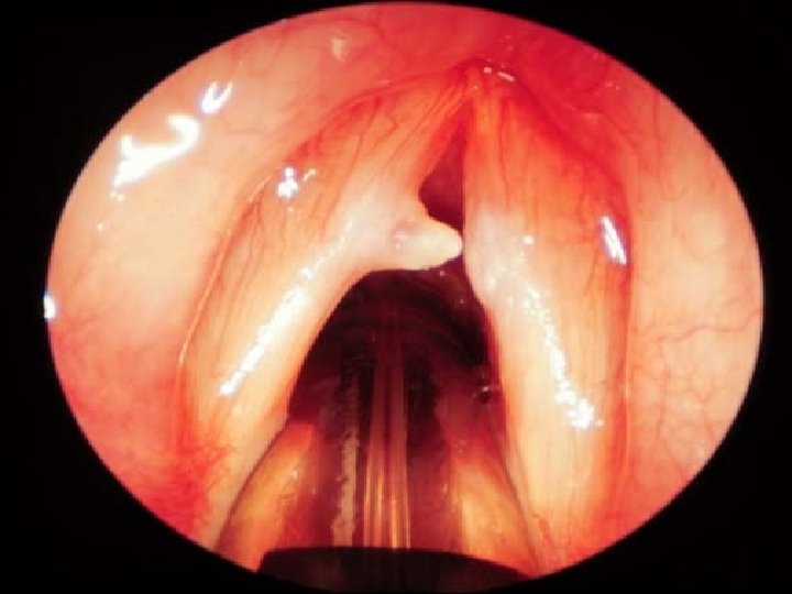
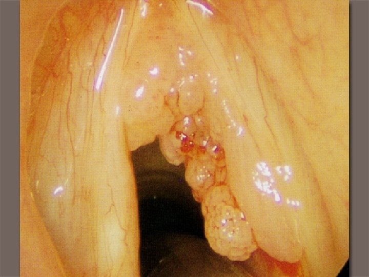
Laryngeal papilloma § § Usually on TVC Rarely more than 1 cm Papillomatosis in children (HPV 6, 11) Malignant transformation is rare
Salivary gland neoplasms § relatively uncommon neoplasm of human § 65 -80% arise within parotid § Chance of malignancy increase with decrease in the size of gland § Slight female predominance( except for warthin tumor) § Adult (5% of cases less than 16 y)

§ Benigns: slow growing, well defined § Malignants: rapid growth , ill defined , fixed
Benign neoplasms § Pleomorphic adenoma (50%) § Warthin tumor ( 5 -10%)
Malignant § § § Mucoepidermoid ca. (15%) Adenoca. , NOS (10%) Adenoid cystic ca. (5%) Acinic cell tumor(5%) Malignant mixed tumor (3 -5%)
Pleomorphic adenoma § § § Also called mixed tumor 60% in parotid Radiation exposure increase the risk Treatment: adequate parotidectomy (4%) enucleation (25%) Malignant transformation : increase with duration, common form is adenoca. § Malignant mixed tumor most aggressive salivary gland tumor
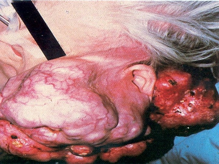
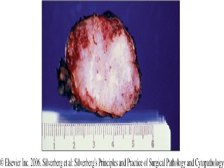
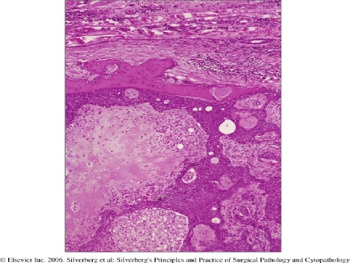
Morphology § Well demarcated <6 cm § Encapsulated( not fully) with tongue like protrusions § Cut surface is gray white and translucent § Epithelial elements (ducts nests of squamous or nests of myoepithelial cells) embedded in myxoid , hyalin or chondroid stroma (both of them from epithelial and myoepithelial cells of ducts)
Warthin tumor § § § Almost always in parotid gland More common in males 40 -60 y 10% multifocal , 10% bilateral Smokers ( 8 times)
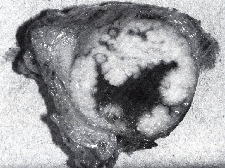
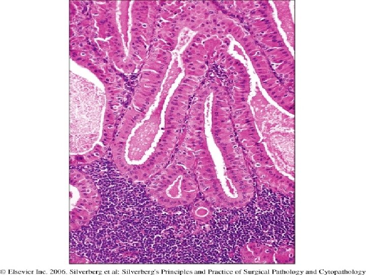
morphology § § § Round , encapsulated , 2 -5 cm Gray , cystic Cysts lined by double layer of epithelium resting on a dense lymphoid stroma § Lining cells are columnar cells with abundant eosinophilic cytoplasms containing many mitochondria( oncocytic cells) upon cuboidal cells
Mucoepidermoid ca. § 60 -70% in parotid § Large fraction of salivary gland neoplasms in other glands § Most common form of malignant tumors § Most common radiation induced neoplasm
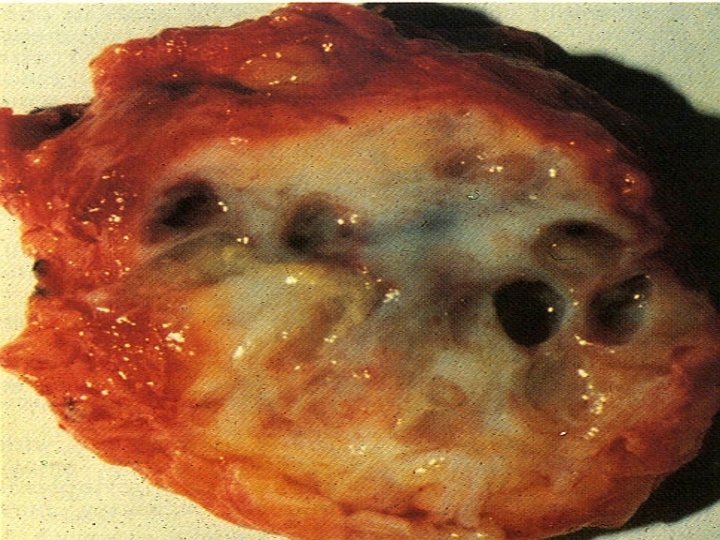
Morphology § § § Lack well defined capsule Infiltrative margin Pale gray white , small cysts Cords , sheets , cystic area Squamous , mucus cells , intermediate (hybrid cells)
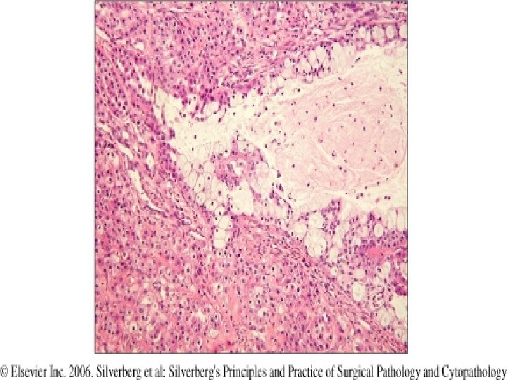
Grade § § Low , intermediate , high Low grade : largly of mucus secreting High grade: largly squamous cells Low , intermediate , high : recur in 15% , 25% , 30% § Low : 90% 5 -year § High : 50% 5 -year
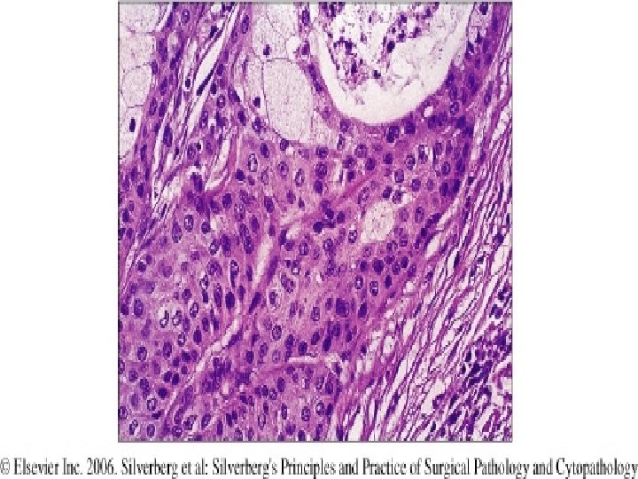
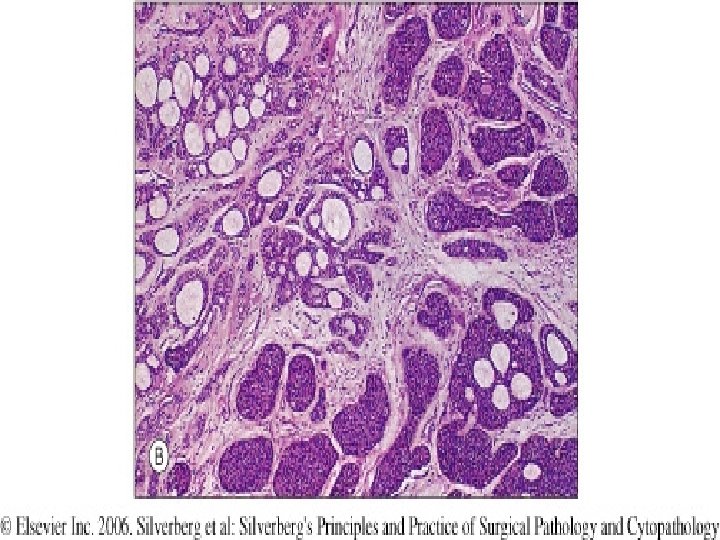
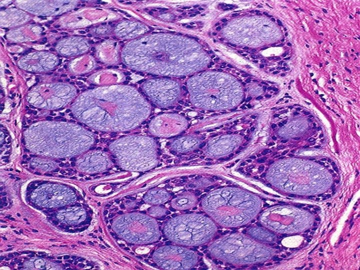
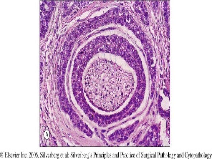
Adenoid cystic ca. § Most common neoplasm in minor salivary gland (mouth , nose , sinuses, upper airway) § Slow growing , unpredictable tumor § Tendency to invade perineural spaces § Most painfull salivary gland neoplasm § 50%disseminate widely to distant sites, sometimes decades later § 60 - 70% 5 -year survival
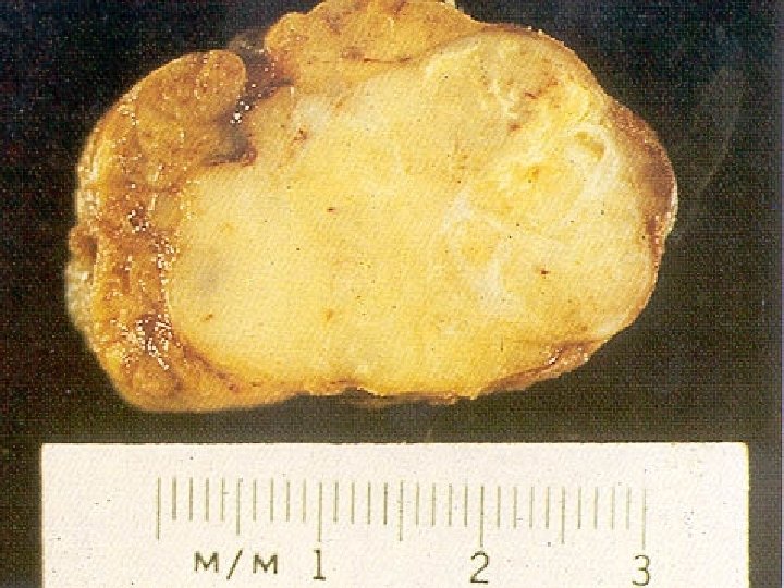
Morphology § Infiltrative , gray pink § Small cells with dark compact nuclei and scant cytoplasms § Forming tubular, solid or cribriform patterns § Histologically is like cylindroma of skin § Gland like structures filled with excess basement membrane
Acinic cell tumor ( ca) § Uncommon § Most in parotid. Rarely in minor salivary glands § Sometimes bilateral or multicentric § Recurrence uncommon § 10 -15% metastasize to LN § Survival: 90% 5 -year
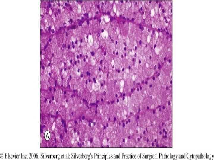
Morphology § § § Generally small , discrete Sheets , glandular and papillary patterns Neoplastic cells have clear cytoplasms like normal acinar cells
- Slides: 84