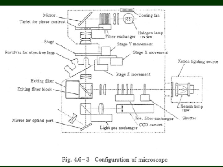
Optical imaging Spectroscopic applications Microscopes conventional optical microscope


 and intensity distribution along the line")


 STED (Stimulated Emission Depletion Microscopy)")


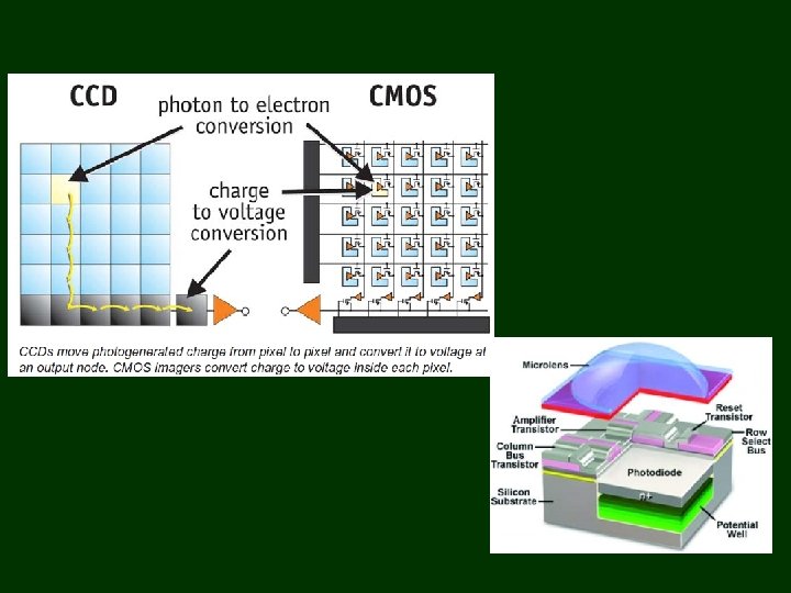
 Typical Quantum Efficiency Curves 100 90 80 70 60 50 40")


- Slides: 33
Optical imaging Spectroscopic applications
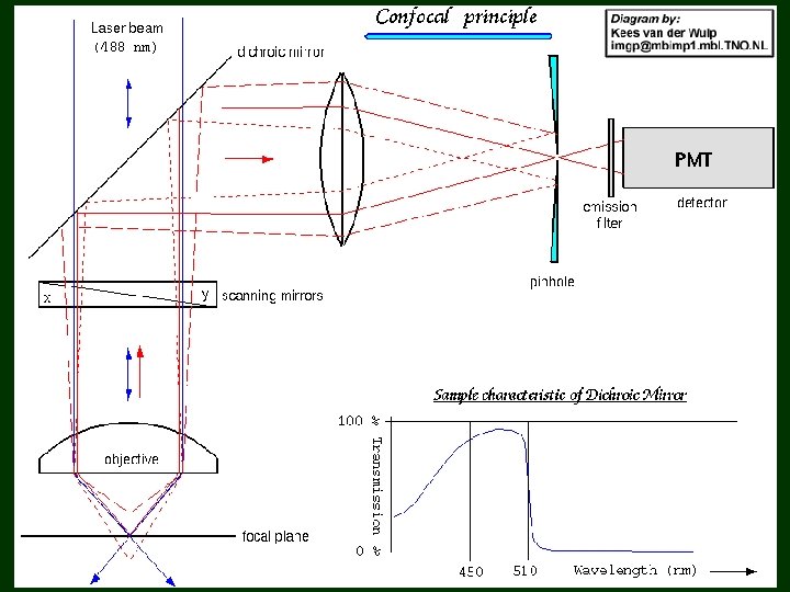
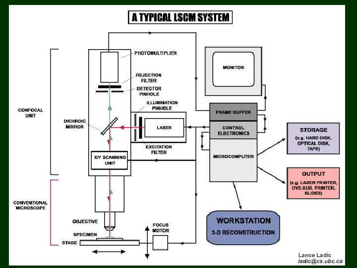
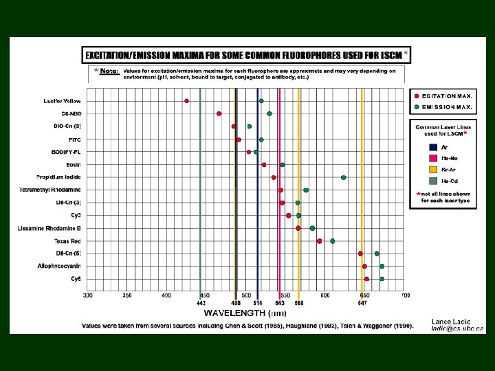
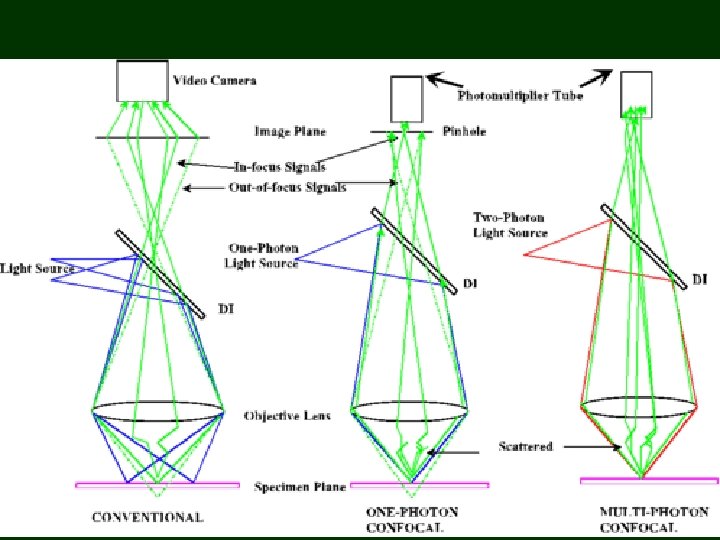
Microscopes conventional optical microscope confocal microscop measurement of fluorescence lifetime Endoscopes, telescopes Photomultipliers, microchannelplate detector gated image intesifiers Streak camera CCD and CMOS cameras
Bazsalikom levél, szekréciós mirigy az epidermisz felszínén Fluorescence from a basil leaf (cell walls fluoresce in the blue-green region, red fluorescence is due to chlorophyll) exc= 351 nm em= 385 -470 nm blue 515 -550 nm green > 650 nm red 100 m
C A 32 m B D Sorghum levél felszine: exc= 351 nm emi= 385 -470 nm kék 515 -550 nm zöld > 650 nm vörös 100 m 32 m A B C D Fluorescence layer by layer from a sorghum leaf On the surface the blue-green fluorescence of cell walls dominates, the chloroplasts lie deeper
Dane. Py infiltrated spinach leaf, at a depth of 15 m em > 650 nm 505 nm < em <550 nm exc = 351, 364 nm, Ar laser
Composite picture (green + red fluorescence) and intensity distribution along the line
Dane. Py infiltrated spinach leaf After 45 minutes photoinhibition treatment: • 2/3 of the photosynthetic activity has been lost, • only 15% protein degradation can be measured • no significant pigment bleach (total pigment) or lipid peroxidation 0’ 15’ exc = 351, 364 nm em > 650 nm 505 nm < em <550 nm 30’ 45’
Principle of multiphoton excitation
Scales in Microscopy
Conventional light microscopy is limited by diffraction
Approaches in super-resolution light microscopy SIM (Structured Illumination Microscopy) STED (Stimulated Emission Depletion Microscopy) SINGLE-MOLECULE IMAGING Schermelleh L, Heintzmann R, . et. al.
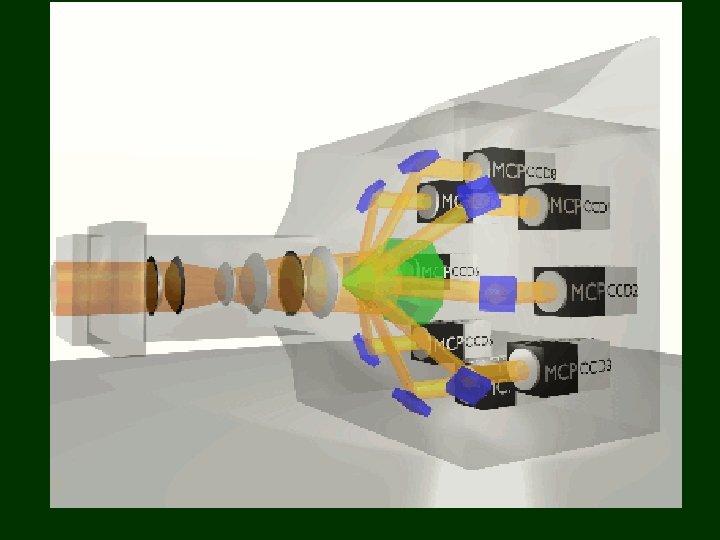
Instrumentation
N-STORM implementation by nikon
Fluorescence lifetime imaging
Measurement of temperature based on phosphorescenc lifetime
Fotoelektronsokszorozók
Fotoelektronsokszorozó működési elve
Microchannel plate detector működési elve
Kapuzható képerősítő működési elve
Példa a kapuzott képerősítő használatára
Streak camera működési elve
Streak camera használata fotokróm spirobenzopirán vizsgálatában
Egy felvétel
Quantum Efficiency (%) Typical Quantum Efficiency Curves 100 90 80 70 60 50 40 30 20 10 0 200 Back-illuminated ‘Virtual Phase’ Front-illuminated Micro. Lens front-illuminated Front-illuminat Gen III+ 300 400 500 600 700 Wavelength (nm) 800 900 1000
Interline CCD
EMCCD enhancing conventional epi-fluorescence microscopy? No Gain EM Gain BODIPY No Gain Texas Red EM Gain