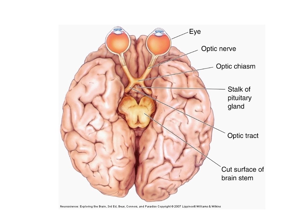
Optic Nerve Projections Optic nerves meet enter the

 Left Visual Field “View from the left eye” http:")


 contains simple and complex")


 Orientation columns of")


 contains simple and complex")
 Action or")


- Slides: 44
Optic Nerve Projections Optic nerves meet, enter the brain, and cross at the optic chiasm. After optic chiasm, the nerve fibers are called the optic tract. Optic nerve from each eye projects partly to contralateral cortex, partly to ipsilateral cortex. Ganglion cell axons are sorted so that: Cells responsive to left visual field (from nose to the left) project to right cortex. Cells responsive to right visual field (from nose to the right) project to left cortex. So damage to left visual cortex causes loss of sight off all of right visual field (from nose to the right).
Self-Portrait by Ernst Mach (1886) Left Visual Field “View from the left eye” http: //publicdomainreview. org/collections/self-portrait-by-ernst-mach-1886/ Right Visual Field
left visual field 150° right visual field nose Left Eye
left visual field right visual field nose
Figure 10. 32
left of nose -> rightside of brain leftside of brain right of nose -> leftside of brain rightside of brain Figure 10. 32
left visual field right visual field left visual field nose right visual cortex
left visual field right visual field nose left visual cortex
Figure 10. 45
looking out left right
looking out left Small lesion in one retina right
looking out left right
looking out left right
looking out left loss of visual cortex right
looking out left right Small stroke in visual cortex
looking out left right
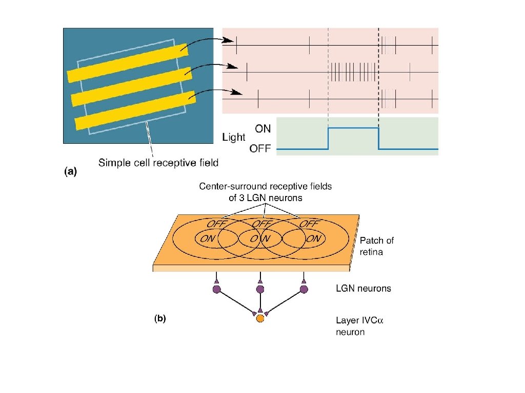
Feature Extraction by Visual Cortex Primary Visual Cortex (V 1) contains simple and complex cells Simple cells respond to orientation of stimulus at a specific spot in visual field; built up from input of ganglion cells Complex cells respond to orientation & direction of movement anywhere in the field; built up from input of simple cells Extrastriate Cortices receives input from visual cortex V 1 Dorsal Pathway (Visual Cortex -> Parietal Cortex) Action or spatial tasks - “where” info Lesions -> can’t pick up or orient objects Ventral Pathway (Visual Cortex -> Temporal Lobe, speech centers) Form recognition - “what info” Lesions -> can’t recognize or describe objects & orientations, but visually guided motor responses okay
More to vision than just edges
More to vision than just edges
Testing the Cortical Response
Receptive Field of a Neuron • Area on the surface of the sense organ which, when stimulated, causes a response in the neuron (activates or inhibits firing). • Usually overlaps with receptive field of other neurons • Variable size at different sites; smaller receptive field gives better acuity (smaller in fovea, bigger at periphery) • Can be mapped at different levels of the nervous system (retinal ganglion cells, LGN, visual cortex) • Often forms a topographically similar map of sense organ surface across surface of neurons. • Determined empirically by probing surface and recording response of a neuron.
Tuning or Selectivity of a Neuron • Analogous to Receptive Field, but instead of spatial dimension, refers to another feature • For example: – color of light, orientation of a bar – texture of an object, temperature of an object – taste quality – molecular feature of an odorant • May be organized into topographic maps of tuning surface across surface of neurons (audition), but not always (taste) • Again, determined empirically
Hubel & Weisel Video Recording from visual cortex of cat while it looks at visual stimulus on projection screen https: //www. youtube. com/watch? v=IOHayh 06 LJ 4 https: //www. youtube. com/watch? v=UU 2 esxyc. MAw
Simple Cell Response to Bar of Light
Simple Cell Response to Bar of Light
Figure 10. 48 Several ganglion cells -> 1 Simple cell
Complex Cell responds to bar of light moving in specific direction
Cortical Architecture 3 overlapping cell types (categorized by their response patterns) Orientation columns of cortex that are arranged cells that respond to orientations Color Blobs groups of cells that respond to one color Ocular dominance columns ribbons of columns that get input from one eye or the other.
Cortical Module – Each module capable of analyzing every aspect of a portion of the visual field
Orientation Columns from surface of cortex
Ocular Dominance Columns
Ocular Dominance Columns Inject one eye with radioactive proline. Transported transynaptically to cortical cells
From Single Neurons to Perception • Visual perception – Identifying & assigning meaning to objects • Hierarchy of complex receptive fields – Retinal ganglion cells: Center-surround structure, Sensitive to contrast, and wavelength of light – Striate cortex: Orientation selectivity, direction selectivity, and binocularity – Extrastriate cortical areas: Selective responsive to complex shapes; e. g. , Faces
Feature Extraction by Visual Cortex Primary Visual Cortex (V 1) contains simple and complex cells Simple cells respond to orientation of stimulus at a specific spot in visual field; built up from input of ganglion cells Complex cells respond to orientation & direction of movement anywhere in the field; built up from input of simple cells Extrastriate Cortices receives input from visual cortex V 1 Dorsal Pathway (Visual Cortex -> Parietal Cortex) Action or spatial tasks - “where” info Lesions -> can’t pick up or orient objects Ventral Pathway (Visual Cortex -> Temporal Lobe, speech centers) Form recognition - “what info” Lesions -> can’t recognize or describe objects & orientations, but visually guided motor responses okay
Extrastriate pathways beyond V 1 for visual info: Dorsal Pathway (Parietal Pathway) Action or spatial tasks - “where” info Lesions -> can’t pick up or orient objects where, how big? what, orientation? Ventral Pathway (Temporal Pathway) Form recognition - “what info” Lesions -> can’t recognize or describe objects & orientations, but motor okay
Visual Areas in Human Brain Copyright © 2016 Wolters Kluwer • All Rights Reserved
Beyond the visual cortex: shape detection in temporal cortex
Beyond the visual cortex: shape detection in temporal cortex
Task -- pick up an object Patients with dorsal lesions cannot use vison to place their fingers in the right place to pick up an object so dorsal pathway required for visual motor skills
Ventral lesion: can’t recognize orientation of card, but can move card to correct orientation ask patient to describe the orientation of a “mail slot” Ventral lesion so ventral pathway required for perception of visual scene
Normal people have these two pathways Ebbinghaus illusion ventral pathway sees two different sizes
“Physical” Ebbinghaus Ask subject to pick up the middle disk, and measure how they seperate their thumb and finger separation anticipates same disc size even though discs “look” different dorsal pathway sees same sizes