Opportunistic infections Opportunistic infections n Decrease in number

Opportunistic infections

Opportunistic infections n Decrease in number of CD 4 lymphocytes is condition for development of opportunistic infections n Risk is started, when number of CD 4 lymphocytes drops to number 500 of CD 4 lymphocytes/mm 3

CD 4 count and opportunistic infection

TUBERCULOSIS - the most important - the most common OI

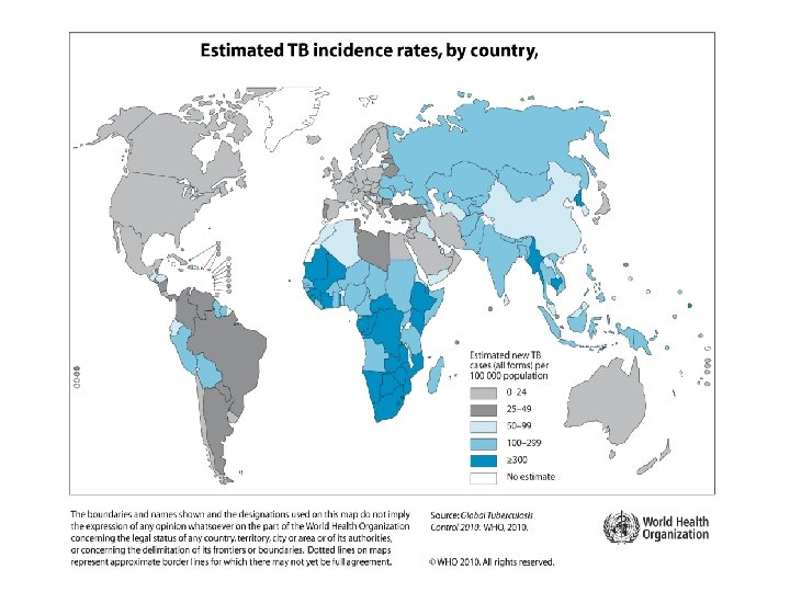
Epidemiology • One-third of the world´s population is infected with TB • HIV infection has had a big impact in increasing the numbers of patients affected with disease caused by TB • TB is the most important severe opportunistic infection among patients with HIV in developing countries
")
TB – estimated new cases (per 100 000)

Tuberculosis • Is a leading cause of HIV-related deaths • • worldwide In some countries with higher HIV preavalence, up to 80% of people with TB test positive for HIV Globally approximately 30% of HIV infected persons are estimated to have latent TB infection

n TB is transmissible to both people with HIV infection u uninfected persons u can be treated and can be prevented
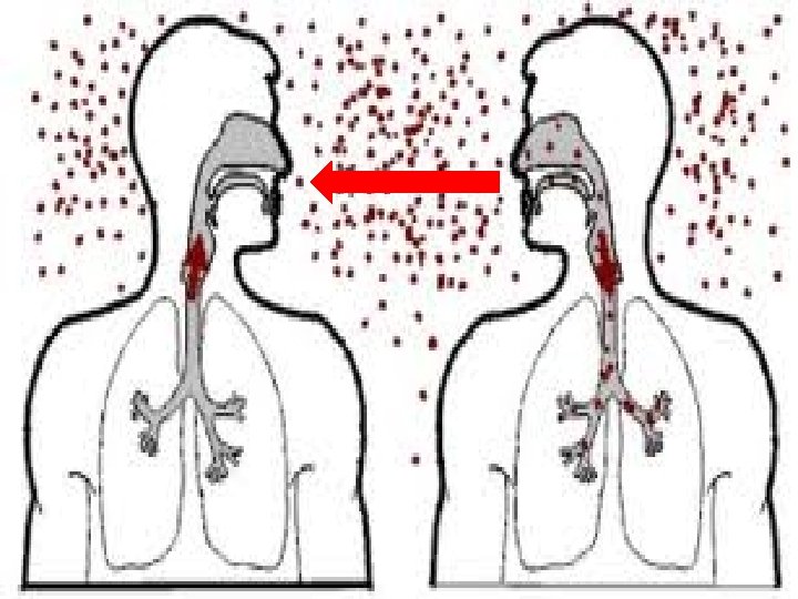

Clinical Manifestations Myco TB Is highly contagious n Leads to a number of serious medical syndromes affecting, at time, most of the organ systems n
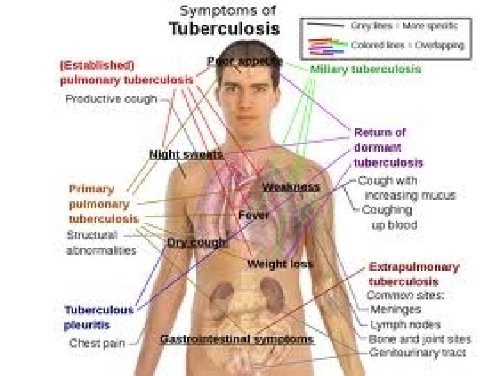

Myco TB can causes: 1. Pulmonary disease Pneumonia t Cavitary disease t
")
Cavities in the lungs (X-ray of thorax)
 § Otitis media § Laryngitis § Miliary TB")
2. Extrapulmonary disease § Adenitis („scrofula“) § Otitis media § Laryngitis § Miliary TB § Meningitis § Skeletal TB § Gastrointestinal TB § Renal TB…

scrofula

TB absces in brain

Skeletal TB - destruction of the lumbar vertebrae - skeleton of the Great Moravian Empire
 is acid-fast, appearing red on a Ziehl-Neelsen stain")
Mycobacterium tuberculosis bacteria (G+) is acid-fast, appearing red on a Ziehl-Neelsen stain

Primary prophylaxis conditions CD 4+ any + TB exposure (when HIV+ individual is in exposure of TB we must start primary prophylaxis) pathogen drug M. tuberculosis isoniazid (+pyridoxin), rifampicin, pyrazinamid, ethambutol Myco TB is highly contagious !!!

Pneumocystis carinii jiroveci Infection

Pneumocystis carinii jiroveci n Is an opportunistic pathogen, the natural habitant of which is the lung n The organism is an important cause of pneumonia in the compromised host n The organism can be found in other organs and tissues

CD 4 count and opportunistic infection

CD 4+ lymphocytes depletion – gradual loss of number CD 4+ count primary HIV infection of CD 4 cells asymptomatic infection A 1 early symptomatic infection B 2 late symptomatic infe C 3 final stadium years

Pneumocystis carinii jiroveci n Has a worldwide distribution n Serologic serveys indicate that already most healthy children have been esposed to the organism n It means that we meet with this organism in early childhood n Taxonomy – the fungal kingdom

Incidence n PCP accounted for of all AIDS-indicator diseases ART 42% before n Incidence of PCP in this population declining (with ART and prophylaxis) n But incidence of extrapulmonary carinii jiroveci is increasing i Pn

Extrapulmonary Pn. carinii jiroveci infection involves in fewer than 3% of cases. n Lymph nodes (in up to 50% of cases) n Spleen n Liver n Bone marrow n GI and genitourinary tracts n Adrenal and thyroid glands n Heart, pancreas, eyes, ears, skin…

Incubation Period n On the basis of animal studies, the incubation period is thought to be from 4 to 8 weeks

Typical Symptoms § Patients with PCP usually develop following: § Dyspnea § Mild fever § Nonproductive cough the

The late signs n Physical findings of PCP include the following: § Tachypnea § Tachycardia § Cyanosis n Lung auscultation is usually unremarkable

Differential Diagnosis The differential diagnosis of PCP is very broad and includes § infectious diseases and also can mimic § noninfectious diseases

Laboratory n There is no reliable way to cultivate the organism in vitro n A definitive is made by histopathologic staining, which selectively stain the wall of Pn. carinii jiroveci, cysts or nuclei n PCR technique which demonstrate nuclei acid

Cysts of Pn. carinii jir. Methenamine silver stain. In smear from bronchoalveolar lavage.
, Giemsa-stained")
Pn. carinii – trophozoites (growth stage), Giemsa-stained

Pn. carinii jiroveci – immunofluorescence with monoclonal antibodies is more sensitive than traditional staining

Laboratory LDH n Elevated serum concentrations of lactate dehydrogenase have been reported but are not specific to Pn. Carinii infection Leucocytes n The white blood cell count is low Oxygen saturation is very low n Is probably the most sensitive noninvasive test for dg. PCP

Arterial blood gases demonstrated §Hypoxia §An increased alveolar-arterial oxygen gradient

Alveolocapillary membrane - characteristic exudate is in the inter alveolar space

Imaging n The classic findings on chest radiography consist of bilateral diffuse infiltrates involving the perihilar regions. n Atypical manifestations also have been reported. n Early in the course of pneumocystosis, the chest radiograph may be normal.

Imaging – HR CT n The most important imaging method shows n White glass picture

CT -White glass picture

Diagnostic/testing procedures Fiberoptic bronchoscopy n With bronchoalveolar lavage remains the mainstay of Pn. Carinii diagnosis Sputum n is a simple, noninvasive technique, but its sensitivity has extremely low Transbronchial biopsy and open lung biopsy n are the most invasive, are reserved for situations in which a diagnosis cannot be made by lavage

Main treatment Trimethoprim-sulfamethoxazol n Is the drug of the first choice for all forms of Pn. Carinii infection n It is administered intravenously (orally) at a dosage 120 mg of TSX/kg/d in four divide doses

Glucocorticoids Administration of glucocorticoids to HIV-infected patients with moderate to severe pneumocystosis can improve the rate of survival n The recommended regimen: 40 mg prednisone PO twice daily, with tapering to a dose of 20 mg/d over a 3 -week period n

Duration of treatment non-HIV-infected patients n Treatment of pneumocystosis should be continued for 14 days (better 21 days) HIV-infected patients n Treatment of pneumocystosis should be continued for 21 days

Alternative treatment Pentamidine 4 mg/kg/d by slow intravenous infusion n Clindamycin n Primaquine avoided in patients with glucose-6 phosphate dehydrogenase deficiency n Trimethoprim + dapson n Atovaquone n

Complications n In the typical case of untreated PCP, progressive respiratory compromise leads to death. n Therapy is most effective when instituted early in the course of the disease, before there is extensive alveolar damage.

Primary prophylaxis n Is indicated for HIV-infected patients at high risk of developing pneumocystosis CD 4+ lymphocyte count < 200/mm 3

Secondary prophylaxis Is indicated for all patients who have recovered from PCP
 per day Alternative regimens n Dapsone")
Prophylactic regimen n Trimethoprim-sulfamethoxazol (160 mg of trimethoprim) per day Alternative regimens n Dapsone (50 mg daily), pyrimethamine (50 mg once per week), and folinic acid (24 mg once per week) n Dapsone (100 mg daily) n Nebulized pentamidine (300 mg once per month via nebulizer)

Primary prophylaxis conditions CD 4+ any + TB exposure CD 4+ < 200/mm 3 pathogen drug M. tuberculosis isoniazid (+pyridoxin), rifampicin, pyrazinamid, ethambutol Pn. carinii jiroveci co-trimoxazol, pentamidine (aerosol), dapson

TOXOPLASMOSIS

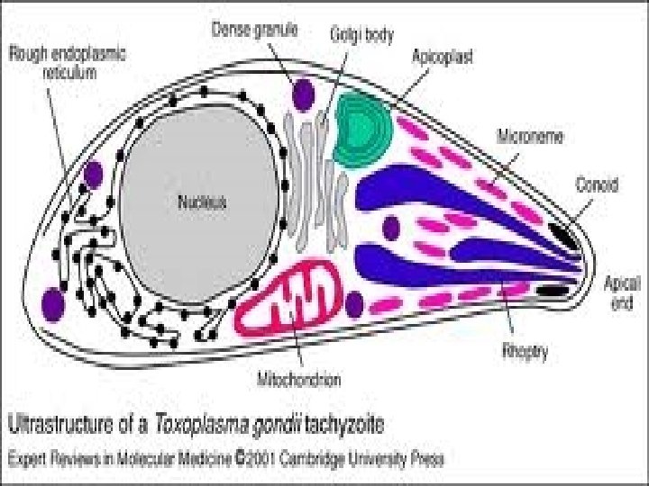
DEFINITION • An acute or chronic infection caused by the obligate intracellular protozoan Toxoplasma gongii • Infection in human is usually asymptomatic • When symptoms occur, they range from a mild, self-limited to a fulminant disseminated disease

SYMPTOMS Usually involve the following: • Central nervous system • Eyes • Skeletal or cardiac muscles • Lymph nodes • Liver • Lungs

DISEMINATED DISEASE Severe infections usually occur • In an immunocompromised patient • By the transplacental passage of parasites from an infected mother to the fetus (congenital toxoplasmosis)

Cysts in tissue

EPIDEMIOLOGY Cases are caused by: • Eating undercooked meat • Contaminated vegetables • Ingestion of oocysts from contaminated soil The seroprevalence depends on geografic location: US – between 3 -67% tropical countries – up to 90%

SYMPTOMS AND SIGNS • Immune responses are able to eliminate most of the tachyzoites • 80 – 90% of cases in immunocompetent persons are asymptomatic

CEREBRAL TOXO Clinical manifestations of CNS infection include the following: • Headache, seizures, weakness • Cranial nerve abnormalities • Visual field defects • Mental status changes • Cerebellar signs

CEREBRAL TOXO • Speech abnormalities • Meningism • Sensory or motor disorders • Disorientation • Hemiparesis • Convulsions • Coma and death

EXTRACEREBRAL TOXO • Less common among patients with HIV inf. • The prevalence is estimated at 1, 5% to 2, 0% • lungs (pneumonitis) • eye (chorioretinitis) • heart Cases of gastrointestinal, liver, skin, or multiorgan involvement also have been reported
 The abscesses of cerebral toxoplasmosis are typically • Multiple")
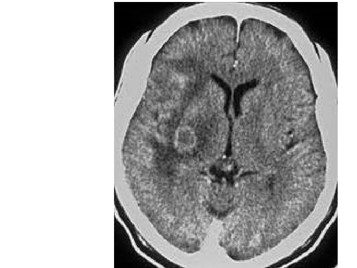
IMAGING On neuroimaging (CT, MRI) The abscesses of cerebral toxoplasmosis are typically • Multiple • Located in the cortex or deep nuclei (thalamus and basal ganglia) • Surrounded by edema • Enhance in a ringlike pattern with contrast

Cerebral toxoplasmosis

SEROLOGY • Approx. 20% of patients • • • have no detectable antibodies Titer of antibodies does not always rise during infection Negative serology does not rule out infection But a rising titer may be of diagnostic significance
 in blood samples suggest that • This")
OTHER LABORATORY METHODS PCR (polymerase chain reaction) in blood samples suggest that • This modality has limited diagnostic value in cases of cerebral toxoplasmosis CSF (cerebrospinal fluid) • Is also nonpathognomonic and reveals elevated protein and mild pleocytosis

EXTRACEREBRAL TOXOPLASMOSIS • Involving other organs among HIV-infected patients is rare • Dg. is usually based on biopsy OCULAR TOXOPLASMOSIS • Is usually based on a suggestive ophthalmoscopic picture • Histopathologic identification of T. gondii in the eye can establish the diagnosis

retinochoroiditis

MAIN TREATMENT The regimen of choice for acute therapy • Pyrimethamine 50 to 75 mg/d + sulfadiazine 4 to 8 g/d • Leucovorin – coadministtered to prevent the folinic acid deficiency and ameliorate the hematologic toxicity of pyrimethamine • Duration of treatment – usually for 6 to 8 weeks

PATIENT FOLLOW-UP After induction treatmen • HIV-infected patients schould receive lifelong suppression therapy pyrimethamine 25 -50 mg/d + sulfadiazine 2 -4 g/d • The doses of TMP/SMX recommended for P. carinii pneumonia appear to be effective
 meat")
PREVENTION FOR INDIVIDUALS AT RISK • Not to eat raw or undercooked („pink“) meat • Wash fruits and vegetables • Wash hands after contact with raw meat • and after contact with soil Wash hands after changing a cat litter box

PRIMARY PROPHYLAXIS conditions pathogen drug CD 4+ any + TB exposure M. tuberculosis isoniazid (+pyridoxin), rifampicin, pyrazinamid, ethambutol CD 4+ < 200/mm 3 Pn. carinii jiroveci co-trimoxazol, pentamidine (aerosol), dapson CD 4+ < 150/mm 3 + antibody to Toxoplasma positive Toxoplasma gondii co-trimoxazol, dapson, pyrimethamin(+folinat)

CONGENITAL TOXOPLASMOSIS • Clinical findings are variable • There may be no sequelae, or sequelae may • • develop at various times after birth Premature infants may present with CNS or ocular disease Full-term infants usually develop milder disease, with hepatosplenomegaly and lyfadenopathy
 1. Retinochoroiditis 2. Hydrocephalus 3. Convulsions")
CONGENITAL TOXOPLASMOSIS Sabin tetrade (classic tetrade of signs) 1. Retinochoroiditis 2. Hydrocephalus 3. Convulsions 4. Intracerebral calcifications
- Slides: 75