
OPHTHALMOLOGY CATARACTS MBCh B 4 Prof P Roux
OPHTHALMOLOGY CATARACTS MBCh. B 4 Prof P Roux 2012
ACQUIRED CATARACT 1. Classification of age-related cataract • Morphological • According to maturity 2. Other causes of cataracts • Diabetes • Myotonic dystrophy • Atopic dermatitis • Trauma • Drugs • Secondary (complicated) 3. Surgery • Large incision extracapsular extraction • Phacoemulsification
Classification of Age-related Cataract According to Morphology 1. Subcapsular • Anterior • Posterior 2. Nuclear 3. Cortical 4. Christmas tree
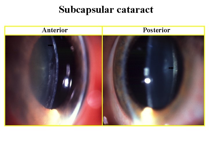
Subcapsular cataract Anterior Posterior
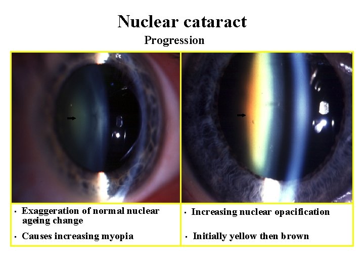
Nuclear cataract Progression • • Exaggeration of normal nuclear ageing change • Increasing nuclear opacification Causes increasing myopia • Initially yellow then brown
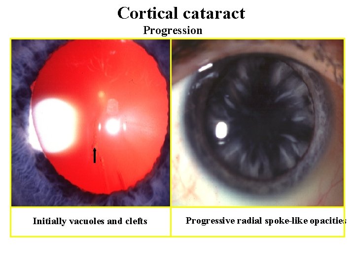
Cortical cataract Progression Initially vacuoles and clefts Progressive radial spoke-like opacities

Christmas tree cataract Polychromatic, needle-like opacities May co-exist with other opacities
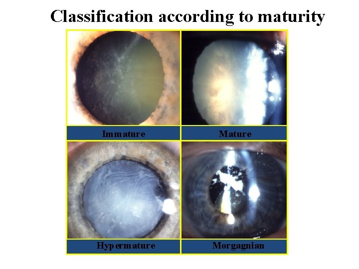
Classification according to maturity Immature Hypermature Morgagnian
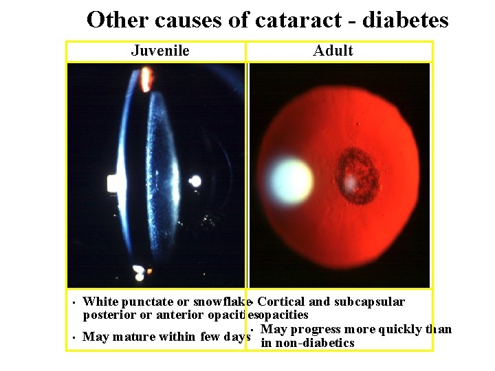
Other causes of cataract - diabetes Juvenile • • Adult White punctate or snowflake • Cortical and subcapsular posterior or anterior opacities • May progress more quickly than May mature within few days in non-diabetics
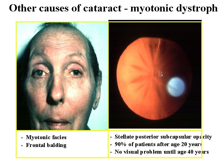
Other causes of cataract - myotonic dystroph • • Myotonic facies Frontal balding • • • Stellate posterior subcapsular opacity 90% of patients after age 20 years No visual problem until age 40 years
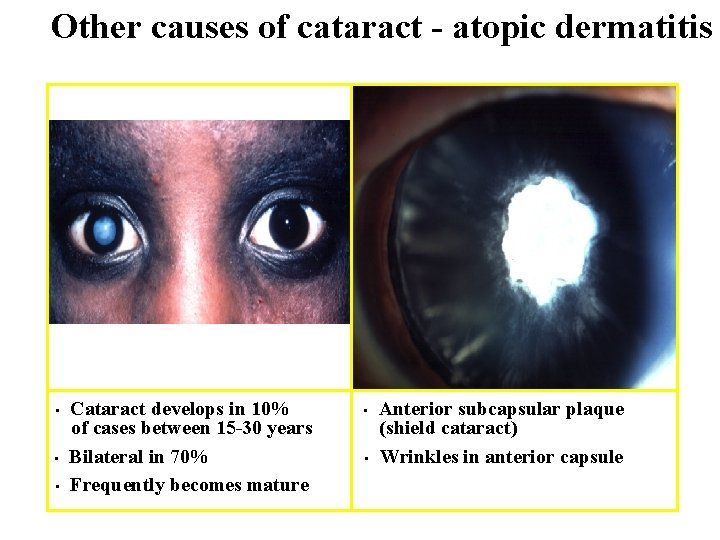
Other causes of cataract - atopic dermatitis • • • Cataract develops in 10% of cases between 15 -30 years Bilateral in 70% Frequently becomes mature • • Anterior subcapsular plaque (shield cataract) Wrinkles in anterior capsule
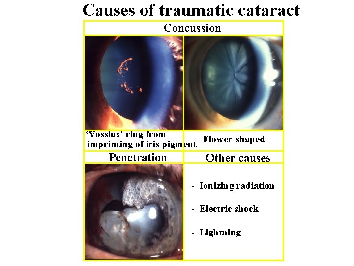
Causes of traumatic cataract Concussion ‘Vossius’ ring from imprinting of iris pigment Flower-shaped Penetration Other causes • Ionizing radiation • Electric shock • Lightning
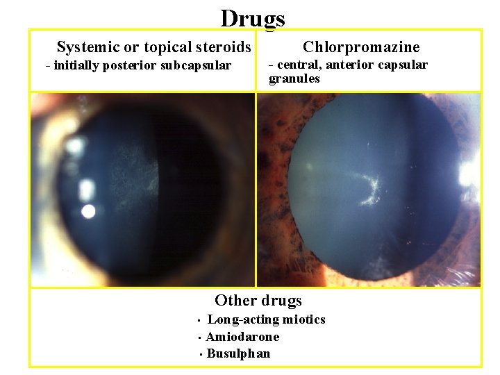
Drugs Systemic or topical steroids - initially posterior subcapsular Chlorpromazine - central, anterior capsular granules Other drugs Long-acting miotics • Amiodarone • Busulphan •
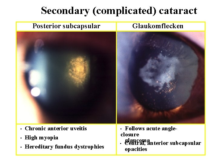
Secondary (complicated) cataract Posterior subcapsular • Chronic anterior uveitis High myopia • Hereditary fundus dystrophies • Glaukomflecken Follows acute angleclosure glaucoma • Central, anterior subcapsular opacities •
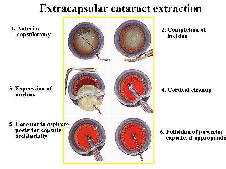
Extracapsular cataract extraction 1. Anterior capsulotomy 2. Completion of incision 3. Expression of nucleus 4. Cortical cleanup 5. Care not to aspirate posterior capsule accidentally 6. Polishing of posterior capsule, if appropriate
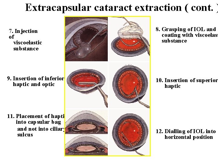
Extracapsular cataract extraction ( cont. ) 7. Injection of viscoelastic substance 8. Grasping of IOL and coating with viscoelast substance 9. Insertion of inferior haptic and optic 10. Insertion of superior haptic 11. Placement of haptics into capsular bag and not into ciliary sulcus 12. Dialling of IOL into horizontal position
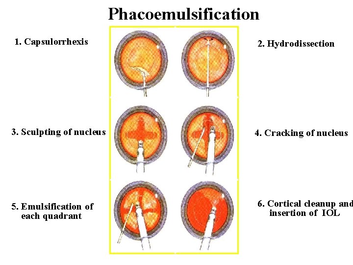
Phacoemulsification 1. Capsulorrhexis 2. Hydrodissection 3. Sculpting of nucleus 4. Cracking of nucleus 5. Emulsification of each quadrant 6. Cortical cleanup and insertion of IOL
- Slides: 17