
Ophthalmic diagnostic tools Tools Instrument drugs By dr
Ophthalmic diagnostic tools Tools &Instrument &drugs By dr. Eman Enweigi
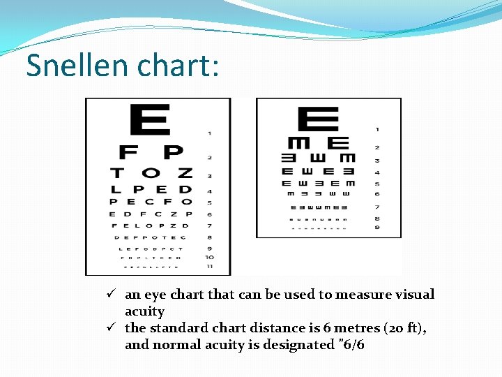
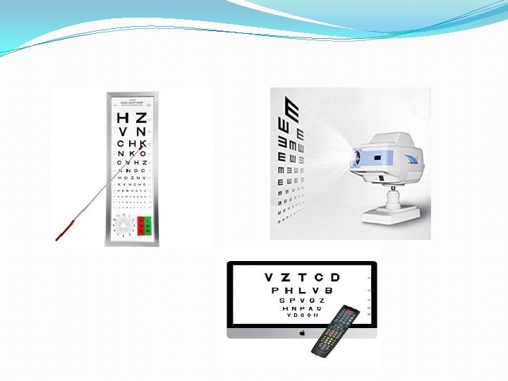
Snellen chart: ü an eye chart that can be used to measure visual acuity ü the standard chart distance is 6 metres (20 ft), and normal acuity is designated "6/6
Retinoscopy ü a technique to obtain an objective measurement of the refractive error of a patient's eyes Manually. ü The examiner uses a retinoscope to shine light into the patient's eye and observes the reflection (reflex) off the patient's retina. ü While moving the streak or spot of light across the pupil the examiner observes the relative movement of the reflex and manually places lenses over the eye (using a trial frame and trial lenses) to "neutralize" the reflex.

ü Retinoscope ü objective measurement of the refractive error of a patient's eyes Manually.
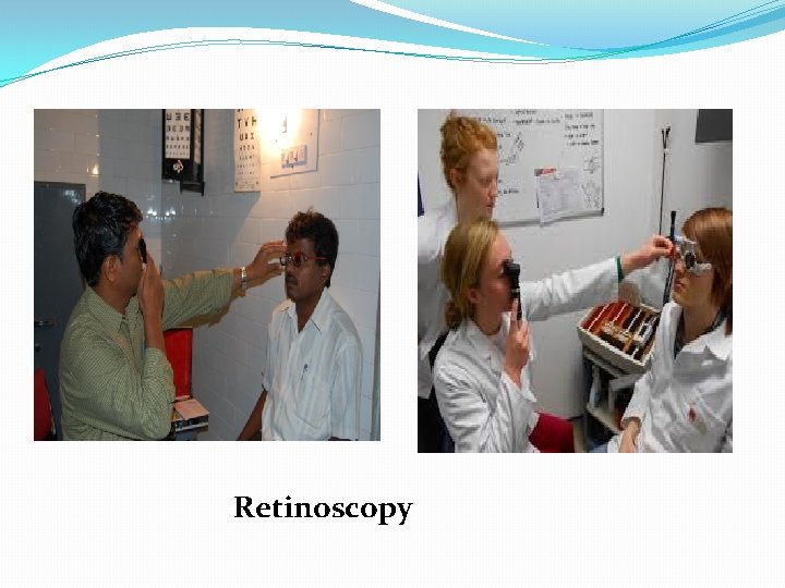
Retinoscopy
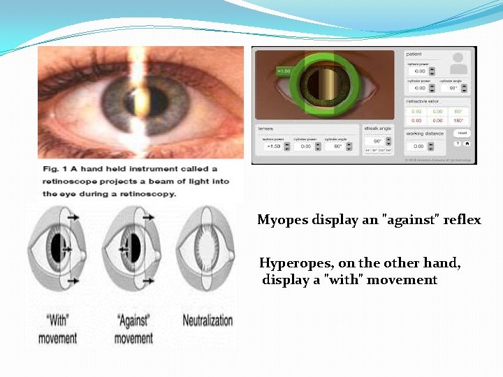
Myopes display an "against" reflex Hyperopes, on the other hand, display a "with" movement
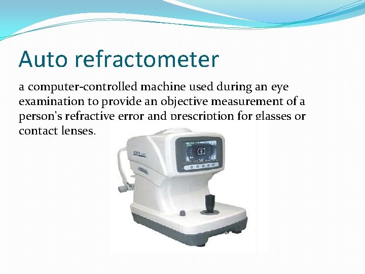
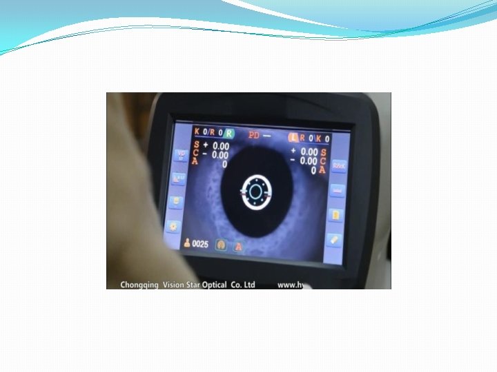
Auto refractometer a computer-controlled machine used during an eye examination to provide an objective measurement of a person's refractive error and prescription for glasses or contact lenses.
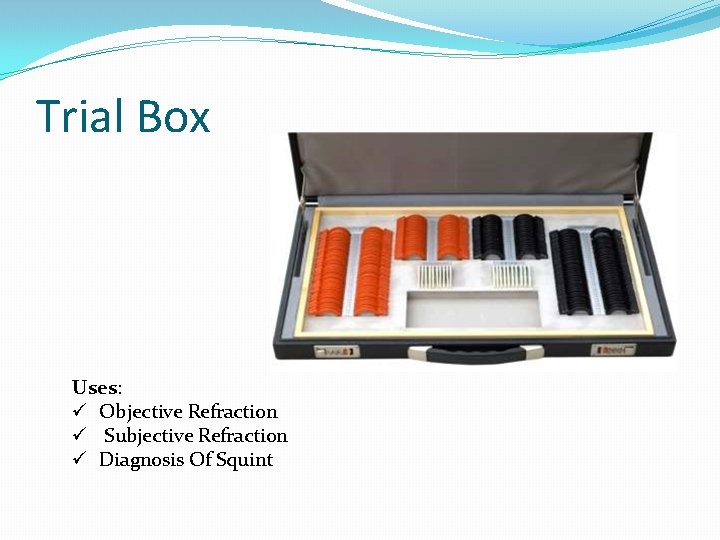
Trial Box Uses: ü Objective Refraction ü Subjective Refraction ü Diagnosis Of Squint
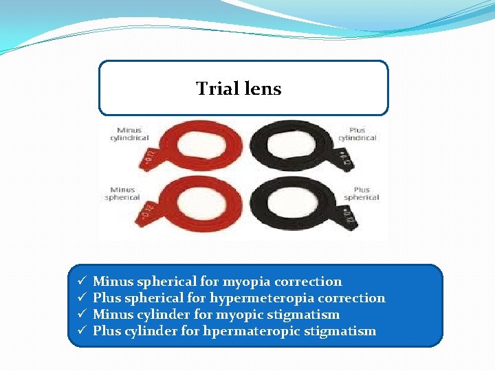
Trial lens ü ü Minus spherical for myopia correction Plus spherical for hypermeteropia correction Minus cylinder for myopic stigmatism Plus cylinder for hpermateropic stigmatism
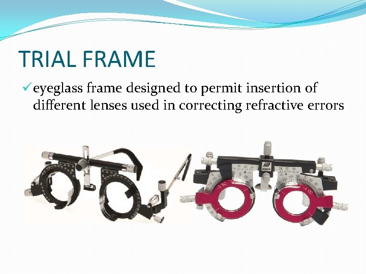
TRIAL FRAME üeyeglass frame designed to permit insertion of different lenses used in correcting refractive errors
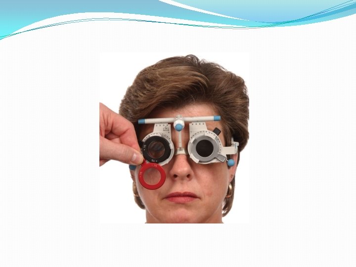
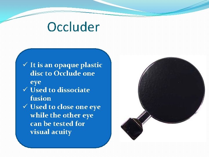
Occluder ü It is an opaque plastic disc to Occlude one eye ü Used to dissociate fusion ü Used to close one eye while the other eye can be tested for visual acuity
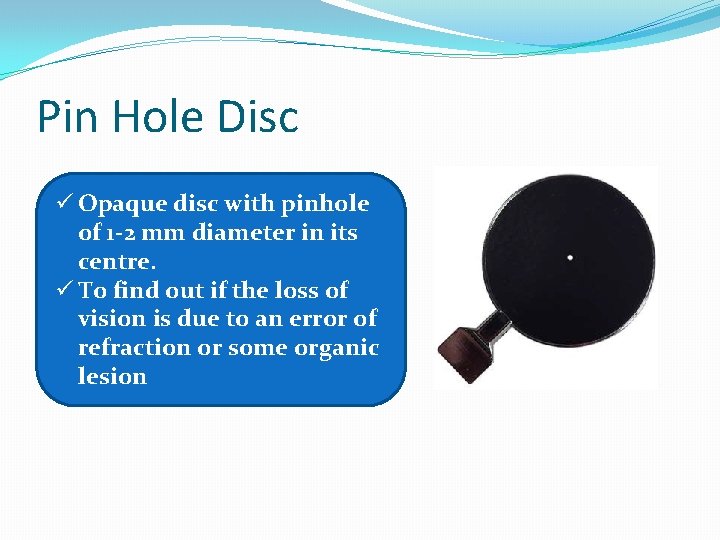
Pin Hole Disc ü Opaque disc with pinhole of 1 -2 mm diameter in its centre. ü To find out if the loss of vision is due to an error of refraction or some organic lesion
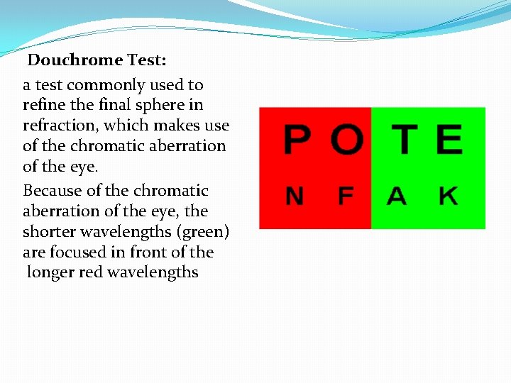
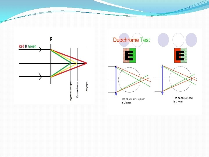
Douchrome Test: a test commonly used to refine the final sphere in refraction, which makes use of the chromatic aberration of the eye. Because of the chromatic aberration of the eye, the shorter wavelengths (green) are focused in front of the longer red wavelengths
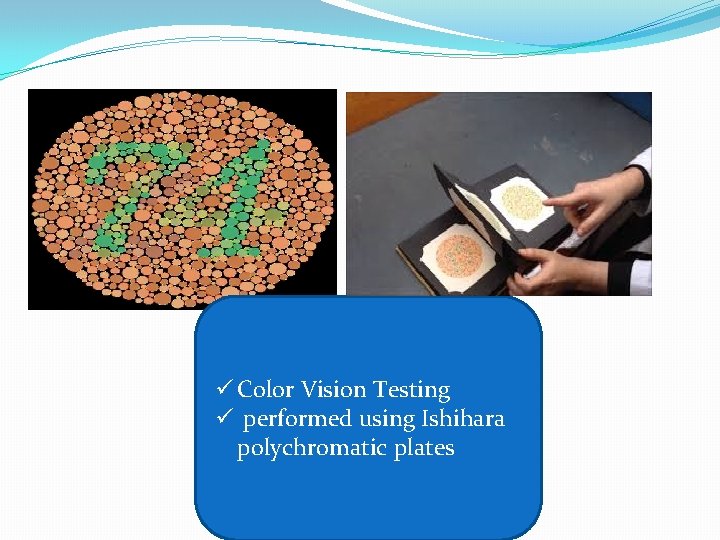
ü Color Vision Testing ü performed using Ishihara polychromatic plates
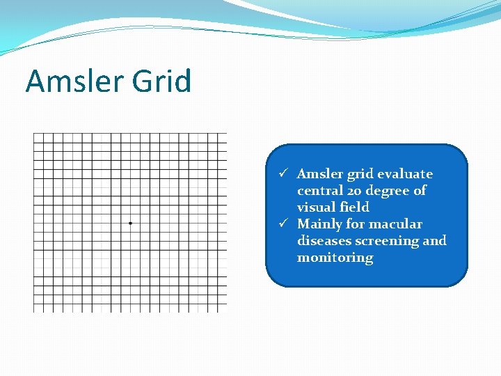
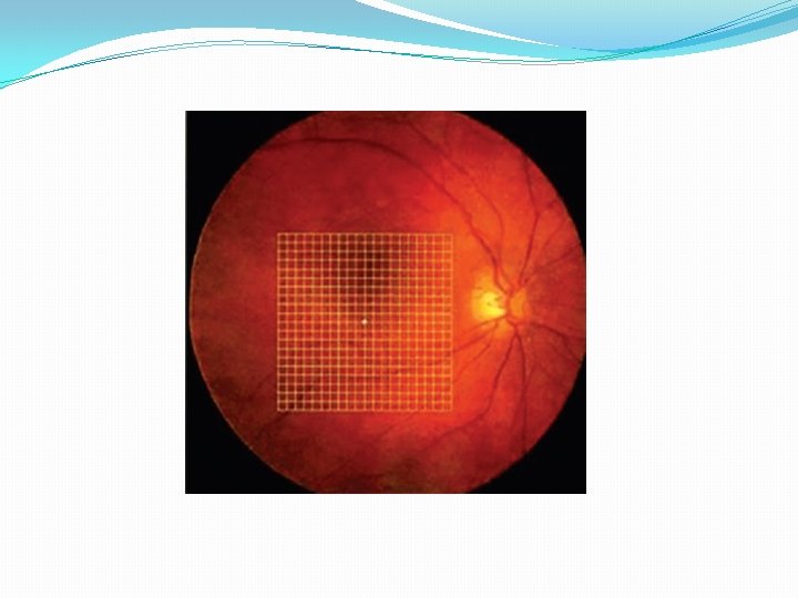
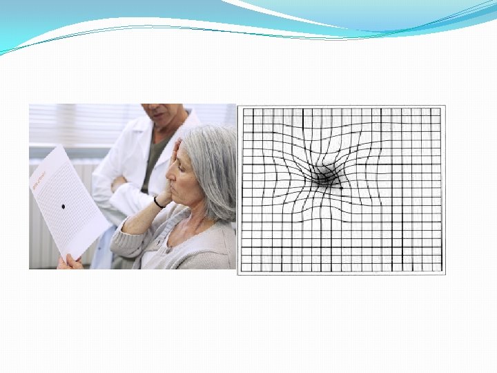
Amsler Grid ü Amsler grid evaluate central 20 degree of visual field ü Mainly for macular diseases screening and monitoring
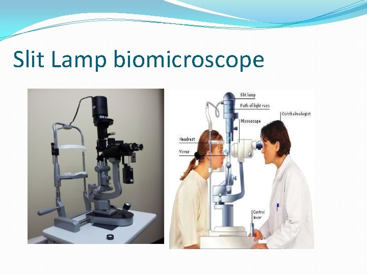
Slit Lamp biomicroscope
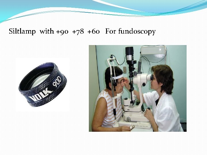
Siltlamp with +90 +78 +60 For fundoscopy
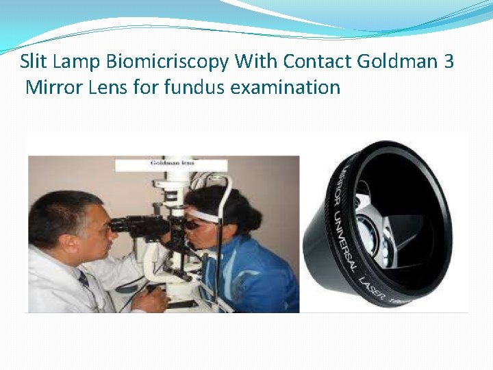
Slit Lamp Biomicriscopy With Contact Goldman 3 Mirror Lens for fundus examination
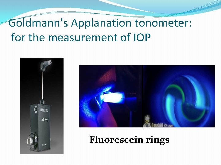
Goldmann’s Applanation tonometer: for the measurement of IOP Fluorescein rings
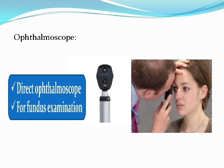
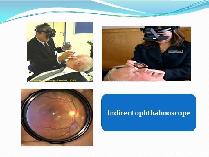
Ophthalmoscope:
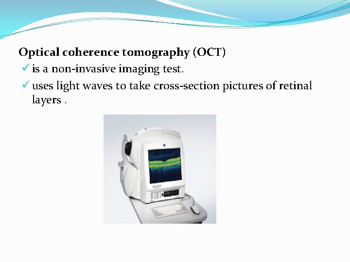
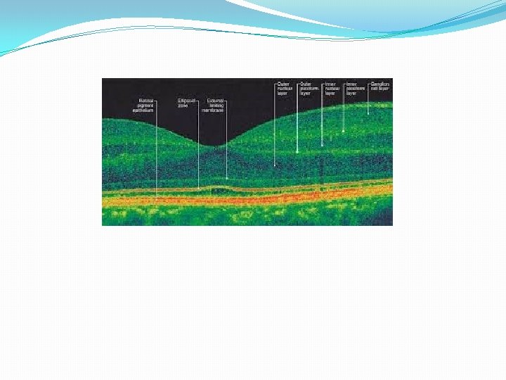
Optical coherence tomography (OCT) ü is a non-invasive imaging test. ü uses light waves to take cross-section pictures of retinal layers.
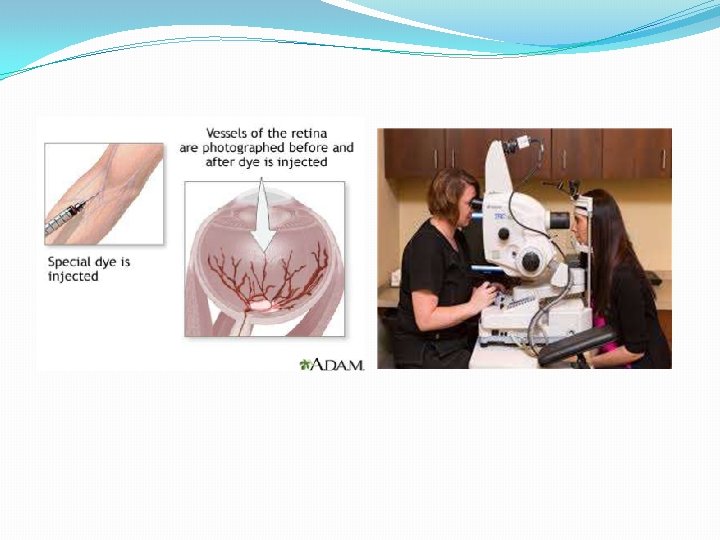
Fluorescein Angiography ü It is an invasive procedure in which fluorescein dye is injected, usually into an antecubital vein. ü Within 10 to 15 seconds, this dye can be seen coursing through the retinal vessels. ü Over a 10 -minute period, serial black-and-white photographs are taken of the retinal vasculature
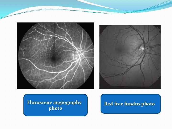
Fluroscene angiography photo Red free fundus photo
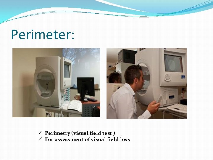
Perimeter: ü Perimetry (visual field test ) ü For assessment of visual field loss
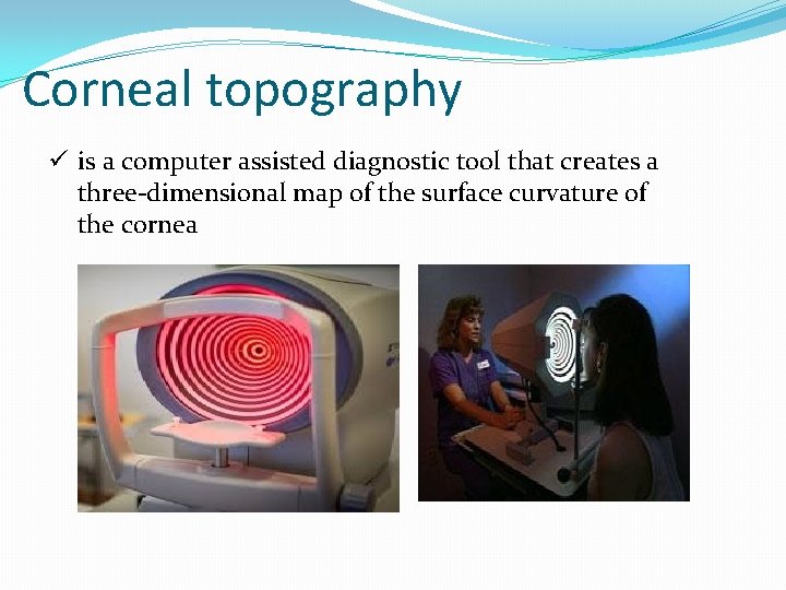
Corneal topography ü is a computer assisted diagnostic tool that creates a three-dimensional map of the surface curvature of the cornea
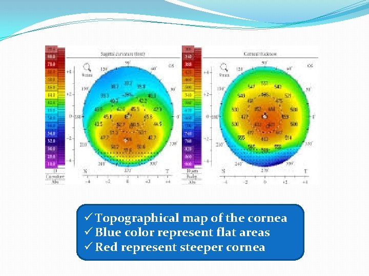
ü Topographical map of the cornea ü Blue color represent flat areas ü Red represent steeper cornea
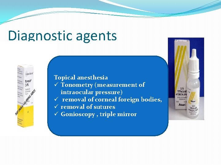
Diagnostic agents Topical anesthesia ü Tonometry (measurement of intraocular pressure) ü removal of corneal foreign bodies, ü removal of sutures ü Gonioscopy , triple mirror
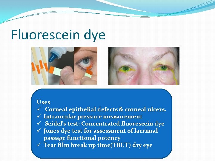
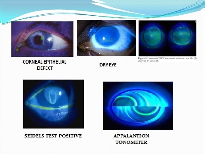
Fluorescein dye Uses ü Corneal epithelial defects & corneal ulcers. ü Intraocular pressure measurement ü Seidel's test: Concentrated fluorescein dye ü Jones dye test for assessment of lacrimal passage functional potency ü Tear film break up time(TBUT) dry eye
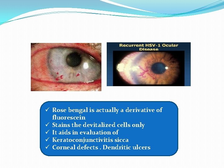
ü Rose bengal is actually a derivative of fluorescein ü Stains the devitalized cells only ü It aids in evaluation of ü Keratoconjunctivitis sicca ü Corneal defects. Dendritic ulcers
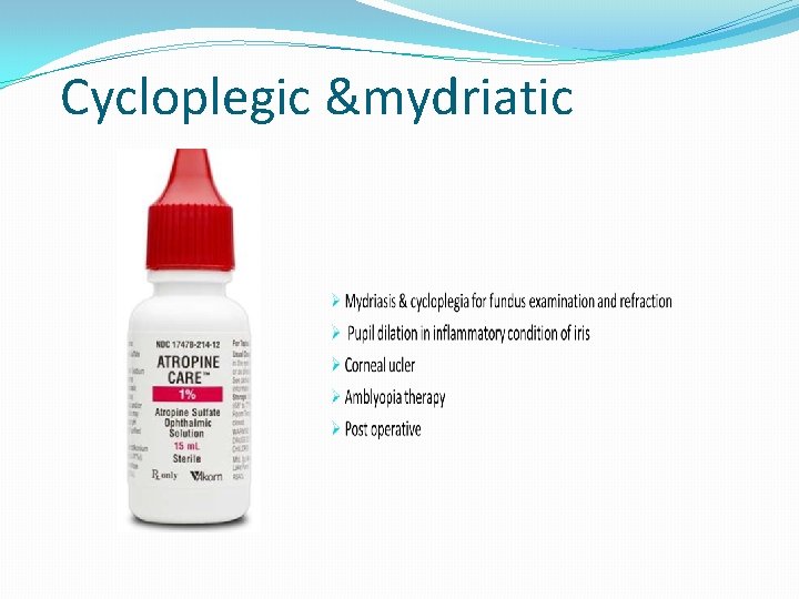
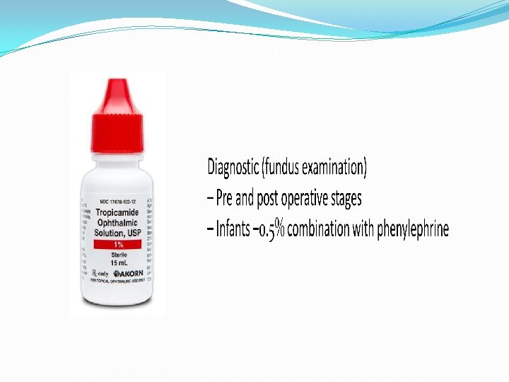
Cycloplegic &mydriatic

- Slides: 43