Open Versus Closed System for Vitrification Pros and


 Cryoloop • (Laneand. Gardner,")

 Rapid-I")


 • Cryotip (Irvine Scientific, CA, USA), • High Security")


 of embryos to the non‐ vitrification")


 Human")


 and single cell genetic analysis has become increasingly")


- Slides: 50
Open Versus Closed System for Vitrification: Pros and Cons Assist. Prof. Dr. Evrim Ünsal
0 utlines… What is the definition of an open versus closed system? Are closed systems as efficient as open systems Friendship of vitrification and PGD/PGS applications
Early history of freezing human gametes and embryos • • • 1949 : First human gamet cryopreservation 1984 : First live birth with FET 1985 : First pregnancies with thawed blastocyst 1986 : First live birth with thawed oocytes. 2007 : The first birth after human blastocysts vitrification of biopsied embryos for PGD (Parriego et al. )
• Over the last five years there has been a dramatic worlwide shift to vitrification from slow freezing. • Since then and up to 2008 it is estimated that between 350, 000 and half a million IVF babies have been born from embryos frozen at a controlled rate; • additionally a few hundred births have been born from vitrified oocytes.
Currently used vitrification techniques differ from each other in many technical details • • • solutions, equilibration and dilution parameters, carrier tools, cooling, storage, warming methods.
The characteristics of cryopreservation methods • exposure time of cells to the different cryoprotectant solutions • their different concentrations, • the rate of formation of extra‐ and intracellular ice crystals,
The wide variety of methods makes selection of the best technique difficult, and causes serious problems when cryopreserved samples are transferred between laboratories.
The use of safe cryopreservation protocol is very important to avoid human cell contamination or cross contamination in common LN 2 tanks. CONTAMINATION The contamination of the sample by freezing or by direct contact with the cooling solution CROSS CONTAMINATION The contamination of the sample within the common container
Possible contamination /cross contamiantion factors • Handling contaminated biological samples (semen, follicular fluid, tissue, etc. ). • Use of contaminated culture media. • Using contaminated LN 2 during the freezing process • İneffective sealing/damage • The air in the room. • Operators. • Use of open devices
Fundamental classification of vitrification system Open system vitrification Closed system vitrification Semi closed system vitrification
30 different carrier tools have been published, 15 versions are commercially available. Cryotop • (Hamawaki et al. , 1999) Cryoloop • (Laneand. Gardner, 2001; Laneetal. , 1999) Open Pulled Straw • (Vajtaetal. , 1998) Most claimed closed system are the results of the modifications of these open systems.
Fully Open Systems Cryotop • (Hamawaki et al. , 1999) Cryoloop • (Laneand. Gardner, 2001; Laneetal. , 1999) Open. Pulled. Straw • (Vajtaetal. , 1998) Cryotech • (Gutnisky et al. , 2013) Cryolock • (Garcia et al. , 2011) Cryoleaf • (Chian et al. , 2009) Vitri-Inga • (Almodulin et al. , 2010)
Semi Closed System • Open cooling and closed storage systems Cryotop • (http: //www. kitazato. co. jp/biopharma/Cryotop 2. htmld) Open Pulled Straw • (Vajtaetal. , 1998) • Avoides contact of the biological sample with the cooling solution • Achieves cooling rates in a high rate
Closed System Cryotop Closed High Security System • (Hamawaki et al. , 1999) Rapid-I • (Larman and Gardner, 2011) Vitrisafe • (Vanderzwalmenetal. , 2009) Cryotip • (Kuwayama et al. , 2005) Cryopette • (Parmegiani et al. , 2012) cells are never in direct contact with liquid nitrogen or vapours thanks to a hermetic seal;
• As liquid nitrogen may contain infective agents, direct contact could theoretically mean a possibility for infection and disease transfer. • Liquid nitrogen, and also vapour phase nitrogen may act as a vector between two contaminated samples. • In reproductive biology, including mammalian and human assisted reproduction, no disease transmission caused by liquid nitrogen mediated cross‐contamination, or other cryopreservation‐ related source, has been reported.
Other contamination factors in IVF • • • collection of semen is not a sterile procedure; oocytes are contaminated with blood during collection; many containers are inappropriately sealed or closed by non‐hermetical methods; the outer surface of straws and vials is always infected; storage tools (canisters, holders) are not sterilized; Openings of dewars mix air with LN 2 vapour and may cause infection; Factory derived LN 2 isusually not transported undera septic conditions, and, accordingly, can not be regarded as sterile, even if during production the infective agents are usually destroyed; contaminated samples (sperm cells, oocytes) cannot be decontaminated; in most IVF laboratories, dewars are not decontaminated regularly; acordingly, LN 2 tanks and LN 2 in tanks should always be regarded as contaminated. Scissors or blades used to cut the straws are usually not sterilized between straws and patients, (Gabor et al 2015)
• No such disease transfer has yet been reported, although an estimated ~1, 000 vitrified embryos or embryos derived from vitrified oocytes by using open systems have been transferred. At present, most embryos and oocytes are vitrified with open systems worldwide, indicating a high overall efficiency and consistency,
The reports describing the use of closed devices for both vitrification and storage of human zygotes and embryos • • Vanderzwalmen et al. , 2012 Kuwayama et al. , 2005; Isachenko et al. , 2007; Stachecki et al. , 2008; • • Vanderzwalmen et al. , 2009, 2010; Liebermann, 2009; Schiewe, 2010; Van Landuyt et al. , 2011. • Vitrification in closed systems is a feasible procedure and can lead to reasonable clinical outcomes. • Nevertheless, the use of closed systems is still not totally accepted by IVF practitioners, under the perception that it leads to reduced clinical outcomes in contrast to their open variations.
• The major drawback of these systems is the reduction in the cooling rate and, in a few cases, the warming rate.
• very high rates of cooling are not mandatory during vitrification • warming rate is of fundamental importance for survival closed devices, even though their insulation results in a lower cooling rate, can perform efficiently if warming rates are properly adjusted
The rapid warming is a crucial point for successful vitrification. • Removal of the sample from the insulating container whilst still submerged in liquid nitrogen and subsequent direct immersion of the cells into the warming solution.
Successful vitrification is an equation with four variables; cooling rate warming rate sample viscosity sample volume
• Cryoloop (Vitrolife, Sweden) • Cryotip (Irvine Scientific, CA, USA), • High Security Vitrification (HSV) straw (Cryo Bio. System, Paris, France),
The open versus closed vitrification systems did not overtly affect the degree of cryo‐injury. The only parameter that appeared to be significantly affected by the type of carrier is the percentage of embryos recovered after warming.
Vitrification of both cleavage and blastocyst stage embryos in the Cryotip resulted in significantly lower recovery rates. The percentage of blastomeres per embryo showing DNA damage was similar between carriers. However, there was a significantly higher rate of DNA damage after vitrification at the blastocyst stage compared to cleavage stage embryos.
• Group I, blastocysts were exposed to two solutions of ethylene glycol/dimethylsulphoxide (10%/10% and 20%/20%), • Group II, blastocysts were pretreated with a solution of lower concentration (5%/5%).
• Although a short exposure (4 min) of embryos to the non‐ vitrification solution (NVS) is enough when applying ultra‐rapid vitrification, it can lead to lower survival and implantation rates when closed conditions are applied. (Vanderzwalmen et al. 2009) • In order to compensate for this reduction, the blastocysts allocated to the closed vitrification group were exposed to an additional solution of lower concentration, aiming at increasing the intracellular amount of the cryoprotectants and the viscosity of the cytoplasm.
A robust cryopreservation set up is mandatory for IVF clinics increase cumulative pregnancy rate lower multiple pregnancies feasible choice for low endometrial receptivity, hyperstimulation e. g. logistic tool in an oocyte‐donation programme expediency for PGD/PGS applications
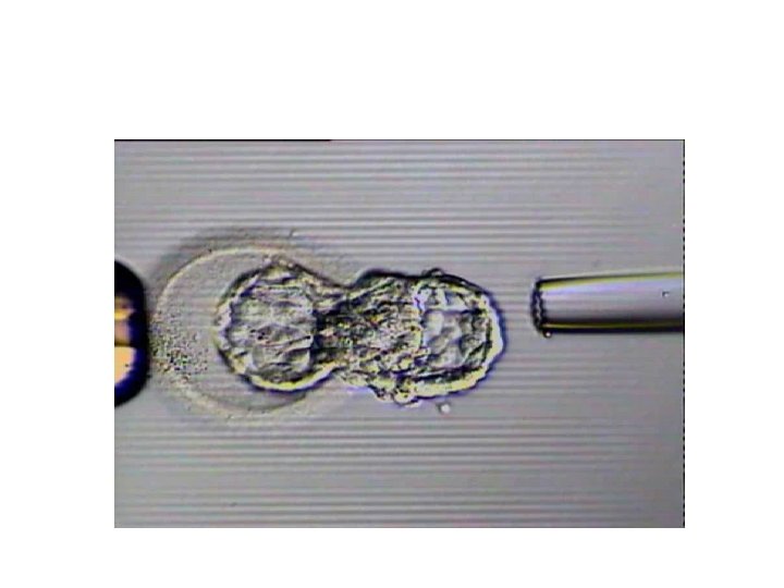
• The first birth after human blastocysts vitrification of biopsied embryos for PGD was reported by Parriego et al (2007). • Bu gelişmenin üzerine genetik testler için zaman sınırlaması ortadan kalktığından PGD ve özellikle de PGS alanında çok önemli gelişmeler gerçekleşti.
Potential Sources of genetic material 1. Polar Body Verlinsky et al. , (1990) Human Reproduction 5: 826‐ 829 2. Polar body Verlinsky et al. , (1990) Human Reproduction 5: 826‐ 829 Blastomeres • Handyside et al. , (1990) Nature 344: 768‐ 770 Trofektoderm Kokkali et al, (2005) Human Reproduction 20: 1855‐ 1859 Mc. Arthur et al. , (2005) Fertility and Sterility 84(6): 1628‐ 36
We only perform blastocyst freezing for biopsied embryos
• • • The survival rate after warming in the non‐biopsied cleavage control group was significantly higher than in the biopsied cleavage group (92. 0% versus 64. 0%, P = 0. 037). At the morula stage, both biopsied and non‐biopsied embryos had similar survival rates. However, a significantly higher survival rate (95. 6%) was observed in the biopsied blastocyst group compared with the control group (81. 3%, P = 0. 035).
Most of the biopsied cleavage stage embryos were destroyed due to blastomeres escaping. • The high osmotic potential of the medium caused the blastomeres to shrink dramatically. • Cell connection loose of blastomeres after biopsy contributes greatly to the unsuccessful vitrification of biopsied cleavage embryos. • 94% survival rate without blastomeres escaping (Zheng et al. , 2005). • Slit opening on the zona, with PZD can block blastomeres escaping.
Biopsy allows better exposure of the expanded blastocoele to the cryoprotectant and results in better dehydration of the blastocoele (Cervera et al 2003) Blastocysts with a larger blastocoelic cavity survived vitrification better when they had partially or completely hatched (Zech et al 2005). Vitrification of blastocysts results in lower DNA damage to the blastomeres following zonal hatching before vitrification (Kader et al. , 2007).
Vitrification at advanced embryo stages is an efficient method for biopsied embryo cryopreservation. • This strategy provides an opportunity to select viable embryos for transfer. • A blastocyst has more cells than at other stages and it has a greater capability of withstanding cell loss in the vitrification procedure. • Each carrier can only contain one embryo, to keep embryo records in a PGD programme, so they are available in fewer numbers and it is much more efficient to vitrify biopsied embryos at the blastocyst stage.
Concurrent PGD for Single Gene Disorders and Aneuploidy on Single Cells • Two blastomere biopsy on day three • Simultaneous biopsy on day 3 and on day 4 • Splitting trofectoderm cells to two pieces • Karyomapping
Simultaneous biopsy on day 3 and on day 4
Splitting trofectoderm cells to two pieces
Fully Hatched Blastocyst Freezing
• It has been shown that high survival rates can be obtained for blastocysts that were biopsied at the cleavage stage using open vitrification. Using the cryoloop system, Keskintepe et al. (2009) reported a higher survival rate (95%) after vitrification than after slow cooling (71%) • Using the cryoloop system, Keskintepe et al. (2009) reported a higher survival rate (95%) after vitrification than after slow cooling (71%)
A total of 837 patients underwent 998 cycles and an embryo transfer
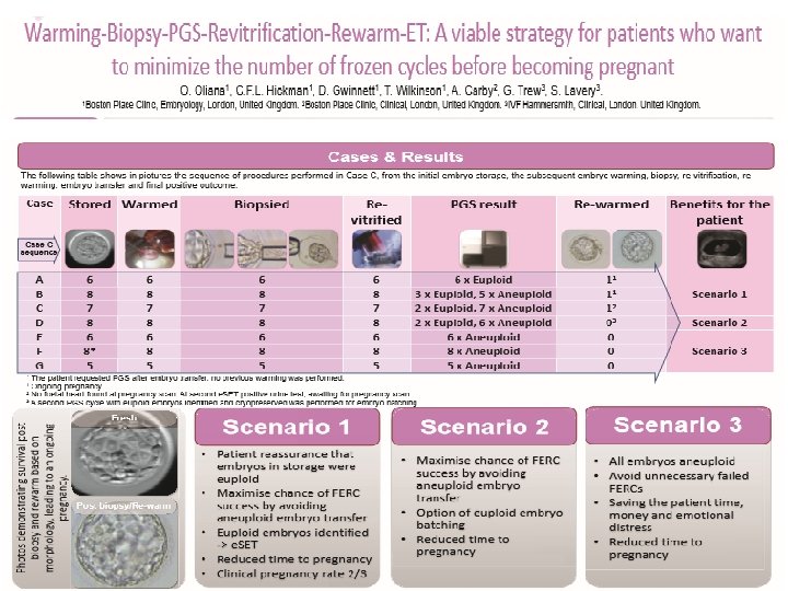
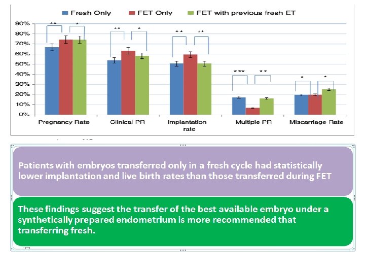
Advantages of FET cycles ver fresh IVF/ET cycles Freeze-all strategy offers the opportunity to control the window of implantation and possibly improve embryo implantation. 1 FET cycles increase the number of biopsied embryos for patients utilizing PGS and streamlines the clinical processes by scheduling single embryo transfer in subsequent cycles 2 Decrease in multiple PRs. This is made possible by the higher single euploid ET rate in an FET cycles. 3 IRs from patients in group “FETwith a previous fresh ET” were the same to those in the “Fresh Only” group (50. 9 vs. 50. 9 %), but significantly lowerthan “FET Only” (50. 9 vs. 59. 5%), Elective embryo cryopreservation has been described as a potential prevention/risk-reducing approach for OHSS 4 5 Your Logo
The application of vitrification for blastocysts and oocytes, opened new perspectives • • • extended embryo culture, Single blastocyst transfer, Blastocyst biopsy, alternative ways for fertility preservation oocyte donation
• Preimplantation genetic diagnosis (PGD) and single cell genetic analysis has become increasingly more popular since its first application (Handyside et al. , 1990) as an alternative to prenatal diagnosis to avoid termination of pregnancy in couples at high risk of transmitting a genetic defect. There are three potential sources of embryonic genetic material for preimplantation genetic analysis: polar bodies (biopsy of the first polar body from oocytes before sperm insemination or biopsy of both polar bodies from oocytes after sperm insemination), blastomeres from cleavage stage embryos and trophectoderm cells from embryos at the blastocyst stage.
• However, due to the discontinuous zona and empty space consequent to the removal of blastomeres, the survival rate of biopsied human embryos was significantly lower than non‐ biopsied embryos when using conventional slow‐freezing in which, the cooling rate was controlled at 0. 2 or 0. 3 C/min after seeding by a cryopreservation machine such as Planer or Minicool (Joris et al. , 1999; Magli et al. , 1999; Ciotti et al. , 2000). I
• Therefore, cryopreservation of biopsied embryos could be performed on days 3, 4 or 5.
The aim of this study to analyse the efficiency of the vitrification of biopsied embryos at the blastocyst stage using closed vitrification and storage. Group 1 : vitrified blastocyst transfer without biopsy Group 2 : fresh blastocyst transfer of biopsied embryos The closed system vitrification is a feasible method for cryopreserving Day 5 and Day 6 blastocysts that were biopsied at the cleavage stage.