Onion Osmosis 1 Before Starting You need to

Onion Osmosis 1

Before Starting… • You need to have – A compound microscope – A diced red onion – A slide and cover slip – Forceps – Some tap water – Some 15% salt solution and a pipette – A piece of paper towel 2

How to: Microscopic Measurement Determine the diameter of your “field of view. ” 1. Begin with a clear ruler. 2. Place on the stage, visible under the lowest magnification for your microscope. 1 mm 3. Determine the diameter of the field of view in mm. 4. 1 mm=1000 Micrometers (μm) 3

How to: FOV under a higher power lens For a higher power, you need to calculate the field of view.
 New FOV 4 x 10 x 4500")
Zoom in Original New Objective FOV (μm) New FOV 4 x 10 x 4500 1800 4 x 40 x 4500 450 4 x 100 x 4500 180 • (Original Obj/New Obj) x Original FOV=New FOV • FOV=Field of View 5

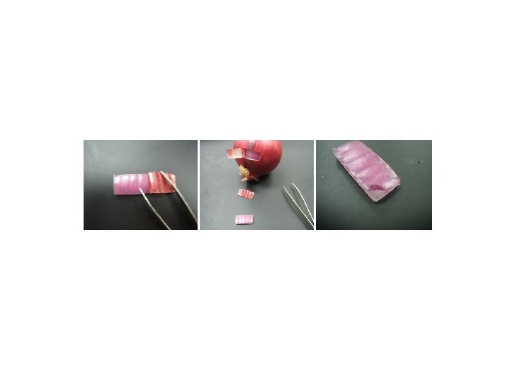
Onion Osmosis

Cut a piece of outer pigmented tissue

Make a wet mount with tap water

Add concentrated salt solution. Hurry…you will want to watch!

Questions? • What if…. . • Can I… • How do I… Jot them down.

Let’s try to answer them. • Can you focus your question to use the materials we have available? • Does your question deal with osmosis? This is the concept our trial focused on. Can you focus your question on osmosis? 12

Modeling • After you form a question, model what you think will happen with model pieces. • Get the OK from your teacher then check it out. • Keep good records. 13

A slide of red onion cells. Onion. Cell Which way will water move if a concentrated sugar solution is added? Show this with your models. 14

Going further: Consider taking a picture through the microscope. Analyze with Image J.

Image J Download for free from NIH.

Determining Percent Change in Area Find your picture file and open it.

To get area and perimeter. . . Select the straight line tool. Drag across your field of view.

Under analyze: Set scale

Pixels are already there. Enter your known distance.

Analyze: Set Measurements

Trace with the freehand tool Go to analyze: Measure Compare before and after
- Slides: 22