One Kidney is Sufficient Just one donated kidney


 Use the information in the tables to describe three differences between blood in")

 Use the information in the tables to describe three differences between")


. • Use of safety goggles,")
- Slides: 23
One Kidney is Sufficient Just one donated kidney is required to substitute two failed kidneys. The life expectation of somebody who donates a kidney is as similar as the general population. There is no increased danger of kidney disease if a wellscreened human being donates a kidney to somebody who is in need of a transplant. http: //www. nhs. uk/Conditions/O rgandonation/Pages/Recommendati ons. aspx
The Nephron Learning Objective: In order to be successful in this lesson you must be able to: Use key words to explain in detail the process of ultrafiltration and selective reabsorption Success…
Objectives • Describe the function of the Kidney • Explain what happens in the Nephron PROGRESS • Use key words to explain in detail the process of ultrafiltration and selective reabsorption
Exam Question
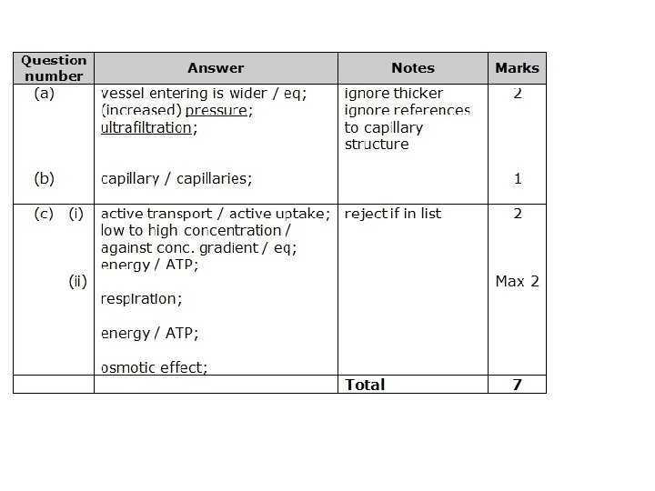
Mark Scheme
State whether the following statements are true or false. True 1. Organs A, B and C remove water from the body. …………. 2. Organ A helps to control body temperature. …………. False 3. Organs B and C remove carbon dioxide from the body. …………. 4. Urine is produced by organ A. True …………. True 5. Water and salts are removed from the body by organ C. ………… 6. Organ A produces urea. 7. Organ A removes urea and glucose from the body. False …………………… (7)
Function of the kidney: • You have two kidneys. • Each receives blood from the renal artery. • Kidneys act like filters, removing urea, excess water and excess salt. • When the urea is diluted with water it is called urine. • Urine is sent to the bladder for storage via the ureter.
Analysis of data: The diagram shows some substances in the blood entering the kidney and in the fluid in the ureter. (a) Name the fluid which passes down the ureter. ………………. . . … (1) (b) What happens to this fluid after it leaves the ureter? ……………………. (2)
(c) Use the information in the tables to describe three differences between blood in the renal artery and fluid in the ureter. (3) (d) Name two substances, from the table, that the kidneys remove from the body. (2) (e) Which other substance makes up most of the fluid in the ureter? (1)
Mark scheme The diagram shows some substances in the blood entering the kidney and in the fluid in the ureter. (a) Name the fluid which passes down the ureter. urine; ………………. . . … (1) (b) What happens to this fluid after it leaves the ureter? stored in the bladder; …………………… until released via the urethra; ………………. (2)
Mark scheme (c) Use the information in the tables to describe three differences between blood in the renal artery and fluid in the ureter. sugar in blood but none in fluid; protein in blood but none in fluid; max 3 more urea in fluid than in blood; more salt in fluid than in blood; (3) (d) Name two substances, from the table, that the kidneys remove from the body. urea; salt; (2) (e) Which other substance makes up most of the fluid in the ureter? water; (1)
Objectives • Describe the function of the Kidney • Explain what happens in the Nephron PROGRESS • Use key words to explain in detail the process of ultrafiltration and selective reabsorption
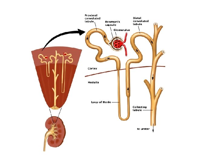
Where do you find the nephrons? Pelvis Renal artery Medulla Renal vein Ureter Cortex
Where do you find the nephrons? Structure of Kidney • Each kidney has four parts: • Cortex – the outer layer jammed pack full of filters called nephrons. Filters the blood. • Medulla – the middle layer which has the tubes carrying filtered wastes to the centre of the kidney. Contains Loop of Henle • Pelvis – area where all collecting ducts come together and connect with ureter. • Ureter – transports urine to the bladder.
Filtered blood leaves the kidney through renal vein the renal vein contains CO 2 and no waste. Blood enters the kidney from renal artery contains O 2 and waste materials Urine leaves kidney through ureter Nephron – the filter
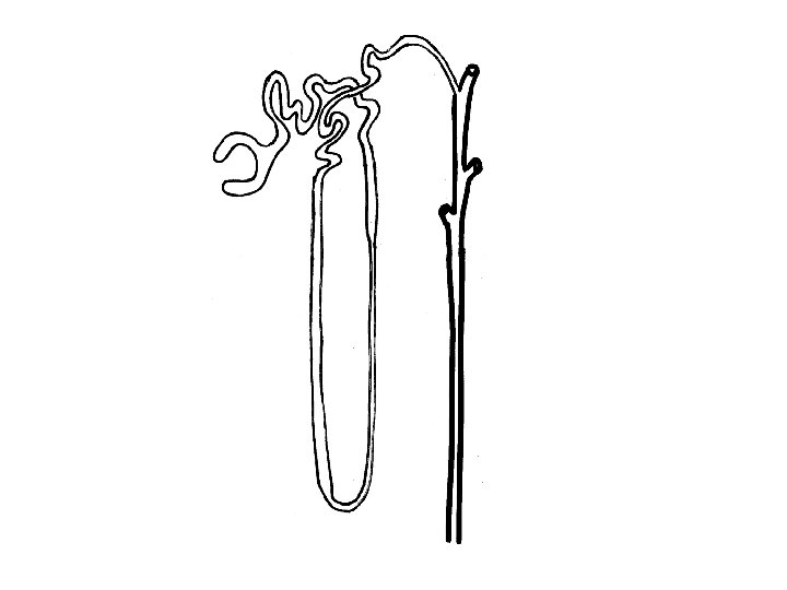
Task 1: Label your diagram of a nephron Task 2: Annotate your diagram to explain what happens in each part of the nephron
Blood is brought to the kidney in the renal artery Small molecules, like urea, glucose, salts and water pass through the filter. Large molecules, like blood proteins are too large to pass through. The blood is filtered as it passes into the glomerulus Urea, waste salts and water are left- this is now urine. It flows down the ureter to the bladder. All the glucose, some salts and much of the water are reabsorbed back into the blood.
Blood is brought to the kidney in the renal artery The blood is filtered as it passes into the glomerulus Small molecules, like urea, glucose, salts and water pass through the filter. Large molecules, like blood proteins are too large to pass through. All the glucose, some salts and much of the water are reabsorbed back into the blood. Urea, waste salts and water are left- this is now urine. It flows down the ureter to the bladder.
Task 3 Ultrafiltration and Selective Student number 2: Reabsorption Student number 1: • • • Describe what ultrafiltration is. Describe where ultrafiltration takes place. Explain what the glomerular filtrate is made up of. • • http: //www. bi ologymad. co m/resources/ kidney. swf • Describe what selective reabsorption is. Describe where selective reabsorption of glucose and water takes place. Describe how water and glucose are selectively reabsorbed
Risk Hazard Control Dissection infection material (treated as contaminated). • Use of safety goggles, gloves and apron at all times. • Teacher reviews experimental procedure with class. • Students informed regarding the hazards. • Biological wastes correctly disposed of i. e. wrapped in newspaper and disposed of by laboratory assistances. • Teacher demonstrates the use of dissecting equipment Sharp, cutting cuts instruments (scalpels). • Teacher distributes scalpels to benches and then removes them from benches. • Scalpels are not to be removed from the bench area i. e. no walking around. • Scalpels always remain on the tops of the benches; are never dropped below the level of the bench. • Students not using the scalpel are to stand opposite the student carrying out the dissection, not beside. • All dissection occurs on the tile placed on newspaper. Students to cut in direction away from hand. • All students wash their hands at the completion of the experiment (before returning to desks).