Oncologic Emergencies Haskell Gill Kirkpatrick M D 92205
 Kirkpatrick M. D. 9/22/05")
 • Affects 5 -10% cancer patients – Most commonly:")


 > 250, 000 may cause vasoocclusive complications • Leukemic")


- Slides: 43
Oncologic Emergencies Haskell (Gill) Kirkpatrick M. D. 9/22/05
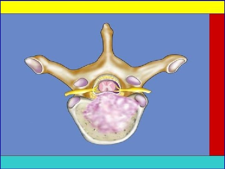
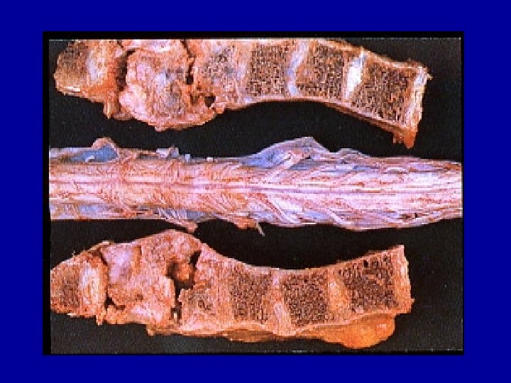
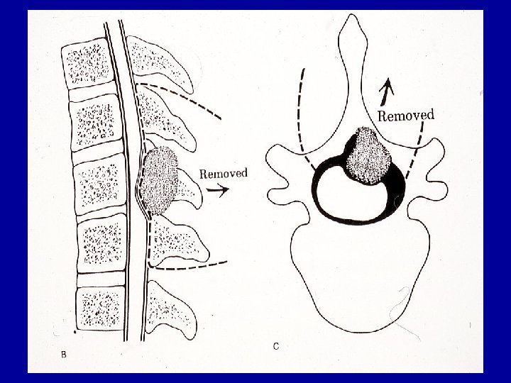
Malignant Spinal Cord Compression (MSCC) • Affects 5 -10% cancer patients – Most commonly: breast, prostate, lung, lymphoma and multiple myeloma • 20% MSCC cases are initial presentation • Bone (axial skeleton) common site of metastasis – Vertebral and epidural venous plexus (Batson’s plexus) • Most common mechanisms – Hematogenous met to vertebral body extending into epidural space – Pathologic fracture of vertebral body (infiltrated with tumor) resulting in cord injury from bone fragmentation or instability • 65% cases affect thoracic spine – 20% cases lumbar spine (colon and prostate predilection) – Cervical and sacral involvement rare
Clinical Presentation of MSCC • Back pain: In certain cancer patients should be considered metastatic origin until proven otherwise • Periostium richly innervated – Vertebral body tender to palpation/percussion • • Pain worse with recumbancy Usually precedes neurologic symptoms (1 -2 months) Radicular pain most common with lumbosacral lesions Thoracic radicular pain usually bilateral, band-like
Clinical Presentation of MSCC • Progression of motor findings: weakness, loss of gait, paralysis • Majority of compressions at thoracic level: paraparesis • Upper lumbar spine: conus medullaris syndrome – Distal lower extremity weakness, saddle paraesthesias and overflow leakage from bowel and bladder • Loss of bladder and bowel function generally a late finding • Majority of patients not ambulatory at time of diagnosis
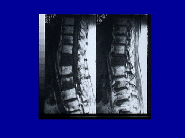
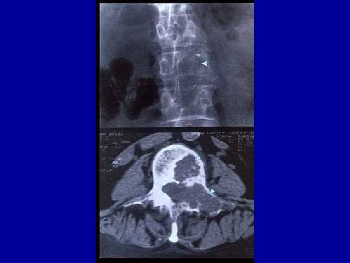
Diagnosis of MSCC • Average time from onset symptoms to diagnosis: 3 months • MRI of whole spine is most sensitive test • Decision to use modality based on history of back pain – Suspicion for pain secondary to Degenerative disease • mostly affects lower cervical and lower lumbar spine • Waxes and wanes • Responds to NSAIDs and bed rest – Suspicion for pain secondary to MSCC • Thoracic spine • Progresses despite conservative treatments • Aggravated by supine position
Treatment of MSCC • Corticosteroids: Optimal dose? – “High dose” studied in only randomized trial (+/- XRT) • 96 mg IV bolus then 24 mg 4 X /day (tapered over 10 days) • Serious side effects (GI perforations and bleeding) – Most common regimen: • 10 mg bolus then 16 mg/day (divided over 4 doses) • Radiation therapy – Relieves pain in most patients – Pre-treatment neurologic fxn strong predictor of response – Underlying tumor type also predictor • Aggressive surgery – New data shows that all patients should be considered for decompressive radical resection
Patchell et al, ASCO 2003
Patchell et al, ASCO 2003
Patchell et al, ASCO 2003
Febrile Neutropenia • Should be considered an emergency – Early studies have shown high mortality when delay initiation of appropriate antibiotics – Before era of empiric antibiotics infection accounted for up to 75% of deaths associated w/ chemotherapy • Definitions: – Fever: single temp > 38. 3°C (101. 3°F) or 38. 0°C (100. 4°F) sustained greater than 1 hour – Neutropenia: usually ANC < 500 • Absolute neutrophil count (ANC)=total WBC X (%neutrophils + %bands)
Infection as Cause of Death in Cancer Patients Bodey GP et al, Ann Intern Med 1966; 64: 328
Organisms Causing Infection During Chemotherapy of Acute Leukemia Bodey GP et al, Ann Intern Med 1966; 64: 328
Infections
Febrile Neutropenia • Seeding of the bloodstream from endogenous flora in the GI tract most common cause • Commonly cultured bacterial pathogens – Gram neg (Pseudomonas, E Coli, Klebsiella etc. . ) – Gram pos (Coag-neg staph, staph aureus, streptococcus etc…) • Commonly cultured fungal pathogens – Candida species, Aspergillus – usually arise later as a secondary infection in patients with prolonged neutropenia and antibiotic use • Viral pathogens – HSV, VZV
Treatment of Febrile Neutropenia • Empiric Antibiotics – Appropriate coverage of known or suspected infection based on history/exam findings/radiographic studies • Monotherapy: – ceftazidime, imipenem, meropenem, or cefepime • Double coverage: – beta-lactam and an aminoglycoside • Awareness of institutional resistance patterns • Addition of empiric Vancomycin – Skin or catheter site infection, hypotensive, hx of MRSA colonization, mucositis, quinolone prophylaxis
Causes of Fever in Patients with Prolonged Neutropenia Who Are Receiving Broad-Spectrum Antibiotics Corey, L. et al. N Engl J Med 2002; 346: 222 -224
Treatment of Febrile Neutropenia • Empiric anti-fungal coverage with persistent fever on broad-spectrum antibiotics and prolonged neutropenia – Amphotericin B (liposomal), caspofungin, voriconazole • Colony stimulating factors – Should not be used routinely – Appropriate for critically ill patients
General Principles for the Management of Fever in Patients with Neutropenia Pizzo, P. A. N Engl J Med 1993; 328: 1323 -1332
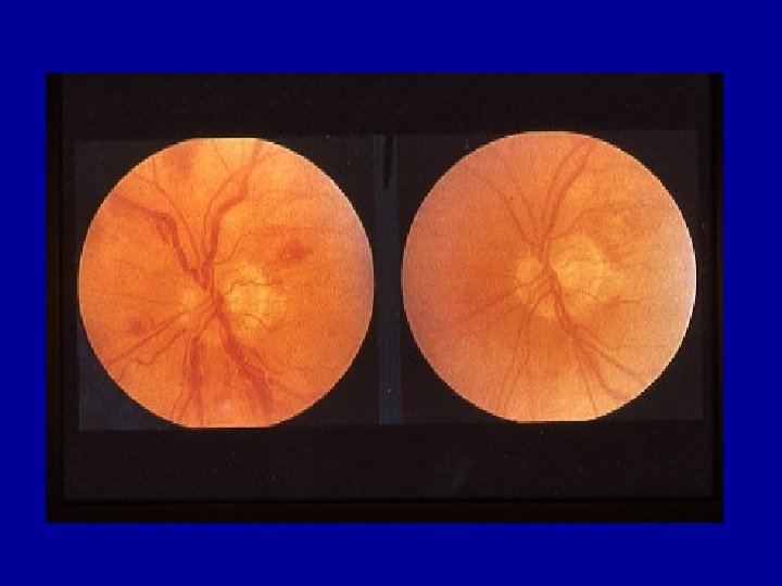
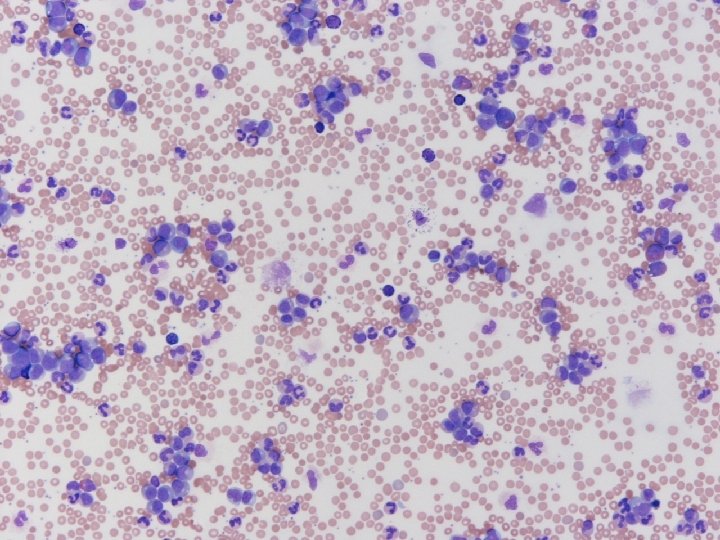
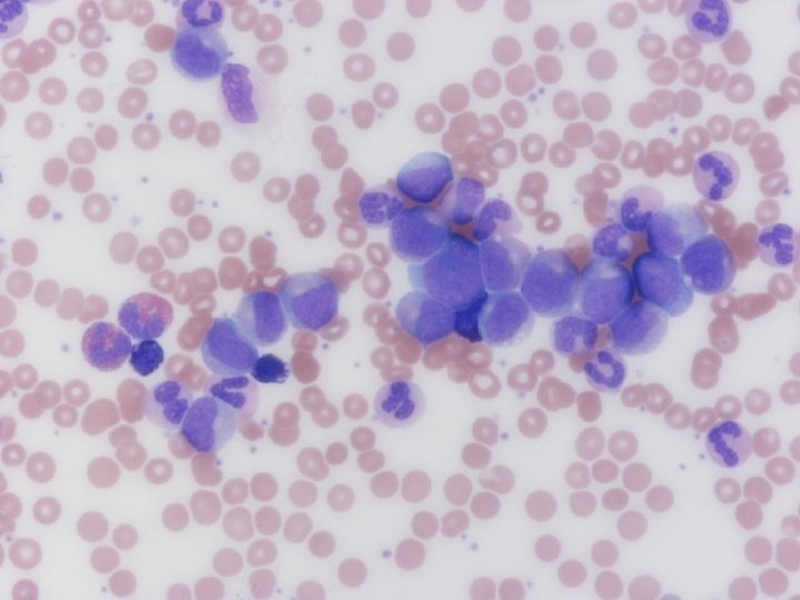
Hyperleukocytosis • Neutrophil count (CML) > 250, 000 may cause vasoocclusive complications • Leukemic blasts (AML) are nondeformable – Cause hyperviscosity at lower counts ( 70, 000 +) • Leukostasis in microvasculature leads to clinical symptoms – Pulmonary: hypoxemia – CNS: headaches, vision changes/loss, focal deficits • Symptomatic hyperleukocytosis and AML associated with initial high mortality
Treatment of Hyperleukocytosis • Emergent leukophoresis can be used – Should be used as adjunct to chemotherapy – Temporizing measure • Initiate cytoreductive therapy ASAP – Blasts are rapidly accumulating – Can result in another oncologic emergency…
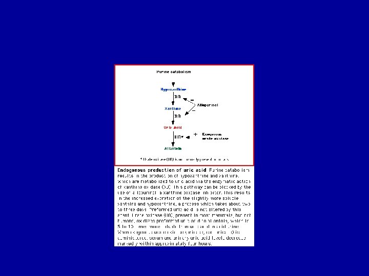
Tumor Lysis Syndrome • Rapid cell death in face of high tumor burden – Large amounts of intracellular metabolites released • Uric acid, potassium, phosphate. . • Most commonly associated with poorly differentiated lymphomas and leukemias – Burkitt’s – ALL (more commonly than AML) • Uric acid can deposit in kidney leading to ARF • Dialysis can support patient • Rasburicase or Elitek (urate oxidase): oxidizes uric acid to allantoin which is water soluble
Prevention of tumor lysis syndrome • Vigorous hydration • Allopurinol 300 -900 mg/day – Ideally 2 days before cytotoxic therapy • Role of alkalinizing urine debatable – Increases the solubility of uric acid and decreases tendency for precipitation but… – Alkalinizing could promote calcium-phosphate deposition – Animal studies have shown that increased tubular flow rate is most important protective measure – Vigorous hydration with saline is likely as effective
SVCS: Primary Pathologic Diagnoses
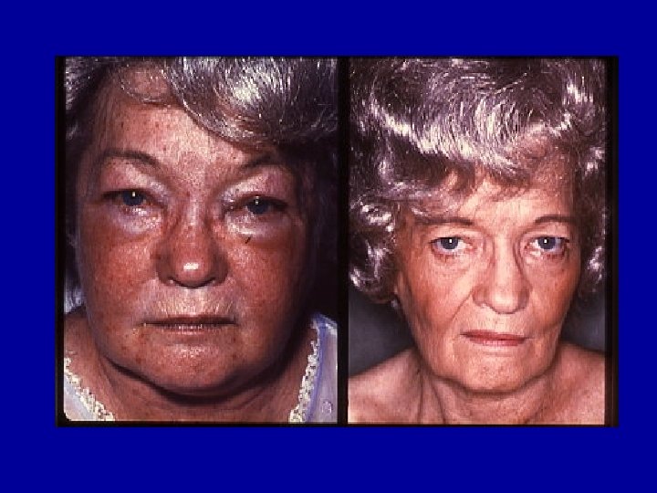
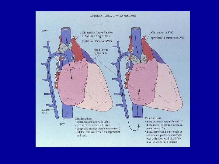
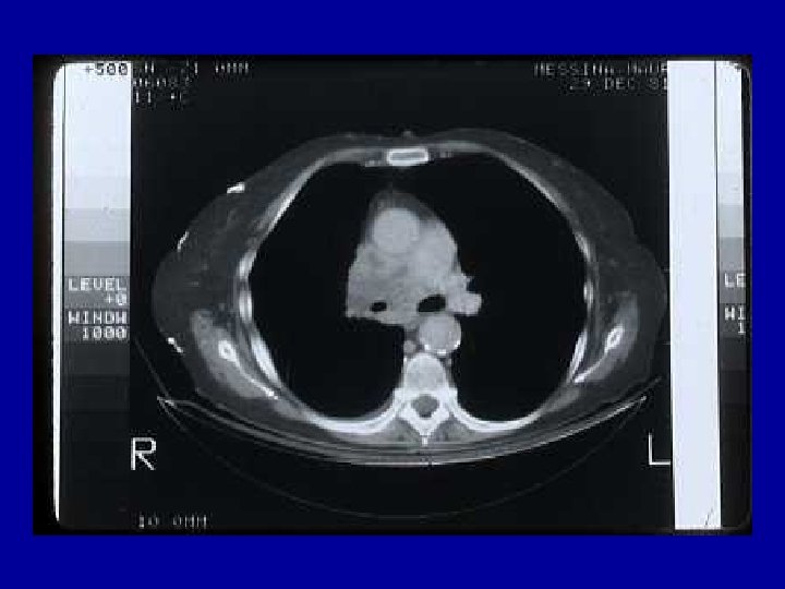
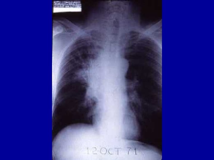
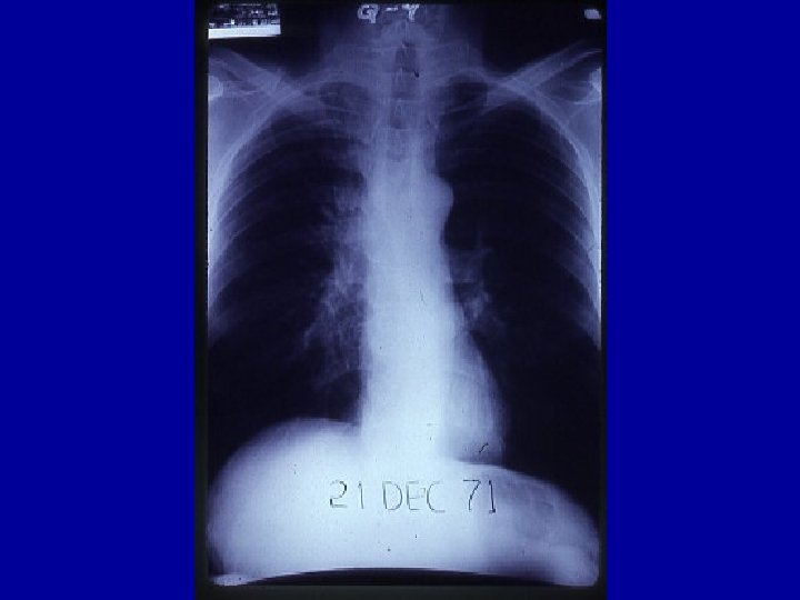
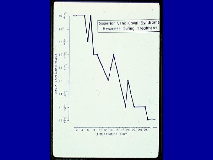
Superior Vena Cava Syndrome • Invasion or external compression of SVC • Malignant tumors responsible for 80% cases – Infection and thrombosis account for most of the rest • Symptoms – Dyspnea – Facial swelling, arm edema, cyanosis • Signs – Venous distension on neck and chest wall – Facial edema
Superior Vena Cava Syndrome • 60% cases due to malignancy present without known diagnosis • CT preferred diagnostic tool • Importance of biospy – Short delay not compromise outcome most cases – Histology helps determine treatment and prognosis • Treatment responsive tumors: SCLC, germ cell tumors, NHL • Role for intraluminal stents?