
ONCOCYTOMA AML ONCOCYTOMA n n Recent diagnosis since
- Slides: 35
ONCOCYTOMA & AML
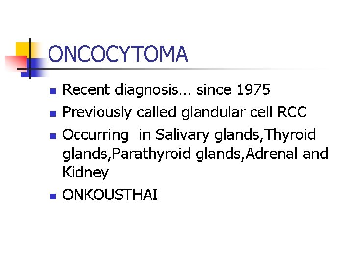
ONCOCYTOMA n n Recent diagnosis… since 1975 Previously called glandular cell RCC Occurring in Salivary glands, Thyroid glands, Parathyroid glands, Adrenal and Kidney ONKOUSTHAI
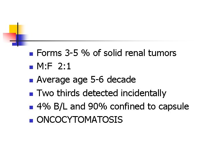
n n n Forms 3 -5 % of solid renal tumors M: F 2: 1 Average 5 -6 decade Two thirds detected incidentally 4% B/L and 90% confined to capsule ONCOCYTOMATOSIS
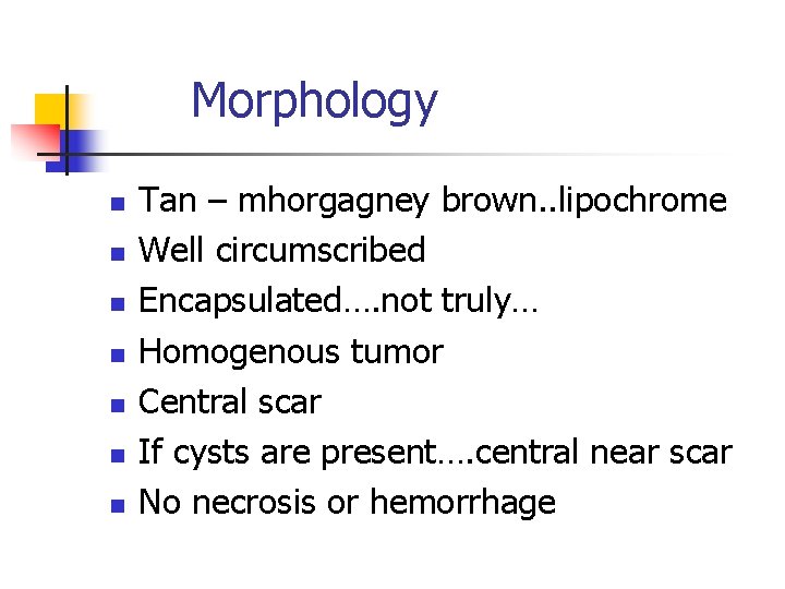
Morphology n n n n Tan – mhorgagney brown. . lipochrome Well circumscribed Encapsulated…. not truly… Homogenous tumor Central scar If cysts are present…. central near scar No necrosis or hemorrhage
Oncocyte n n n n Oncocyte……origin from itercalated cells of CD Large polygonal epithelial cell Eosinophillic/acidophillic Abundant mitochondria expanding the cytoplasm( swollen & densely packed mito) Organoid/tubulocystic/solid/mixed pattern Less microvillus & absent brush border Low nuclear grade I-II Archipelagic architexture
Atypical features n n 10 -20% can have perinephric fat extension 20 -30% can have hemorrhage 7 -32% can have co-exsistant RCC 4 -13% can be B/L or multiple.
Genetic defects n n n CH…. I CH…. 11 CH…. . 14 CH…. Y No changes in CH…. 3
Investigations n n n ANGIO : spoke wheel pattern, orderly vascular arrangement, absent AV shunting, no vascular puddling, peritumoral halo (lucent-rim sign). CT : homogenous mass, stellate scar, MRI : well defined loww intensity mass on T 2 image SESTAMIBI Tec 99 Stellate scar : 1/3 by CT and ¼ by USG
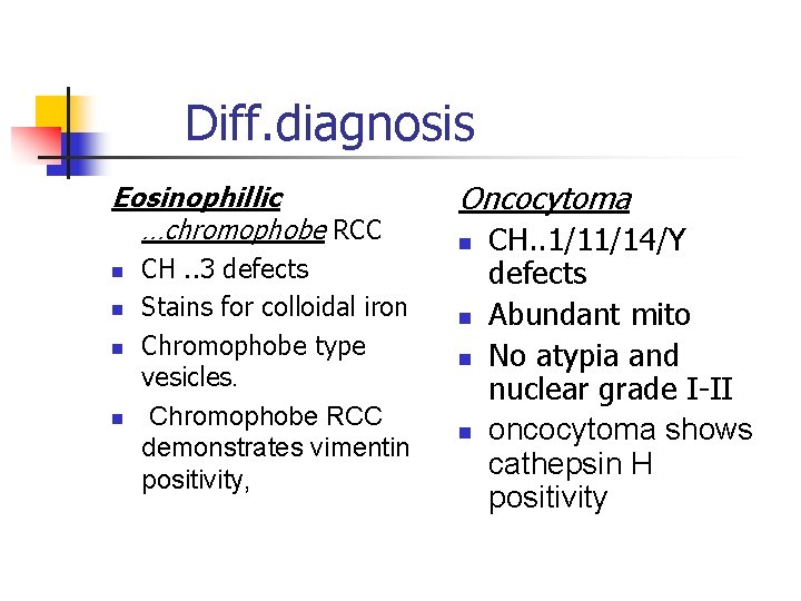
Diff. diagnosis Eosinophillic …chromophobe RCC n n CH. . 3 defects Stains for colloidal iron Chromophobe type vesicles. Chromophobe RCC demonstrates vimentin positivity, Oncocytoma n n CH. . 1/11/14/Y defects Abundant mito No atypia and nuclear grade I-II oncocytoma shows cathepsin H positivity
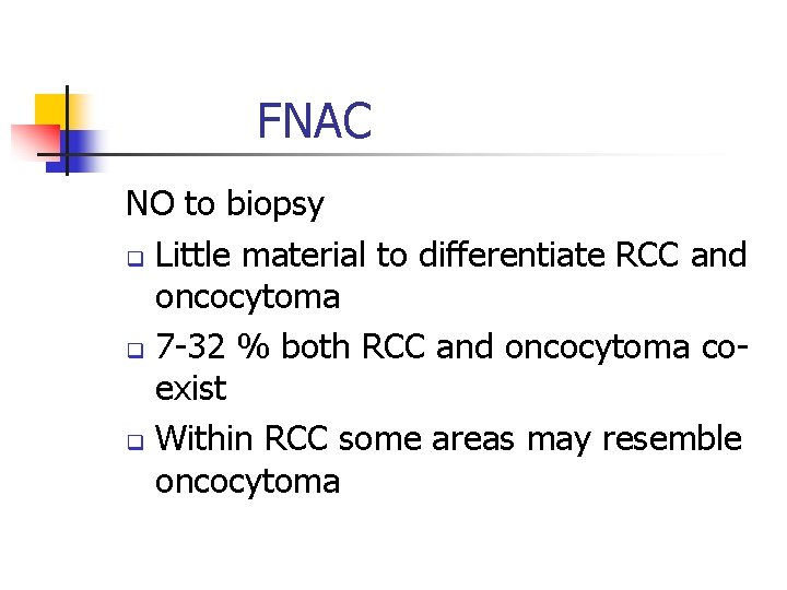
FNAC NO to biopsy q Little material to differentiate RCC and oncocytoma q 7 -32 % both RCC and oncocytoma coexist q Within RCC some areas may resemble oncocytoma
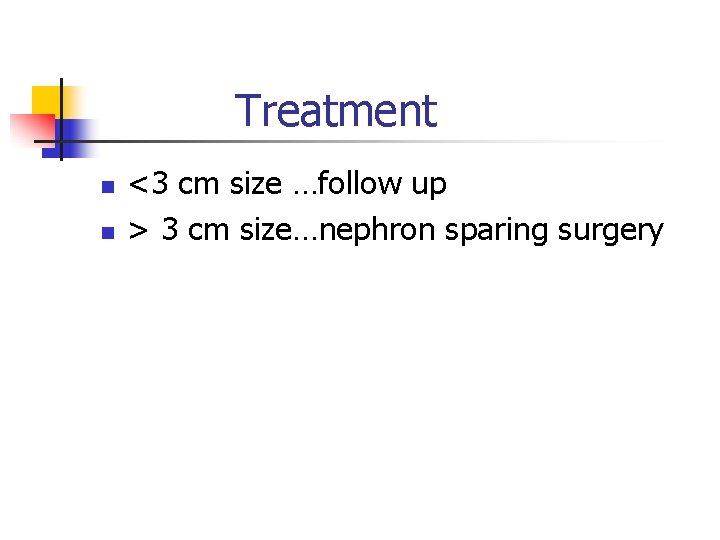
Treatment n n <3 cm size …follow up > 3 cm size…nephron sparing surgery
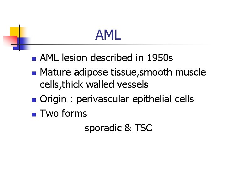
AML n n AML lesion described in 1950 s Mature adipose tissue, smooth muscle cells, thick walled vessels Origin : perivascular epithelial cells Two forms sporadic & TSC
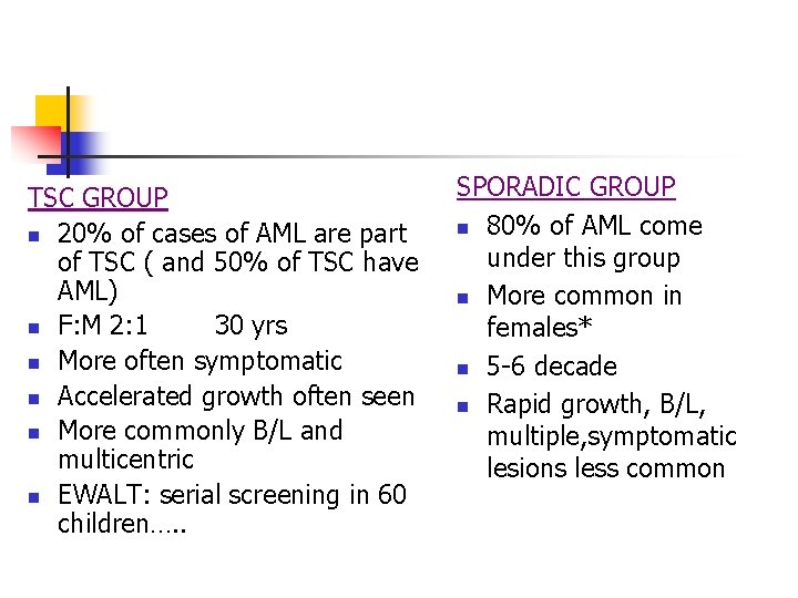
TSC GROUP n 20% of cases of AML are part of TSC ( and 50% of TSC have AML) n F: M 2: 1 30 yrs n More often symptomatic n Accelerated growth often seen n More commonly B/L and multicentric n EWALT: serial screening in 60 children…. . SPORADIC GROUP n 80% of AML come under this group n More common in females* n 5 -6 decade n Rapid growth, B/L, multiple, symptomatic lesions less common
Wunderlich syndrome…. 10% cases n n n Retroperitoneal hemorrhage/flank pain/hematuria/tender mass/shock Next to RCC…. commonest cause of retro peritoneal bleed Thick blood vessel. . no inner intimal layer and elastic layer replaced by fibrous tissue.
PRESENTATION n n n TRIAD…pain/tender mass/gross hematuria Other symptoms : anemia/HT/renal failure/nausea/vomiting/loin pain/renal failure 50% asymptomatic
HISTOLOGY n n n FAT: immature – mature fat cells Vessels : thick walled easily friable Smooth muscles: spindle/round epitheloid cells around vessels
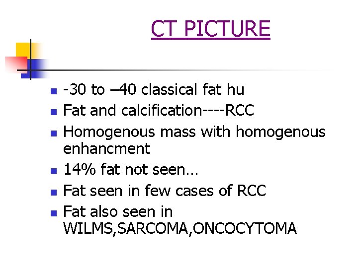
CT PICTURE n n n -30 to – 40 classical fat hu Fat and calcification----RCC Homogenous mass with homogenous enhancment 14% fat not seen… Fat seen in few cases of RCC Fat also seen in WILMS, SARCOMA, ONCOCYTOMA
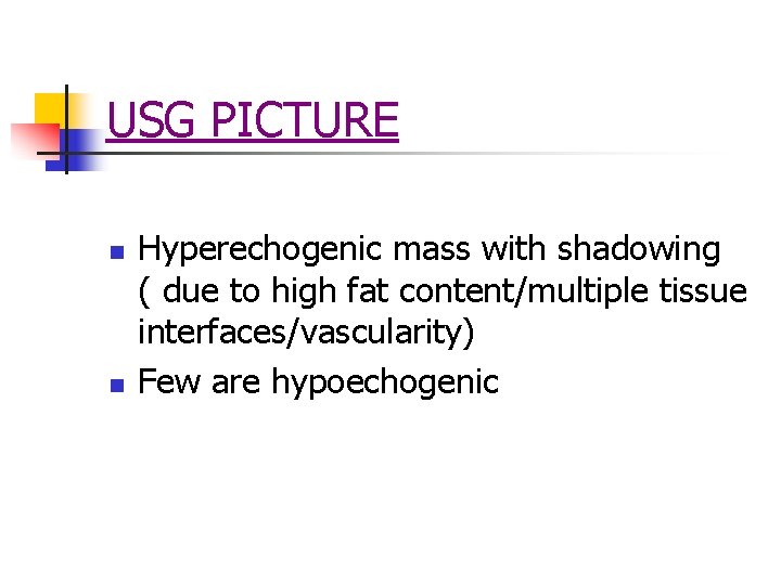
USG PICTURE n n Hyperechogenic mass with shadowing ( due to high fat content/multiple tissue interfaces/vascularity) Few are hypoechogenic
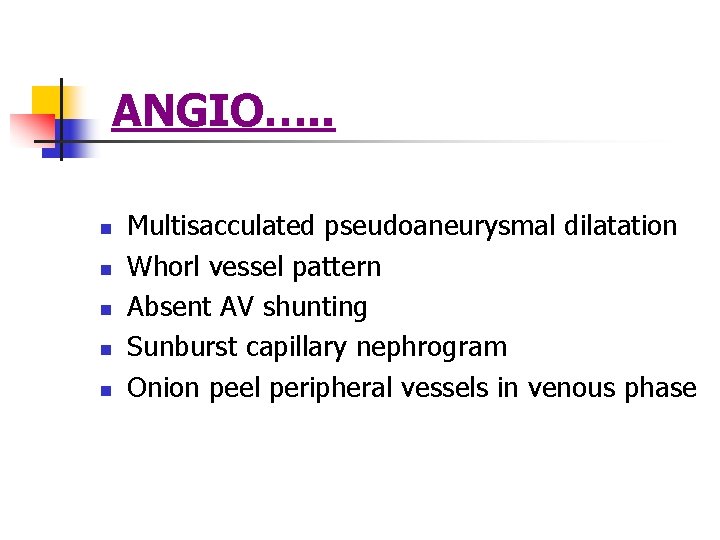
ANGIO…. . n n n Multisacculated pseudoaneurysmal dilatation Whorl vessel pattern Absent AV shunting Sunburst capillary nephrogram Onion peel peripheral vessels in venous phase
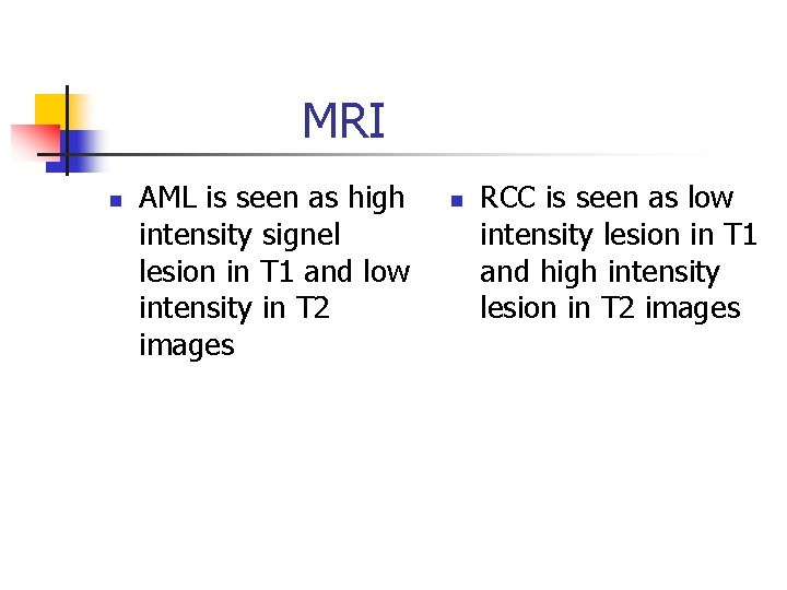
MRI n AML is seen as high intensity signel lesion in T 1 and low intensity in T 2 images n RCC is seen as low intensity lesion in T 1 and high intensity lesion in T 2 images
n n Extra renal…. hilar LN, retroperitonium, liver ER/PR present HMB-45 Ab…. melanoma associated antigen( also seen in melanoma, cardiac rhabdomyoma, cerebral tubers) EPITHELOID AML…malignant variant can have local invasion-distant mets. this has abundant epitheliod polygonal muscle cells.
n AML size >4 cm…… 80% symptomatic n Sporadic AML …growth rate 5% n TSC/MULTICENTRIC……growth rate 20% n Biopsy ……not advised Bleeding/seedling/negative biopsy dilemma
TREATMENT n n Angio embolisation( gelatin/absolute alcohol/iodised oil/polyvinyl alcohol/coils)… 5% abscess, 3% pleural effusion Post embolization syndrome Nephron sparing surgery
Nephrectomy n n Whole kidney replaced by tumor Solitay large tumor at hilum Suspicion of malignancy Large retro peritoneal bleed
n n AML with renal vein tumor thrombus AML with RCC…. 1 -3% of TSC…. young women Recurrent AML and pregnency ? Spontanous rupture common ? prophylactical removal >4 cm size
TSC n n n Bourneville classical description VOGT triad. . MR, seizures, adenoma sebaceum FORME FROSTE…. TCS without MR Gene……TSC 1 & TSC 2 more severe disease more renal involvement
TYPES TSC gene 1 n Protein HAMARTIN for cell to ceel adhersion Viz actin and plasma membrane binding protien TSC gene 2 n Encodes for TUBERIN which regulates cytoplasmic vesicle transport…defect leads to accumulation of microvesicles in smooth muscle…( GTPase based)
Neurological manifestations Cortical tubers n Subependymal nodules n Subependymal giant cell astrocytoma n Seizure disorders n Cognative and behavioral disturbances ……………vigabratin n
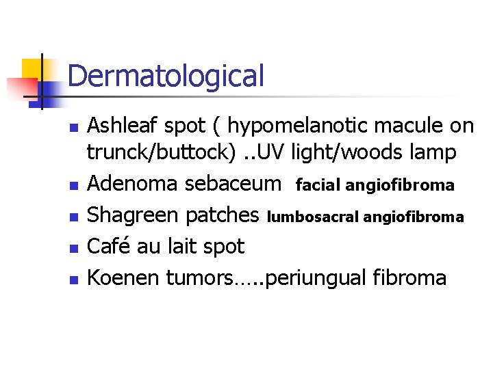
Dermatological n n n Ashleaf spot ( hypomelanotic macule on trunck/buttock). . UV light/woods lamp Adenoma sebaceum facial angiofibroma Shagreen patches lumbosacral angiofibroma Café au lait spot Koenen tumors…. . periungual fibroma
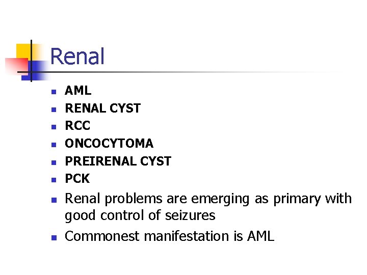
Renal n n n n AML RENAL CYST RCC ONCOCYTOMA PREIRENAL CYST PCK Renal problems are emerging as primary with good control of seizures Commonest manifestation is AML
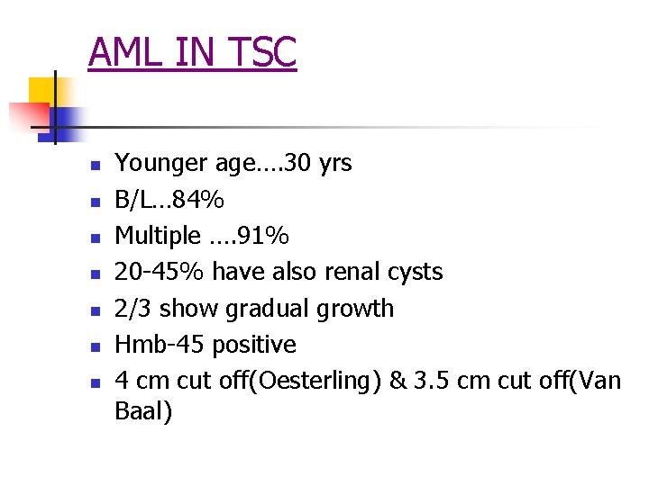
AML IN TSC n n n n Younger age…. 30 yrs B/L… 84% Multiple …. 91% 20 -45% have also renal cysts 2/3 show gradual growth Hmb-45 positive 4 cm cut off(Oesterling) & 3. 5 cm cut off(Van Baal)
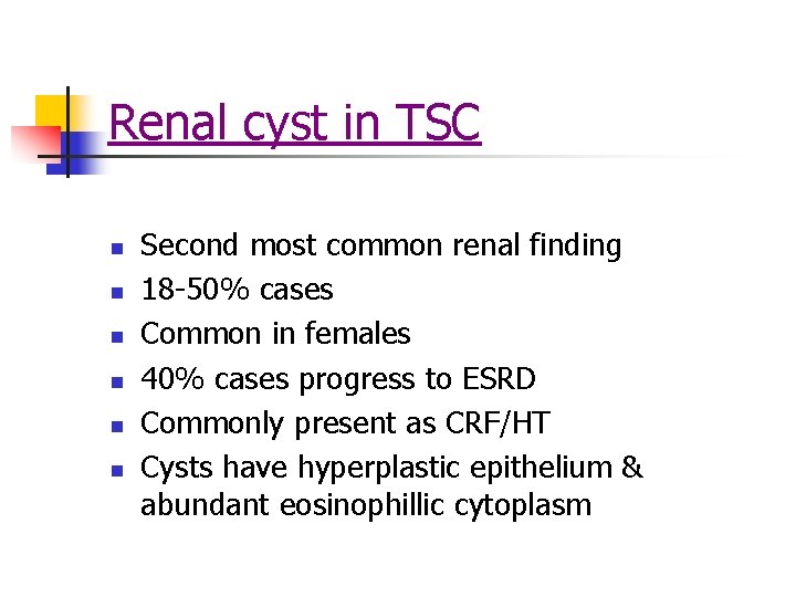
Renal cyst in TSC n n n Second most common renal finding 18 -50% cases Common in females 40% cases progress to ESRD Commonly present as CRF/HT Cysts have hyperplastic epithelium & abundant eosinophillic cytoplasm
RCC in TSC n n n Seen in 2 -4 % Age average is 28 yrs 80% in females 40% B/L Usually low grade/low metastatic potentialcommonly clear cell type
n n CVS & TSC : cardiac rhabdomyoma, peripheral arterial aneurysm RS & TSC : lymphangioleiomyomatosis OCULAR : retinal hamartoma AML IN LIVER
EVALUATION OF TSC n n n CT brain CT/MRI abdomen CXR Echo Woods lamp for skin macules