Odontogenic infection pathway Odontogenic infections are caused by

Odontogenic infection pathway

Odontogenic infections are caused by oral pathogens that inhabit the surface of the teeth and oral mucous membranes and are also found in the gingival sulcus and saliva

Microorganisms ivolved in mixed bacterial infections of the oral cavity
 § progressive dental caries")
Infection in oral cavity can be: Dental origin (primary infection) § progressive dental caries § extensive periodontal disease § trauma caused by dental procedures Nonodontogenic source (secondary infect. ) § an infection surrounding the oral cavity as the skin, tonsils, ears or sinusitis

Dental infection normally produce the classic signs of infection: Rubor - due to vasodilatation effect of inflammation Tumor - caused by pus accumulation and oedema Calor - caused by accelerated local metabolism Dolor - results from pressure on sensory nerve caused by edema or infection Functio laesa - problems with mastication, trismus, dysphagia, and respiratory impairment

Spread of dental infection The various pathways of spread with odontogenic infections: 1. per continuitatem 2. The path of least resistance - by spaces in the 3. head and neck 4. 2. by vascular system 5. 3. by lymphatic system

1. Spread of dental infection per continuitatem

Spread of apical infection § periodontal gap § alveolar process

§ The type and virulence of the microorganisms involved and the immunological condition influence the degree of spread of infection § Infection may be: - localized (abscess) - diffused (infection tends to spread rapidly through the tissues along the line of least resistence into the anatomically demarcated tissue spaces)

Abscess A closed tissue space with supuration from a dental infection Periapical - progressive carries, pathogens invade the pulp and spread apically Periodontal - caused by spread from an infected gum (usually in adults) Pericoronal - around an erupting third molar

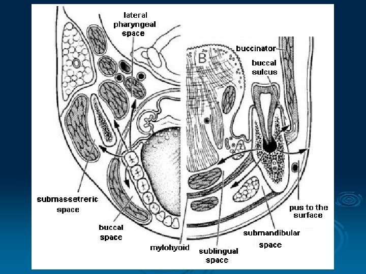
Local abscess can spread along the anatomically demarcated tissue spaces An barrier is the fascia and the muscle attachments to the bones buccinator muscle mylohyoid muscle

Vestibular Abscess § abscess perforate bone on the vestibular plate of the alveolar process § the roots of all teeth of upper and lower jaw § if the roots are localized upon the muscle insertion (lower jaw) or below muscle insertion (upper jaw)

Palatal Abscess § the roots of the upper lateral incisors or the first premolars and molars (roots often incline palatally) § usually no spraed over palatine raphe

The submucosal portion of the hard palate contains neurovascular bundle, minor salivary glands a lymfoid tissue § the rich innervation of the periosteum - painful ! § the course of the palatine artery - bleeding !

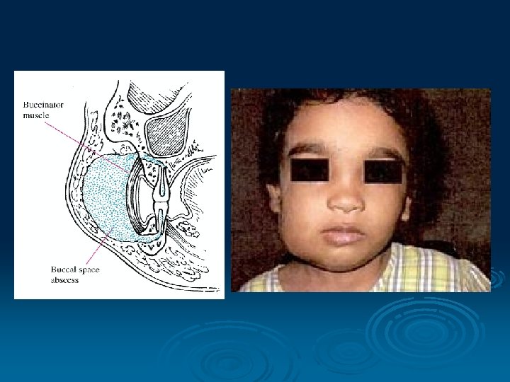
Buccal Space § premolars and molars both jaws § if the roots are localized above the buccinator muscle insertion (upper jaw) or below insertion (lower jaw) § infection spread into the soft tissues of the cheek → along anatomical planes toward the infratemporal or pterygopalatine fossa (pterygomandibular raphe!)

Infratemporal Space § molars of upper jaws § infection may ascend into the cavernous sinus (through venous plexus in the ovale and spinosum foramen), orbita, temporal fossa, pterygopalatine fossa Infratemporal space

Temporal Space § between the temporal fascia and the temporal bone § inferiorly communicate with infratemporal space Temporal space

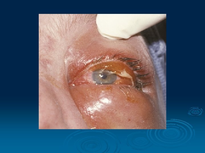
Infraorbital Space § usually anterior superior teeth, less often the premolars § between the levator anguli oris and the levator labii superioris muscles § possible infection via the angular vein → opthalmic vein → spread into the cavernous sinus § collateral oedema often includes the upper lip and lower eyelid

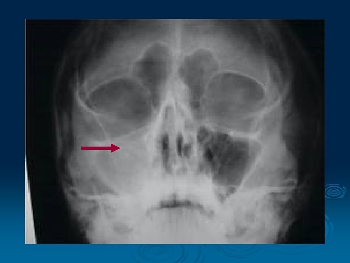
Maxillary Sinus § occasionally of dental origin, more often by respiratory infection § buccal and sometimes palatine root of first or second molar, second premolar that perforate the sinus floor § the floor of nasal cavity is infected from the anterior teeth

Submental Space § mandibular anterior teeth § the root of teeth lay below the muscles insertion (mental + depressor labii inf. muscles) § spread beneath the mylohyoid muscle into the submandibular area

Submandibular Space § mandibular posterior teeth § the root of teeth lay root apices lay below the buccinator muscle insertion § spread beneath the mylohyoid muscle into the submandibular area

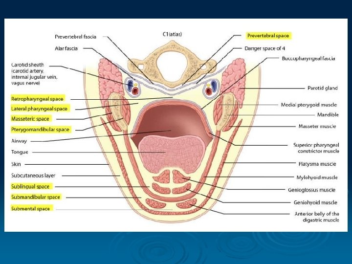
Sublingual Space § mandibular posterior teeth § spread to the sublingual space - between the mouth floor and mylohyoid muscle § CAVE! Ludwig´s angina § spread along submandibular duct into submandibular space

Ludwig´s angina = the right and left submandibular, sublingual and submental spaces are infected A fulminant infection can spread rapidly to pharyngeal and retropharyngeal space

Sublingual space Submental space Submandibular space

Masseteric Space l: parotideomasseteric fascia m: ramus of the mandible s: zygomatic arch i: insertio of the masseter muscle § posterior teeth of the lower jaw § expand laterally to the pterygomandib. space § oedema of the overlying masseter muscle

Masseteric space

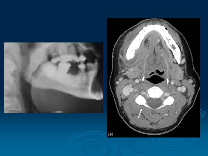
Pterygomandibular Space § carious, partially erupted mandibutal third molar or needle tract infection of anesthetize of inferior alveolar nerve § infection may spread into infratemporal space

Pterygomandibular space Alveolar inferior artery, vein and nerve !

Lateral Pharyngeal Space § peritonsillar infection penetrate the pharyngeal constrictor muscles → lateral pharyngeal space § shaped like an inverted pyramid, base at the base of the skull and its apex at the hyoid bone § space is divided into prestyloid and poststyloid compartments (by aponeurosis of Zuckerkandl and Testut, joining the styloid process to the tensor veli palatini)

Lateral pharyngeal space

The Neck Spaces Visceral space Visceral Paravisceral Retrovisceral Pretracheal space Prevertebral space

Abscess § Subcutaneous - between the superficial cervical fascia and platysma § Suprasternal - between the superficial and middle cervical fascia § Pretracheal § Parapharyngeal § Retropharyngeal

Retropharyngeal abscess

2. Spread of dental infection by blood system

§ Bacteremia - bacteria traveling in the blood § Infected thrombus - dislodge from the inner blood vessel wall and travel as an embolus → dural venous sinuses → brain or internal jugular vein → thrombophlebitis In general, veins of the head and neck lack valves, so blood can flow into and out of the cranial cavity !

Anterior pathway ophtalmic v. infraorb. v. deep facial v. Posterior pathway pterygoid plx. → oval or spinosum for.

3. Spread of dental infection by lymphatic system Repetition of the 2 nd semester

Buccal Vestibular Palatal Buccinator muscle Vestibular Sublingual Buccal Mylohyoid muscle Submandibular

Spatium basale intermusculare linguae Sublingual space st-gl hy-gl g-gl Gl. s-li g-hyo myl ohy oid. Gl. s-m dig. Submental space a. lingualis n. lingualis dct. submand. n. XII. Submandib. space
- Slides: 45