Odessa National Medical University Department of Normal and


- Slides: 144

Odessa National Medical University Department of Normal and Pathological Clinical Anatomy Lecture on the topic: General doctrine of tumors. Oncogenesis. Anatomical and microscopic features and types of growth of benign and malignant tumors. Morphological characteristics of the main stages of development of malignant tumors. Clinical and morphological nomenclature of tumors. Benign and malignant non-epithelial (mesenchymal) tumors. Sarcoma: features of development and metastasis. Epithelial tumors: benign organspecific epithelial tumors, cancer (features of development, metastasis, histological forms).

Tumours § Tumour, neoplasm or blastoma is a pathological process which is characterized by uncontrollable proliferation of cells. The replication of tumour cells differs dramatically from the other kinds of growth and proliferation of cells, which are observed in other pathological processes. The proliferation of cells in tumour does not submit to the regulative influences of human body.

§ Atypism of cells is the second important typical feature of a tumour. It concerns the structure, metabolism, function, replication and differentiation. § Anaplasia or cataplasia is acquiring new features by tumour cells, which are not typical for normal cells.

§ § § There are the following types of atypism: 1. morphological 2. biochemical 3. histochemical 4. antigenous atypism of a tumour cell. § Oncology is a special branch of medicine dealing with the study of tumours.

Tumour structure and features of a tumour cell § The outward appearance of a tumour can be different. It can have the shape of a node, a mushroom cap. Tumour surface can be smooth, rough, uneven. The tumour can be located within the organ or on its surface. Sometimes it diffusely invades all the organ. The tumour, located on the surface of an organ or the mucous membrane, is called a polyp. This tumour can often be connected with it by stem.

§ Tumour can often ulcerate. In this case it is called cancerous ulcer. On section tumour is homogenous, usually whitish-grey or greyish -white tissue. But frequently the section surface is motley, because there are hemorrhages, centers of necrosis. Tumour can have fibrous structure. Tumour of some organs (ovary) consists of separate cavities, cysts, which are filled with fluid or mucus.

§ Spleen with metastasis of melanoma

§ Liposarcoma

§ Cystadenoma of ovary

§ Teratoma

§ Tumour sizes are different. It depends on the quickness of its growth, origin and location. Consistency depends on the prevalence either of parenchyma or stroma. In the first case the tumour is soft, in the second it is compact, dense. § The tumour secondary changes are: inflammation, centers of necrosis, covering by mucus, calcification.

§ Microscopic structure of tumours significantly varies. However all tumours have some common structural features. Every tumour consists of parenhyma and stroma. But their ratio can be very different. In some tumours parenchyma prevails, in others stroma is prevalent. Sometimes parenchyma and stroma are distributed evenly.

§ The tumour parenchyma is formed by the cells which characterize this kind of tumour and determine its morphological specificity. The tumour stroma is formed from the connective tissue of the organ, in which it develops. It contains vessels and nervous fibers. § Most of the tumours resemble an organ by the structure that is they have both parenchyma and stroma. These tumours are called organoma. Sometimes stroma is developed weakly and consists of thinwalled vessels and capillaries only.

§ Such tumours are called histioid tumours. They usually develop quickly and are early exposed to necrosis. § A tumour when its structure conforms to the organ (or tissue) structure where it develops, is called homologous. Homologous tumours are mature and differentiated. If the cellular structure of the tumour differs from that of the organ (or tissue it has grown on), we speak about heterologous tumour. Such tumours are immature and undiffirentiated or rarely lowely differentiated.

§ Such tumours which develop due to heterotopia, i. e. they have a germ layer displacement (ectopia) are called heterotopic or ectopic (for example, osseous tissue tumour in the lung). § Morphological atypism of a tumour can be tissue and cellular. § Tissue atypism is characterised by impairment of tissue interrelatedness typical for this organ, i. e. reflects the impairment of organotypical and histotypical differentiation.

§ We speak of the discrepancy in the shape and size of epithelial structures; in the ratio of parenchyma and stroma in epithelial (especially glandular) tumours; in the different thickness of fibrous structures and in their chaotic location in tumours of mesenchymal origin. Tissue atypism is characteristic of mature, benign tumours. § Cellular atypism, as a manifestation of the tumour growth on the cellular level, reflects the disturbance of cytotypical determination.

§ It is expressed in polymorphism or monomorphism of cells, nuclei, nucleoli, in hyperchromatism of nuclei; plyploidy; changes of nucleo-cytoplasmatic index in favour of the nuclei due to their enlargement and appearance of great number of mitoses (karyokinesis). § Cellular atypism (or atypia) can be manifested differently. Mitotic pathology is an important manifestation of morphological atypism of a tumour cell.

§ Adenocarcinoma

§ Pathology of mitosis in a tumour cell confirms the idea that the factors evoking tumour influence the generic apparatus of the cell which determines uncontrollable growth. § Cellular atypism is typical for immature malignant tumours. § We also can determine atypism of the ultrastructural organization of a tumour cell and its specific differentiation qualities with the help of an electronic microscope.

§ Ultrastructural atypism is manifested by increasing of number of ribosomes. There are changes in the shape, size and location of the mitochondria. The nucleus is enlarged with diffuse or marginal location of chromatin. Numerous membrane contacts of the nucleus, mitochondria and endoplasmatic retina appear. These contacts are rare for the normal cell. Hybrid cells are also a manifestation of cellular atypism at the ultrastructural level.

§ Ultrastructural atypism occurs in undifferentiated cells, among which there may be stem cells and precursos cells. § Specific differentiation of tumour cells can be manifested to a different extent - high, moderate and low.

§ A group of differentiated tumour cells is also heterogenous as to the extent to manifestation of the specific ultrasturctural signs, the signs of differentiated. Some tumour cells do not differ from normal elements of the same type, others have some specific traits, allowing us to speak of the reference of a certain tumour cell to a definite type.

§ The ultrastructural analysis of tumour cells proves the fact that undifferentiated cells prevail in an immature tumour with high grade of malignancy. An increase in number of differentiated cells in the tumour testifies the growth of the tumour maturity and lowering the grade of its malignancy. § Biochemical atypism of tumour tissue is determined by a number of peculiarities in metabolism, which differ from the normal ones.

§ Tumour tissue is rich in cholesterol, glycogen and nucleic acids. Glycolitic processes prevail over the oxidizing ones. There is a small number of aerobic enzymatic systems, i. e. cytochromoxydaze, catalase. A manifested glycolysis is accompanied by accumulation of lactic acid in the tissues.

§ Histochemical atypism is characterized by changes in metabolism of proteins and particularly of its functional groups in a tumour cell; by accumulation of nucleoproteids, glycogen, lipids, glycosaminoglycanes; changes of oxidizingrestoration processes.

§ Antigenous atypism of tumour is manifested in that the tumour has a number of specific antigens, pertaining only to it. The tumour antigens are: § 1. viral antigens § 2. tumour carcinogen - specific antigens § 3. graft-specific antigens § 4. embryonic antigens § 5. heteroorganic antigens

§ There is antigenic simplification in undifferetiated malignant tumours. This factor and the appearance of embryonic antigens is a reflection of the tumour cell anaplasia. § Functional characteristics of a tumour cell reflecting tissue and organ specificity depend on the extent of morphological and biochemical (histochemical) cataplasia. More differentiated tumours keep up functional peculiarities of the original tissue cells.

§ For example, tumours of adrenal glands excrete a great amount of certain hormones and give typical clinical symptoms, allowing to come to the conclusion that there is a tumour lesion of these glands. Surgical removal of these tumours terminate these symptoms. § Insignificantly differentiated or undifferentiated tumour cells can loose their ability to perform the function of the original tissue.

§ But sometimes the formation and excretion of mucus remains in the acutely anaplastic tumour cells, for example, of the stomach. § The behaviour of tumour cells, their ability to the uncontrollable growth, ability to develop and replicate beyond the location of the main locus, absence of maturation tendency, the ability to invade the tissues and to destroy them, the ability to implantation and seeding in new sites indicate that tumour cells acquire new qualities. These qualities become hereditary.

§ But the maturation of insignificantly (low) differentiated tumours can occur too. Then that cells acquire external likness with the cells of the original tissue. That is why it should be said that the tumour is under influence of the body where it develops. But at the same time the tumour influences the human body as well. That is why we should not consider that tumour is autonomous formation.

Tumour growth § There are three kinds of tumour growth depending on grouping and grading of differentiations. They are: § Effusive § Appositive § Infiltrative (invasive)

§ 1. Effusive growth. The tumour grows "as from itself", moving aside the surrounding tissue. Parenchymatous elements of the tumour adjacent to the tissues get atrophied. Then a collapse of stroma develops and the tumour is surrounded by a capsule (pseudocapsule). The effusive growth of a tumour is slow. It is typical for mature, benign tumours. But some malignant tumours (like cancer or the kidney) can grow effusively.

§ Leiomyoma of uterus

§ 2. Appositive growth occurs due to the neoplastic transformation of normal cells into the tumour ones. This is observed in tumour site. § 3. Infiltrative or invasive growth is characterized by the tumour tissues and their subsequent destruction (destructive growth). The invasion is usually in the direction of the less resistance along intertissue fissures, along the distribution of the nervous fibers, blood and lymphatic vessels.

§ Scyrrhus

§ The complexes of tumour cells destroy the vascular walls, penetrate the flow of blood and lymph, grow into the friable connective tissue. But if an organ capsule, membrane and other firm tissues are met on the way of the invading tumour cells, first they spread over the organ surface, then they penetrate the capsule, membranes and deep into the organ. One should understand that the tumour boundary is not clear but obliterated in invasive growth.

§ Infiltrative growth of a tumour is fast. It is typical for immature malignant tumours. § There are monocentric (one focus) and multicentric (many foci) growth depending on the number of the tumour foci. § The tumour growth can be endophytic and exophytic as to the lumen of the hollow organs. Endophytic growth is an infiltrative growth of tumour into the mass of the organ wall. This tumour can nearly be imperceptible on the surface of mucous membrane (for example, of the stomach).

§ But on the section one can see that it is invaded with the tumour. § Exophytic growth is an effusive growth of tumour in the organ cavity (for example, of the stomach, too). The tumour can completely fill the cavity, being connected to the wall by a small stem (pedicle).

§ Exophytic growth of tumour of stomach

§ Leiomyoma of uterus

§ Osteosarcoma of skull

Benign and malignant tumours § Tumours are different in the clinical aspect. That is why they are divided into three groups: § § § Benign Malignant Tumours with a locally destructive growth

§ Benign or mature tumours consists of differentiated cells to a degree when it is still possible to determine the type of the tissue from which they have grown (homologous tumours). Only the organotypical and histotypical differentiation is impatied. These tumours are characterized by tissue atypism, its growth is effusive and slow. This tumour does not influence the body destructively. It does not give metastases, as a rule. A benign tumour can malignize i. e. turn into a malignant one.

§ Malignant or immature tumours consist of poorly differentiated undifferentiated cells. They loose the likeness with the original tissue (heterologous tumours). Not only the organotypical, hystotipical differentiation is impaired but also the cytotypical differentiation. This type of tumour is characterised by cellular atypism, combining with the tissue one. The tumour growth is infiltrative and fast.

§ Squamous cell carcinoma in situ

§ Squamous cell carcinoma with keratinization

§ Malignant tumours having insignificant stroma grow quickly, while those having considerable stroma grow more slowly, but faster than the benign tumours. Sometimes malignant tumours grow unevenly: their growth becomes more intensive during the development of an inflammation in the tumour region. § There are differentiated (highly, moderately and poorly differentiated) tumours which are less malignant low grade and undifferentiated ones which are malignant tumours of high grade.

§ Malignant tumours give metastases, occur and exert not only local but also general influence on the human body. § Metastasing is a process when tumour cells get into the blood and lymphatic vessels, form tumour emboli, then they are carried away by the blood and lymph flow from the main site and proliferate there. This way the metastases or secondary tumour sites appear in the lymphatic nodes, liver, lungs, brain and other organs.

§ There are hematogenous, lymphogenous, implantational and mixed metastases. § Very often the tumour in a metastasis has the same structural organization as in the main site. However the tumour cells can differentiate and become more mature on the contrary, can become cataplastic to a higher degree as compared with the primary tumour site. § Secondary changes appear very often in the metastasis. Metastatic sites usually grow faster than the main site of the tumour.

§ The time which is necessary for the metastasis development is variable. Sometimes metastasis appears very quickly immediately after the formation of the main site, in other cases it develops in 1 -2 years. It is possible that in separate cases there can be latent or delayed metastasis, which appears after many (7 -10) years after radical surgery of the primary tumour site. § The tumour is recurrent if it appears at the same site from which it was excised surgically or by means of radiation therapy.

§ This tumour develops from separate tumour cells, which were left in the tumour zone. The tumour recurs from the nearest lymphogenous metastasis, which was not removed during the operation. § The tumour influence on the human body can be local and general. § Tumour local influence depends on the tumour specificity: if it is a benign tumour it only squeezes adjacent tissues and neighbouring organs, the malignant one destroys them.

§ The general influence is especially typical for the malignant tumours. It is manifested in the distrubance of metabolism and in the development of cachexia. § Tumours with locally destructive growth have somewhat intermediate position between the benign and malignant tumours. They have the signs of infiltrative growth, but they do not metastasize.

Tumour morphogenesis § The tumour morphogenesis or mechanism of its development in morphological aspect can be divided into the stage of precancerous changes and the stage of tumour formation and growth. § The stage of precancerous changes is an obligatory stage of the tumour development.

§ Among the precancerous changes morphologists point out the so called background changes and hyperplasia and metaplasia, manifested by dystrophy and atrophy. All these changes leading to restructurization of organs and tissues, become the basis for appearance of hyperplasia and dysplasia sites, which are considered as the precancerous stages proper.

§ Cellular dysplasia has the main significance in the precancerous changes as it is considered in present. This is a process of increase of cell atypism in connection with the coordination impairment between their proliferation and differentiation. There are several degrees of cellular dysplasia. And it is difficult to distinguish the extreme degree of dysplasia from tumour. Taking into consideration that precancer conditions obligatorily turn into cancer while others do not turn into it, they are subdivided into the obligatory and optional precancerous stages.

§ Obligatory precancer stage ends up obligatorily by the cancer development. It is usually connected with hereditary predisposition (for example), neurofibromatosis). The optional precancer stages are: hyperplastic - dysplastic processes and some dysembryoplastic ones. § The so called latent period of cancer, i. e. the time period from the precancer stage to the full-blown cancer, is different for the tumours of different localization.

§ The tumour formation, its transition from the precancerous stage into cancer stage has been studied insufficiently. According to the experimental data one can suppose the following scheme of the tumour development: § a) the disturbance of regenerative process; § b) precancerous changes, characterized by hyperplasia and dysplasia; § c) malignisation of proliferating cells by stages; § d) appearance of tumour primary site; § e) tumour progressive development.

Histogenesis of tumours § The tumour histogenesis is the process of determining its tissue origin. § Histogenesis of a tumour and histological structure of a tumour are different concepts. Tumour can have similarity with some issue or other to histological structure, but histogenetically it may not be connected with this tissue.

§ One can easily determine the histogenesis of tumours, consisting of differentiated cells as there is a great similarity of tumour cells with the cells of the original organ or tissue. But it is very difficult to determine the histogenesis of tumours which consist of undifferentiated cells which have lost the similarity with the cells of the original organ or tissue. That is why there is a number of tumours with unestablished histogenesis.

§ Usually the tumour appears in those parts of tissue and organs where is a more intensive proliferation of cells in the course of regeneration. These are so called proliferative sites of growth. Here the less differentiated cells - precursor cells - are found, and more frequently there arise conditions for the devleopment of cellular dysplasia with its subsequent transformation into a tumour.

§ Such centers can be found in perivascular tissue, in the basal zone of stratified squamous epithelium, in the mucous membrane crypts. The sourse of tumour can be parts of epithelial metaplasia. In this case the appearing undifferetiated cells are subjected to cataplasia. Sometimes tumour appears from the torn off tissue - embryos tissue dystopias in embryogenesis.

§ Tumours are divided into endo-, exo-, and mesodermal according to the origin from the derivatives of different embryonic layers. Some tumours consisting of derivatives of two or three embryonic layers are called mixed and refer to the teratoma and teratoblastoma group. The law of specific productivity of tissues is retained in tumour genesis, that is the epithelial tumour develops only from epithelium, muscular from non-striated and striated muscles, nervous from different cells of the nervous system, etc.

Etiology of tumours § There are four main theories as to the various points of view on the etiology of tumours. They are: § 1. viral genetic § 2. physicochemical § 3. dysontogenetic § 4. polyetiological

§ Viral genetic theory (or viral oncogene theory). Its essence is a conception about the integration of genomes of the virus and normal cell, i. e. in the fusion of the viral nucleic acid with the genetic apparatus in the cell turning into a canceros cell. According to this theory, the process of carcinogenesis consists of two stages, in each the virus role is different. § 1. Penetration of viruses into cellular genome and transformation of cells into a tumour ones

§ 2. Replication of the newly formed tumour cells when the virus does not play any role. § Physico-chemical theory considers that tumours occur under the action of various physical and chemical substances. Now a great group of tumours is known which is termed professional cancer. For example, there is lung cancer appearing due to pollution by dust containing carcinogenous substances (carcinogeus) at cobalt mines.

§ Chemical carcinogeus attract special attention at present. They influence the genetic code of the cell. Dyshormonal oncogenesis is close to the chemical one. It has been shown that hormonal disbalance influences the appearance and stimulation of tumour growth. Dysbalance of tropic hormones is considered to be a triggering mechanism of carcinogenesis.

§ Dysontogenetic theory. According to this theory tumours appear from embryonic cellular-tussue shifts and pathologically developed tissues under the influence of a number of provoking factors. § Polyetiological theory emphasizes the significance of various factors in the appearance of tumours (such as chemical, physical, viral, parasitogenic, dyshormonal, etc). It is considered that a combination of these factors can lead to a severe dysfunction in metabolism in the cells and the appearance of clones of tumour cells.

Classification of tumours § Modern classification of the tumours is built on the histogenetic principle according to their morphological structure, localization, peculiarities of structure in separate organs (organospecifically), to their being benign or malignant, according to this classification. There are sever tumour groups and their total number is over 200 names.

§ 1. Epithelial tumours (cancers) without specific localization (organo-non-specific). § 2. Tumours of exocrine and endocrine glands as well as of the epithelial layers (organospecific) § 3. Mesenchymal tumours § 4. Melanogenic tissue tumours § 5. Tumours of nervous system and cerebral membranes § 6. Tumours of the blood system § 7. Teratomas

Mesenchymatose tumours § Tumour does not come into human body from outside, it develops out of cells and tissues of a person. Tumourcan be developed out of all cells of the organism. The lower the differentiation of a tissue, the more often tumour is developed in it. There are the following groups of tumours of mesenchymatous origin:

§ § § § Tumours of the mesoderm: 1. Tumours of the connective tissue 2. Tumours of the muscular tissue 3. Tumours of vessels 4. Tumours of the blood-forming tissue 5. Tumours of the pigment tissue 6. Tumours of the nervous tissue § Tumours originated from epithelium: ectoendoderms

§ They can be benign and malignant. However, relatively of the notion of nonmalignant can be seen while studying particular forms of tumours. The connective tissues is the least differentiated and tumours of it are the most frequent. There exist benign tumours which look like mother tissue, and malignant tumours - out of immature connective tissue which are called sarcomata.

§ The typical representative of benign tumours out of the connective tissue is the fibroma. All the elements of ripe connective tissue from fibroma: cells, fibers, vessels, intercellular substance. They all can be in fibroma in different number and form, and as a result different types of fibromas can occur. As far as their structures are concerned, the fibroma looks like the connective tissue, distinguished from it by non-typical features (thickness of fibro-vascular nodes, course of fibers and number of cells).

§ The fibroma consists of nodes of collagen's fibers of different thicknes that interlace in different directions. There are oval, sprout, and spindle-like cells among them. § One should mention two types of fibromas: § 1. Hard fibroma - it is a node of dense consistence, the cross-section is whitich, the structure is foliated. The fibers are in the form of thick homogenous gialinous nodes. § 2. Soft fibroma - it is elastic. It resembles edematic tissue, whitich, without clear foliated surface of cross-section. Its size is 10 -15 cm in diameter.

§ If there are many growing vessels in fibroma, it is called angiofibroma. There are combinations - fibrolipoma, fibroadenoma. Benign fibroma has a capsuly, grows expansively, slowly, has a stem, has hystiogenic structure, is located in the skin and mucous membrane. It may be in the form of nodes in the skin or in subcutaneous cellular tissue, the skin is not changed. Over large fibromas the skin is usually of crimson colour.

§ Fibroma of ovary

§ Fibroma of ovary on cross section

§ Dermatofibroma microscopically

§ Scar fibroid - is an extensive growing of fibrous tissue in the skin in a zone of trauma, especially after burns. Under microscopic examination the growing of the scar tissue with hyalinosis of nodes of collagen fibers.

§ Fibromas of the mucous membrane - look like limited overgrowth on a stem, noded or pear-shaped, of soft contistence (sometimes dense). Very often they resemble the formations of the connective tissue, can be angiofibromas - on mucous membranes of the nose (polypus), they are soft, edematic, clammy, turn into mixofibroma.

§ Nasopharyngeal fibroma or youthful angiofibroma of nasopharynx is characterized by rapid progressive growing, sore bones, grows through the eye socket, the cavity of the skull, sinuses of the nose, the ethmoid bone. It is typical for males not older than 16. Localization: vault of nasopharynx, it looks like polypus or can be located in para- or retropharynx spaces. It is of very dense cartilaginous consistence. Under microscopic examination it can be attributed to the thick fibroma.

§ Fibromas can be located in the larynx, trachea, can narrow clear space and cause asphyxia. If they are located in larynx on vocal cords, they can block voice.

§ Fibrosarcoma should be attributed to malignant tumours of fiber connective tissue. § Under macroscopic examination it resembles a clear contoured nod, surrounded by a capsule; in other cases it has typical infiltration growing. Under microscopic examination it consists of fibroblastous cells and collagenous fibers, the number of which can differ. Depending on interrelations of fibers and cells and the degree of kataplasia, differentiated and low differentiated fibrosarcomas are distinguished.

§ Differentiated fibrosarcoma is characterized by the abundance of collagegenous fibers, regulated localization of cells and fibers. The cells look like normal fibroblasts and are distinguished by hyperchromatosis of nucleus. § Low differentiated fibrosarcoma is distinguished by predominance of cells over fibers. Hyperchromatosis of nuclei, their polymorphism and the abundance of wrong mitosis should be mentioned. Such tumours are malignant.

§ Dermatofibrosarcoma

§ Fibrosarcoma

§ Lipoma is a ripe tumour of the adipose tissue, its basis is formed from the connective tissue and forms cells of different size or limits fat lobules. These lobules are irregular, represented by lipoblasts. The tumour is limited by capsule. The size of the tumour can differ - from small to huge.

§ In a cross-section it is of yellowish colour, has lobules. There are soft and hard lipomas. Besides those there are fibrolipomas. Degenerative changes are developed in fibromiona - petrification, ossification, and clammity. Lipomas are developed in the places with adipose tissue, that is in the skin, the subserous tissue, the epiploon, mammary glands, intermuscular tissue, retroperitoneal cellular tissue. Sometimes in places without adipose tissue - cortex of the kidneys, the cranial cavity, the cerebral membrane, the uterus.

§ These are heterotopic lipomas on grounds of hamartia. § Lipoma of the skin is developed in adults on any part of the skin. They can be single, multiple, their size can be from pea-sized up to 16 kg. The form is spherical on a stem or baggy, dough-like, or thick. The skin is of usual colour or yellowish. Under exhaustion lipoma does lose fat, but continues to accumulate it.

§ Lipoma of small intestine

§ Lipoma of small intestine

§ Lipoma of small intestine

§ Some cases of lipoma of the heart are described. They grow in the form of nodes of subepycardial fat. Retroperitoneal lipomas can be very large. At the institute of oncology of the National Academy of Sciences of the former USSR a lipoma with weight of 51 kg was operated. Within a year lipoma can grow up to 7 kg. Benigh lipoma can also cause death as a result of mechanical violation of blood circulation in the internal organs.

§ Liposarcoma is a malignant tumour of the adipose tissue, its structure resembling the structure of the adipose tissue. That is the reason of polymorphism. The tumour occurs more often among men of any age. The localization is same as in lipomas. The thigh, fossa poplitea, shin, buttocks, retroperitoneal cellular tissues are more often affected by this tumour. It grows relatively slowly and can reach huge sizes. It can have a mass of 3 kg.

§ It does not give metastases for a long time. Under microscopic examination it has the form of a node or conglomeration of fused nodes, it has a thick consistence, the surface of a cross-section is motley, the tumour is sappy, white, resembles "fish meat". Very often places of necrosis and hemorrhage can be seen. Under microscopic investigation it is motley.

§ Mixoidual liposarcoma resembles adipose tissue of an embryo and under macroscopic examination it resembles mixoma. It consists of a number of fused nodes. It is characterized by the abundance of capillaries, among which amorphous mixomatous masses with star-shaped and spindle-like cells occur.

§ Liposarcoma

§ Liposarcoma

§ Liposarcoma

§ Round-cell sarcoma is characterized by predominance of lipocytes of the round form with a nucleus in the center of them. Polymorphic liposarcoma is characterised by the polymorphic cell composition.

Tumours of the muscular tissue § They are divided into tumours of smooth muscle (so-called leyomyomas) and of diametrical striped muscles (rhabdomiomas) and their malignant analogues (malignant leyomyoma and malignant rhabdomioma). One should differentiate a myoma of myoblasts (Abrikosov's tumour), its histogenesis is questionable.

§ Leiomyoma of uterus

§ Leiomyoma of uterus

§ Leiomyoma of uterus

§ The source of smooth muscle cells in soft tissues are the walls of vessels, perivascular cambial elements of mesenchymal origin. Diametrical striped muscles have mesodermal origin and arise of myomata. § Leyomyoma is the tumour of the smooth muscles of the inner organs (the alimentary canal, the bronchi, the uterus, the ovaries). The tumour arises at any age among people of both sexes, more often at the age of 3050).

§ It is a clearly limited node of dense consistence, surrounded by sclerotic tissues, its size can be 4 -5 cm in diameter and more. They can be multiple. Under microscopic examination it consits of tumour cells of spindle form that are gathered in nodes in different directions. In myomas the violation of circulation can occur. It causes edema, necrobiosis of tissues with formation of the seats of necrosis and cysts.

§ Malignant leyomyoma occurs in extremities very often in connection with large vessels and in retroperitoneal cellular tissue. Its development is rather malignant with early hematometastases. The relapses can happen very seldom. It has shape of a node 15 -20 cm in diameter. The surface of a cross-section is motley, hemorrhage, places of necrosis and melting tissue can be seen.

§ Leiomyosarcoma of uterus

§ Leiomyosarcoma of uterus

§ Leiomyosarcoma of uterus

§ Leiomyosarcoma of uterus

§ Rhabdomyoma is a ripe benign tumour of diametrical striped muscular tissue. It was described by von Recklinghausen in 1862. In total 50 cases of rhabdomyoma of the heart have been described. The tumour can be occasionally found in the heart in children aged from some months to some years postmortem. Children die because of low functional ability of myocardium. The tumour resembles a number of nodes. It can be as large as a pin head or as a walnut, more often in ventricles. The nodes are white, and clearly seen in the thick of the myocardium.

§ Rhabdomioma of heart

§ Rhabdomyoma of heart

§ Under microscopic examination it consists of fused fibers in disorder, among which there are oval cells with vacuoles in the protoplasm can be seen. § Rhabdomyosarcoma is a malignant analogue of rhabdomyoma. It is found very seldom. It is rather malignant. Sex and age are of no importance. The duration of life of the sick person is not more than 2, 5 years. It forms multiple metastases, has no relapse after extraction. Metastases are hematogenic into the lungs, skin, bones, liver, and other organs.

§ Rhabdomyosarcoma

§ It is a node of soft consistence, not clearly bordered from the surrounding tissues, on the surface of cross-section it is white or pink, has the appearance of "fish meat". Hemorrhages and necrosis are very frequent, large polymorphism of structures is a characteristic feature of the tumour. Tumour cells resemble embryo muscular cells at different stages of embryogenesis. There are distinguished embryonic, alveolar, pleomorphic, and mixed forms of tumours.

Tumours of vessels (angiomas) § They affect arteries, veins, capillaries, and lymphatic vessels. For the development of tumour dysembryoplasia has great importance. In embryogenic period it is proliferated, forming irregularly shaped vessels of various structures.

§ Benign hemangioendemyoma develops in early childhood on the skin and subcutaneous cellular tissue of the head and neck. Under microscopic examination it consists of a number of endothelial cells. § Capillary angioma consists of a large number of vessels, the walls of which have some rows of the endothelium, which fills in the lumen of capillaries. It occurs more often on the skin, in the subcutaneous cellular tissue, in the muscles. It is usually smooth or a bit salient.

§ Hemangioma

§ Cavernous angioma consists of large cavities full of blood. The cavities are built like venous sinuses of cavernous bodies. Cavernoma arises on the face, skin, in the liver, in muscles, bones, in the brain in the form of nodes dark red in colour, sized from millet seed to a fist. The surface is uneven, soft, it disfigures the face. It can be easily wounded, bleeds, on the cross-section it is of spongy structure. Sometimes in can form thrombi, which are organized.

§ Hemangioma of liver

§ Glomus-angioma (tumour of Barre-Masson). It is benign, develops from myoarterial glomus more often among elderly people of both sexes, is located on the hands and feet, on fingers, in the zone of the nail bed. it is very painful on the skin. It is a bundle of 0, 3 cm in diameter, of soft consistence, of gray-pink colour. Under microscopic examination it consists of small glottisshaped vessels covered by endothelium and surrounded by epithelial cells.

§ Lymphangioma is a tumour of lymphatic vessels, grows in the form of bundles, resembles boiled sago, semitransparent, pink, elastic. Its structure is spongy, cavities are covered with the endothelium, arises on the skin, the subcutaneous cellular tissue, on the serous and mucous membrane. It affectes the face, nose, some cases of hemangioma of the spleen were described.

§ Congenital lymphangioma

§ Congenital lymphangioma

§ Congenital lymphangioma

§ Malignant hemangioendothelioma is the most malignant, built of atypical anastomosis vessels, and distinguished by rapid growth and early abundant metastases. It is a node of 8 -10 cm in diameter, sappy, soft, red in colour. Its stroma is scanty, consists of polymorphic oval cells with hyperchromic nuclei.

§ Malignant hemangiopericytoma is located mainly in the soft tissues of extremities, the retroperitoneal cellular tissue, is characterized by aggressive growth, often develops metastases. It is characterized by polymorphism of cells with predominance of spindle-shaped forms with figures of division among them.

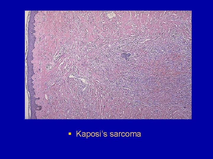
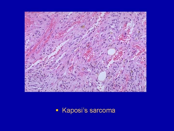
Kaposi's sarcoma § At one time thought to be an uncommon neoplasm of elderly, mostly Jewish, men of European extratcion, Kaposi's sarcoma (KS) has come into prominence because several variant forms have been identified. The form that affects elderly men is referred to as classic KS. Another pattern, called African or endemic KS, is endemic among black African young men and children. A third variant occurs in immunosuppressed transplantation patients, and yet another highly virulent variant, called epidemic KS, is particularly common in acquired immonodeficiency syndrome (AIDS) patients.

Morphology § § 3 stages can be indentified: patch plaque nodule

§ Microscopic examination discloses only dilated blood vessels, lined by normal-looking epithelial cells, with an interspersed infiltrate of lymphocytes, plasma cells, and macrophages (sometimes containing hemosiderin). Such lesions are difficult to distinguish from granulation tissue. Over time lesions in the classic disease spread proximately and usually convert into larger, violaceous, raised plaques that reveal dermal, dilated, jagged vascular channels lined by somewhat plump spindle cells accompanied by perivascular aggregates of similar spindled cells.

§ Kaposi’s sarcoma

§ Kaposi’s sarcoma

§ Kaposi’s sarcoma

§ Kaposi’s sarcoma

Tumours of vague of questionable histogenesis § Mixoma is built of mucous tissue, through which the vessels pass. It resembles gelatine, pink in colour, in the form of polypus or a node on the mucous tissue of the bladder or the stomach. It is a cellular structure, it is branchy inside and consists of star-shaped cells. Mixomatous tissue is sprinkled in the form of nodes into fibroma, osteoma, chondroma (for the second time -

§ Mucous tissue exists only in the embryonic period. The formal genesis mixoma is the remnants of the embryonic mucous tissue. § Chrondroma is a tumour of cartilaginous tissue like a hyalinous cartilage. It is developed at early age, often can be multiple. It grows out of the perichondrium, at first forming indifferent cells of embryonic type, later chondroblasts. The tumour is developed in different parts of the skeleton, more often in the parts where growing is more distinguished - on the borderline of the dyaphisys and epyphisys.

§ It develops in sufferers from rickets under the wrong formation of the cartilage. Sometimes it is formed outside its connection with the bones - embryonic dystopia in lungs, mammary glands, and kidneys. § Under microscopic examination it resembles normal cartilaginous tissue which consists of mesenchymal substance and cartilaginous capsules containing some cartilaginous cells.

§ There are some layers of the connective tissue in the cartilage and they give lobule structure to the tumour. The distinctive features of chondroma and cartilage are: different sizes of cartilaginous capsules and cells. The tumour grows slowly, expansiverly, sometimes developing metastases.

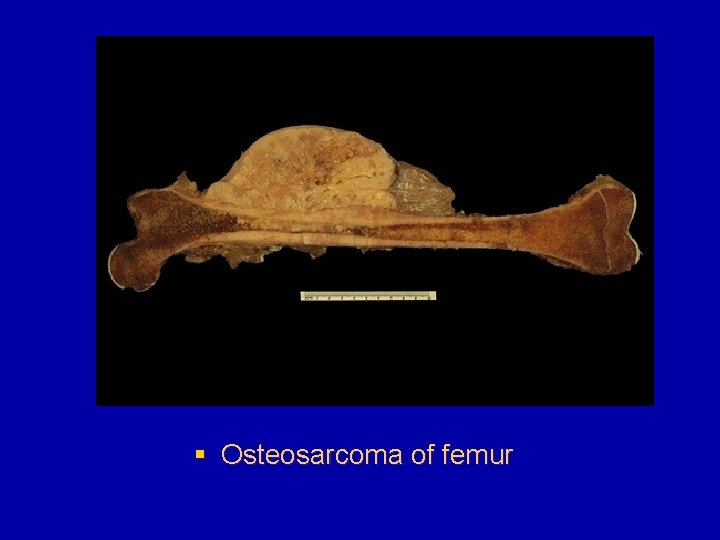
§ Osteosarcoma of femur

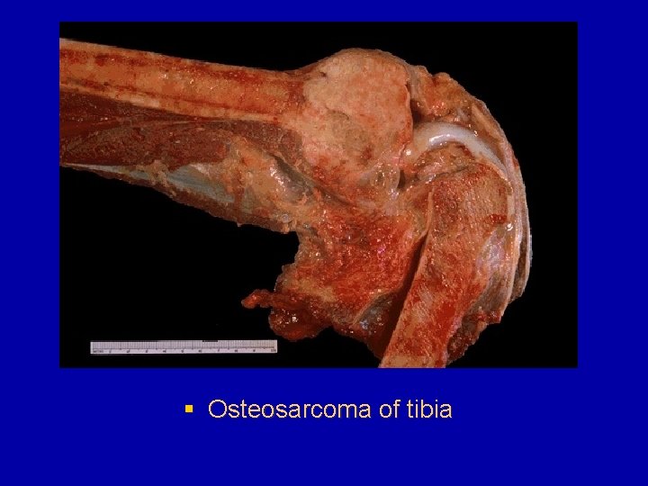
§ Osteosarcoma of tibia

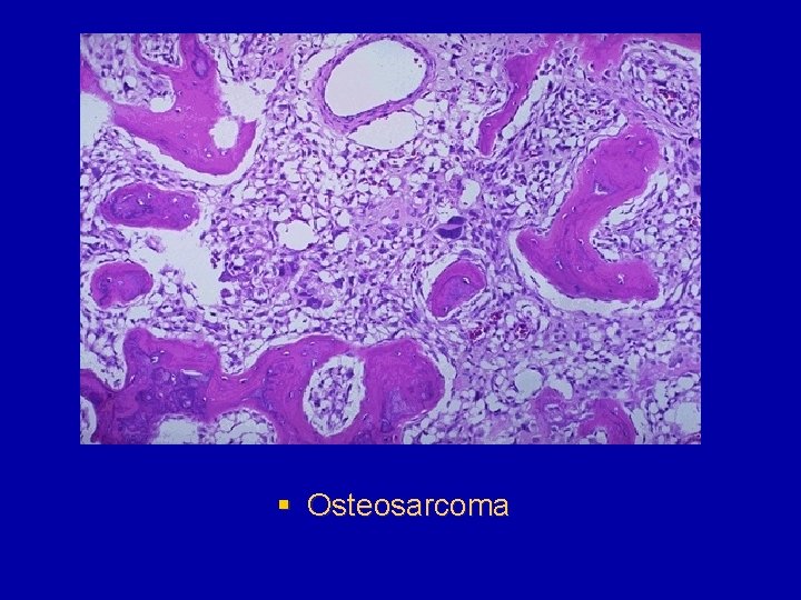
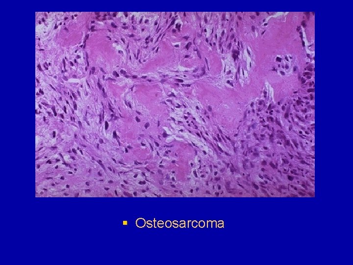
§ Osteosarcoma

§ Osteosarcoma