Ocular tumours Dr Raja S Bengharbia The conjunctiva

Ocular tumours Dr. Raja S. Bengharbia

The conjunctiva

THE IRIS

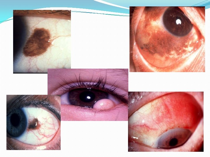
Iris naevus Typical Diffuse Obscures iris crypts • Pigmented, flat or slightly elevated • May cause ipsilateral hyperchromic Diameter usually less than 3 • heterochromia Occasionally mild distortionmm of pupil • May be associated with Cogan- • and ectropion uvea Reese syndrome

Lesions suggestive of melanoma: -Ectropion uvea. -Interinsic vessels -Sectoreal lens opacity. -Rapid growth. -Old age with fair colour -Glaucoma.

Iris Melanoma 1. Very rare - 3% of uveal melanomas 2. Presentation - fifth to sixth decades 3. Very slow growth 4. Low malignancy 5. Excellent prognosis

presentation Usually pigmented nodule at least 3 mm in diameter Invariably in inferior half of iris Angle involvement may cause glaucoma Occasionally nonpigmented Surface vascularization Pupillary distortion, ectropion uveae and cataract

Differential diagnosis of iris melanoma Large iris naevus distorting pupil Leiomyoma Primary iris cyst Ciliary body melanoma eroding iris root Adenoma of pigment epithelium Metastasis to iris

Treatment of iris melanoma Small tumour - broad iridectomy Non-resectable tumour Angle invasion by - radiotherapy or enucleation tumour - iridocyclectomy

CILIARY BODY TUMOUR

Ciliary body melanoma Rare - 5% of uveal melanomas Presentation - 6 th decade May be discovered by chance Prognosis - guarded

Signs of ciliary body melanoma Sentinel vessels Erosion through iris root Extraocular extension Lens subluxation or cataract, RD
 4 -biopsy.")
Investigations: 1 - triple mirror contact lens. 2 -transillumination. 3 -ultrasonography (US) 4 -biopsy.

Treatment options of ciliary body melanoma 1. Iridocyclectomy - small or medium tumours 2. Enucleation - large tumours 3. Radiotherapy - selected cases

TUMOUR OF THE CHOROID

Typical choroidal naevus -5 -10% of caucasians Round slate-grey with indistinct margins Associated with NF 1 Flat or slightly elevated Diameter less than 5 mm Location – usually at post. equatorial Asymptomatic

Investigation: -flourescein angiography: hypofluorescence Iodocyanine green angiography US. . flat or slightly elevated lesion

Suspicious choroidal naevus Diameter more than 5 mm • Elevation 2 mm or more • Surface lipofuscin Posterior margin within 3 mm of disc May have symptoms due to serous fluid

Choroidal melanoma Most common primary intraocular tumour in adults Most common uveal melanoma - 90% of uveal melanomas Presentation - sixth decade Prognosis - usually good

Clinical features: 1 - asymptomatic for small size tumour 2 -decrease V/A, metamorphopsia, visual field loss, floaters , photopsia

Brown, elevated, subretinal mass Secondary retinal detachment Occasionally amelanotic Double circulation Choroidal folds
 is common Mushroom-shaped if breaks through Bruch’s membrane Ultrasound -")
Surface orange pigment (lipofuscin) is common Mushroom-shaped if breaks through Bruch’s membrane Ultrasound - acoustic hollowness, choroidal excavation and orbital shadowing

Treatment of choroidal melanoma 1. Brachytherapy - less than 10 mm elevation and 20 mm diameter 2. Charged particle irradiation - if unsuitable for brachytherapy 3. Transpupillary thermotherapy - selected small tumours 4. Trans-scleral local resection - carefully selected tumours less than 16 mm in diameter ﻭ 5. Enucleation - very large tumours, particularly if useful vision lost 6. Exenteration - extraocular extension

Poor Prognostic Factors of Uveal Melanomas 1. Histological Epithelioid cells • Closed vascular loops • Lymphocytic infiltration • 2. Large size 3. Extrascleral extension 4. Anterior location 5. Age over 65 years

Differential diagnosis of choroidal melanoma Large choroidal naevus Metastatic tumour Localized choroidal haemangioma Choroidal detachment Choroidal granuloma Dense sub-retinal or sub-RPE haemorrhage

Choroidal haemangioma 1 - circumscribed choroidal haemangioma 2 -diffuse choroidal haemangioma

Circumscribed choroidalhaemangioma Presentation - adult life Dome-shaped or placoid, red-orange mass Commonly at posterior pole Between 3 and 9 mm in diameter May blanch with external globe pressure Surface cystoid retinal degeneration Exudative retinal detachment Treatment - radiotherapy if vision threatened

Diffuse choroidal haemangioma Typically affects patients with Sturge-Weber syndrome Can be missed unless compared with normal fellow eye as shown here Diffuse thickening, most marked at posterior pole

Choroidal metastatic carcinoma Most frequent primary site is breast in women and bronchus in both sexes Fast-growing, creamy-white, placoid lesion Most frequently at posterior pole Deposits may be multiple Bilateral in 10 -30%

Melanocytoma Affects dark skinned individuals Usually asymptomatic Most frequently affects optic nerve head Black lesion with feathery edges

TUMOURS OF THE RETINA

Differential diagnosis of leukocoria Congenital cataract Unilateral or bilateral Coats disease Unilateral Persistent hyperplastic primary vitreous Unilateral Posterior pole toxocara granuloma Unilateral Inflammatory cyclitic membrane Unilateral or bilateral Advanced retinopathy of prematurity Always bilateral but may be asymmetrica

Retinoblastoma

1. Most common primary, malignant, intraocular tumour of childhood 2. No sexual predilection 3. Presents before age of 3 years (average 3 months ) 4. Heritable (40%) or non-heritable (60%) 5. Predisposing gene (RPE 1) on 13 q 14 6 - B/L in 25 % of cases.

Presentations of retinoblastoma Leukocoria - 60% Anterior segment invasion Secondary Strabismus - 20% glaucoma Orbital inflammation Orbital invasion
 MR")
Investigations: US to detect calcification and to R/O other cause (Coats disease ) MR to detect optic nerve involvement and extra-optic extension CT

More advanced retinoblastoma Friable white mass Fine surface blood vessels Cottage cheese appearance Vitreous seedings

Multiglobulated white mass with overlying retinal detachment May be difficult to visualize through deep detachment

Treatment Options of Retinoblastoma 1. Small tumours Laser photocoagulation • Transpupillary thermotherapy • Cryotherapy • 2. Medium tumours Brachytherapy • Chemotherapy • External beam radiotherapy • 3. Large tumours Chemotherapy followed by local treatment • Enucleation • 4. Extraocular extension External beam radiotherapy • 5. Metastatic disease Chemotherapy •

Poor Prognostic Factors in Retinoblastoma 1. Optic nerve involvement 2. Choroidal invasion 3. Large tumour 4. Anterior location 5. Poor cellular differentiation 6. Older children

THANK YOU
- Slides: 42