OCULAR EXAMINATION Dr Neeti Gupta Associate Professor Aim


- Slides: 49
OCULAR EXAMINATION Dr. Neeti Gupta Associate Professor
Aim and Objectives • Understand the fundamental principles and perform a systematic eye examination. • On completion of this session you will be able to: - Recognize normal and abnormal anatomy - Systematically examine an eye - Correctly document examination findings
Equipment required to examine an eye • Fine beamed torch • Cotton buds • Local anaesthetic eye drops, eg Paracaine Fluorescein strips • Magnification – slit lamp, indirect ophthalmoscope, loupes
Patient Assessment General Physical Examination Sclera – for jaundice Conjuntiva – Anemia Lids – xanthelesma Puffy eye lids – Nephrotic syndrome, Thyroid eye ds, Hypertension - Lympadenopathy – Tuberculosis, tumor and viral conjuntivitis - Parotid swelling or lacrimal gland swelling • -
Systemic Examination • In view of ocular diseases as ocular signs can be early manifestation of systemic diseases. - Scleritis Dry Eye Peripheral corneal ulcers Uveitis Dislocation of lens
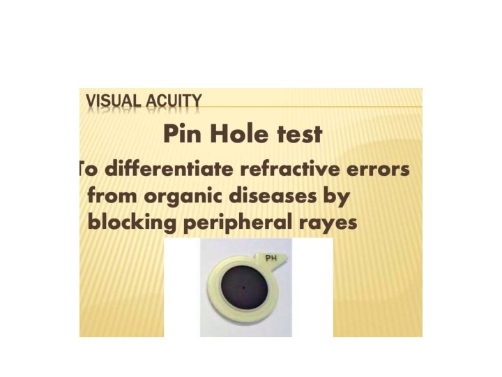
Ocular examination • Vision Assessment – - Visual acuity – Estimation of its ability to discriminate between two points - Snellens chart -
Optokinetic nystagmus
• Projection of rays- PR • Field of vision - Confrontation test - Perimetry Kinetic Static
• Head Posture
• Forehead
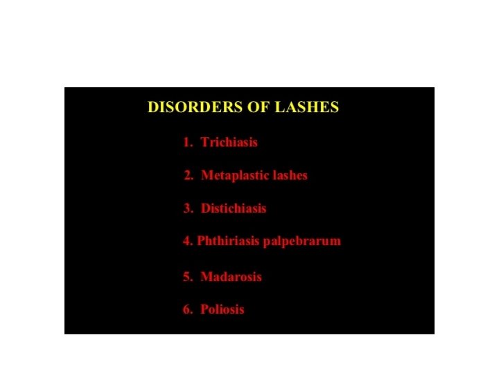
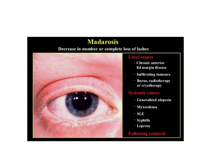
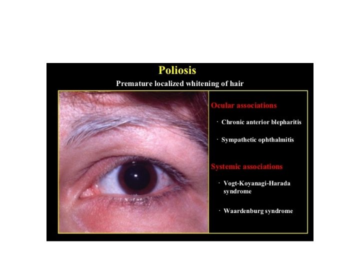
• Eye Brows Level Whitening Absence of lashes
Orbital margins • Trauma • Tumors
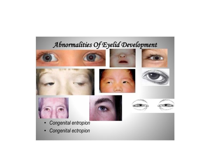
• Lids and Lashes
Lagophthalmos Lid Retraction
Visual Axis – Alignment
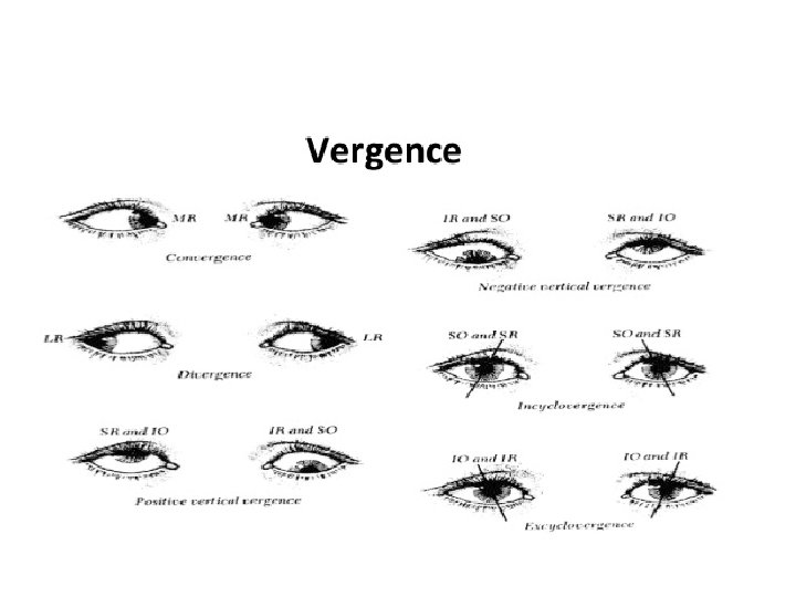
Ocular movements Duction – unilateral movement Version – binocular movement in same direction
• Proptosis • Enophthalmos
• Regurgitation test
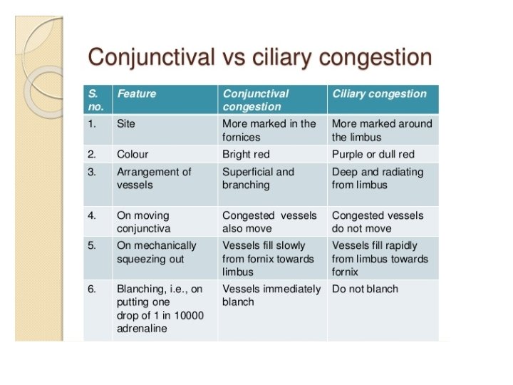
• Conjunctiva - Colour - Chemosis - Congestion
Conjunctiva Covers the inside of eyelids and the sclera – does not pass over the cornea; is vascular. • Normal – translucent, flat, sclera visible beneath • Abnormal – Injected - bloodshot – Chemosis (oedema) – Discharge – Subconjunctival haemorrhage – Lacerations – Lesions
Eversion of lids
Double eversion
Chemosis
Congestion
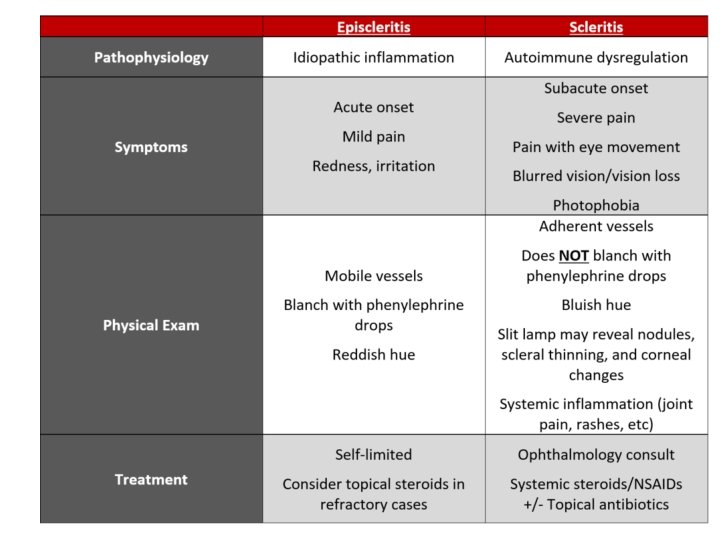
Episcleritis Scleritis
Conjuntival degenerations Pinguecula Pterygium
Vitamin A Deficiency
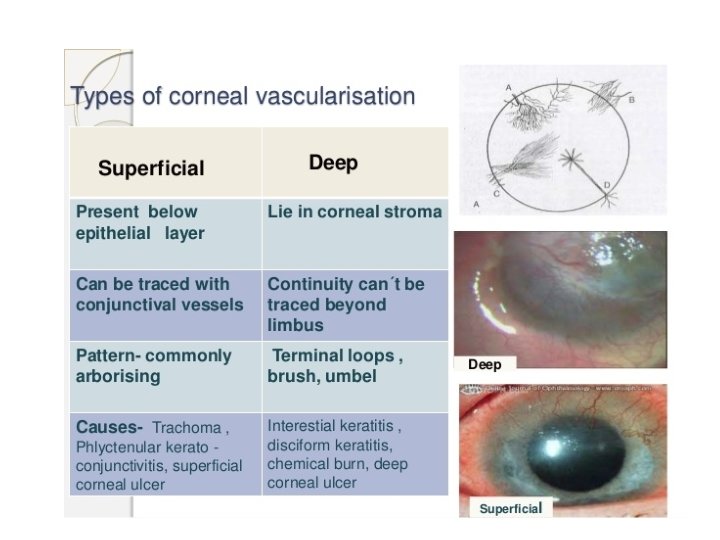
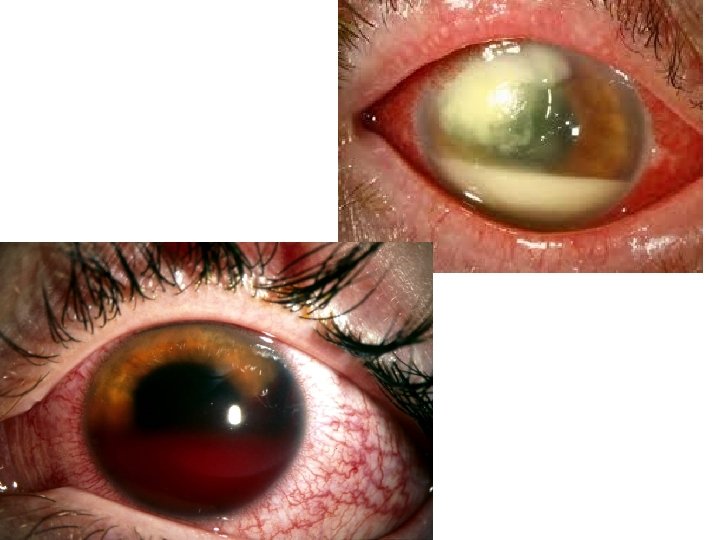
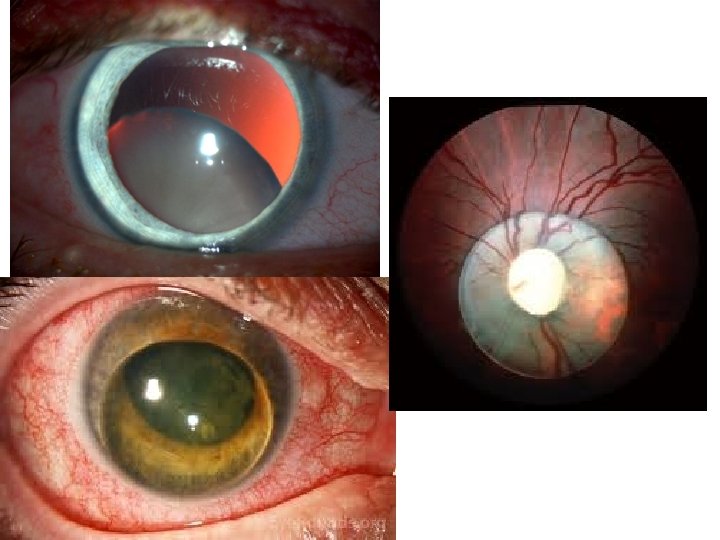
Cornea • Size - 11. 7 mm and 12. 6 mm Microcornea - <10 mm Megalocornea - >13 mm • Shape – watch glass • Surface – • Transparency • Sensitivity • Sheen
Anterior Chamber Shallow – Deep – Irregular – Abnormal Content -
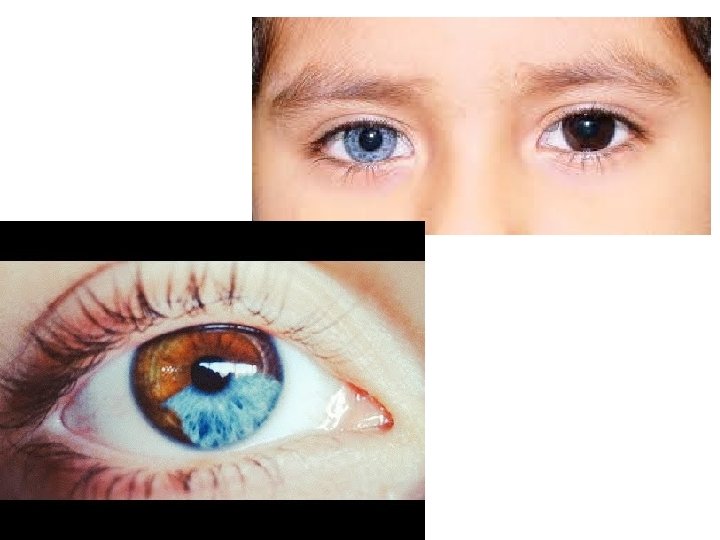
IRIS • Color. Heterochromia iridium- Different colour of two eyes Heterochromia iridis –Different colour of same iris • Iris Pattern • Iris Defects
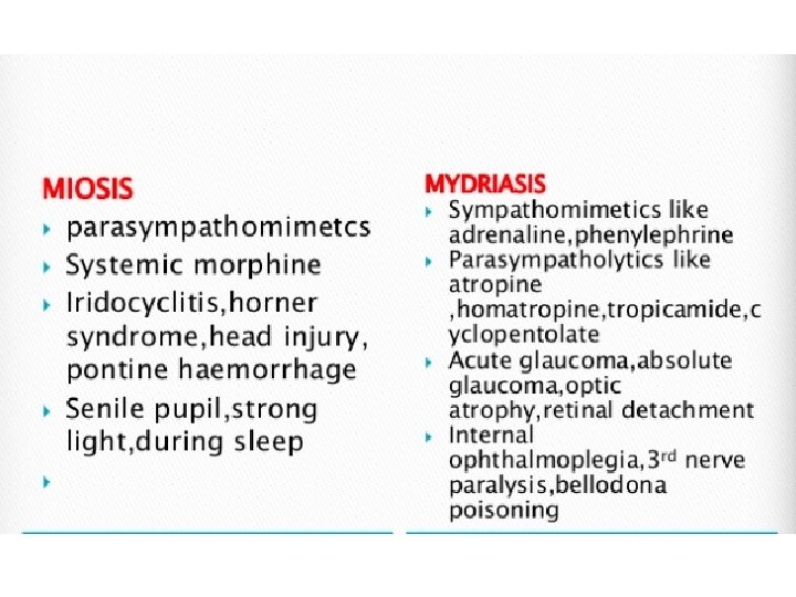
Pupil • Size – Miosis, Mydriasis , Anisocoria • Shape – Irregular, Festooned • Number - polycoria • Reaction – Direct, Indirect, Marcus gunn
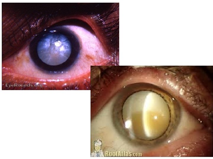
Lens • Position – Subluxation Dislocation • Shape – Biconvex Spherophakia Lenticonus Coloboma • Color – Brown/ Black , Greyish white , Pearly white, Milky White
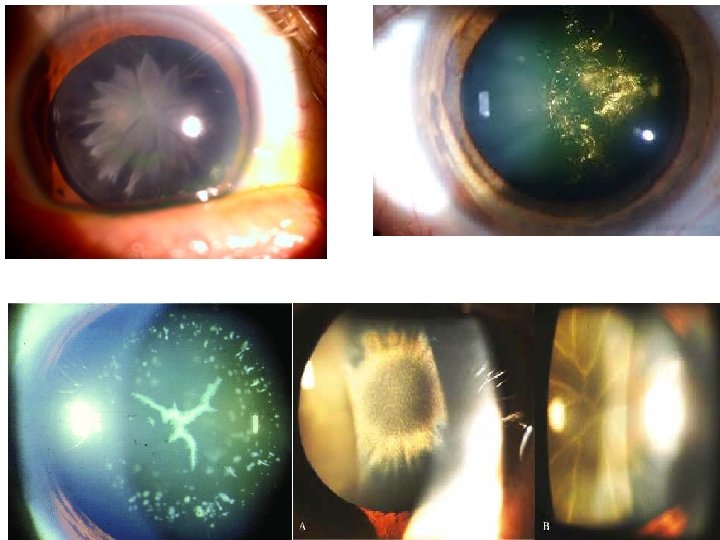
• • Snow flake cataract Sunflower cataract Rosette cataract Christmas tree cataract
Digital Tension