OBSTETRICAL HEMORRHAGE Robert K Silverman MD SUNY Upstate

OBSTETRICAL HEMORRHAGE Robert K. Silverman, MD SUNY Upstate Medical University Department of OB/GYN Division of Maternal-Fetal Medicine Syracuse, New York

Catastrophic Obstetrical Hemorrhage n Educational Objectives – Review hematological changes in pregnancy – Evaluate definitions and classification – Consider etiology and risk factors – Explore effect of mode of delivery – Develop management strategy – Propose conclusions

OB Hemorrhage OB hemorrhage accounts for 50% of all postpartum maternal fatalities n The single most important cause of maternal death worldwide n 88% of deaths from postpartum hemorrhage occur within 4 hours of delivery n Int. J. Gynecol. Obstet 1996; 54: 1 -10
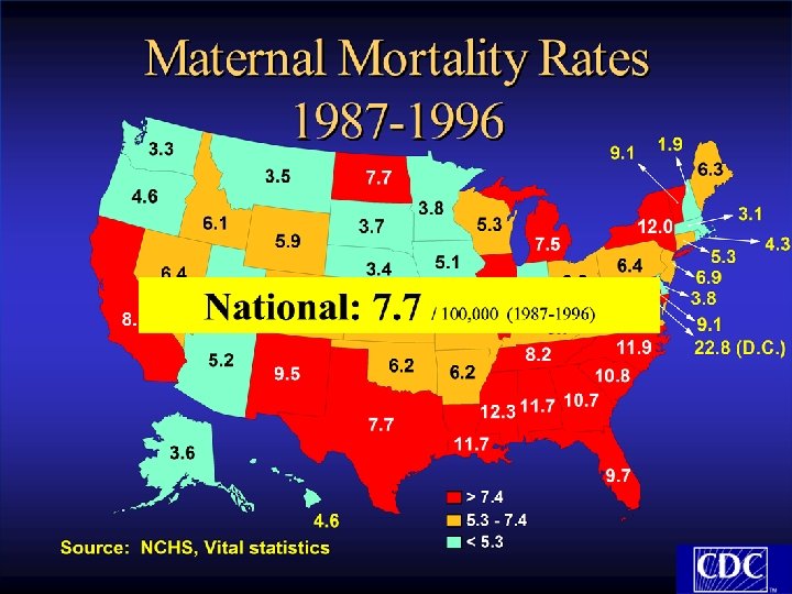
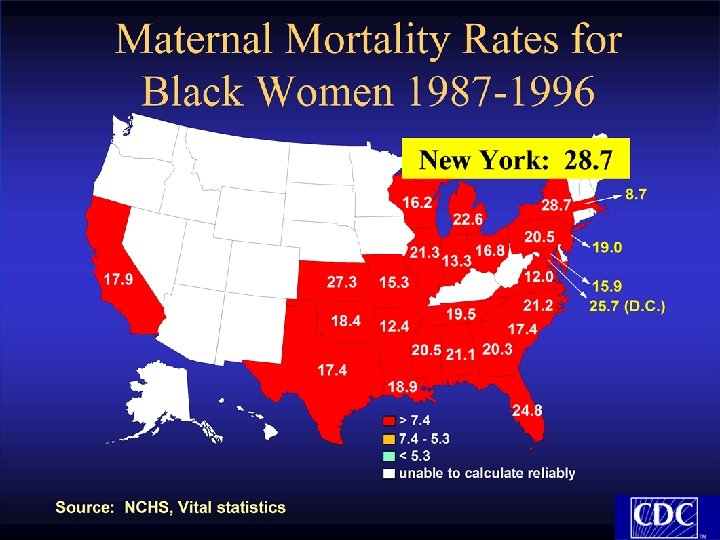

Approximately one-half of maternal deaths are preventable!!

Hematological Changes in Pregnancy n n n 40% expansion of blood volume by 30 weeks 600 ml/min of blood flows through intervillous space Appreciable increase in concentration of Factors I (fibrinogen), VIII, IX, X Plasminogen appreciably increased Plasmin activity decreased Decreased colloid oncotic pressure secondary to 25% reduction in serum albumin

Estimation of Blood Loss n Visual – Underestimates by ½ to 1/3 n Hypotension – May be masked by hypertensive disorders n Tilt-test – False positives (conduction anesthesia) – False negatives (hypervolemia of pregnancy) n Tachycardia – Unreliable n Urine flow – Reflects adequate of perfusion

Reduced Maternal Blood Volume n Small stature n Severe preeclampsia/eclampsia n Early gestational age

Effect of Acute Blood Loss on Hematocrit n Change usually delayed at least 4 hours n Complete compensation takes 24 hours n Above affected by degree of intravenous hydration

Average Blood Loss and Complexity of Delivery Vaginal delivery– 500 ml n Cesarean section– 1000 ml n Repeat cesarean section & TAH– 1500 ml n Emergency hysterectomy– 3500 ml. n Pritchard AJOB 1961 Clark Obstet Gynecol 1984
")
Classification of Hemorrhage in the Pregnant Patient * Hemorrhage Acute Blood Loss Class (ml) Percentage Lost 1 2 3 4 15 20 -25 30 -35 40 900 1200 -1500 1800 -2100 2400

Classification of Hemorrhage in the Pregnant Patient Hemorrhage Signs and Symptoms Class 1 Usually none 2 Tachycardia, tachypnea orthostatic changes, prolonged hypothenar blanching, narrowing of pulse pressure 3 Overt hypotension, marked tachycardia (120 -160 bpm), marked tachypnea (30 -40/mln, cold, clammy skin 4 No discernible blood pressure, oliguria or anuria, absent peripheral pulses

Etiology of Obstetrical Hemorrhage Abnormal placentation n Trauma n Uterine atony n Coagulation defects n

Etiology of Obstetrical Hemorrhage n Trauma – Episiotomy – Vulvar Lacerations – Vaginal lacerations – Cervical lacerations – Cesarean section extensions – Uterine rupture

Risk Factors for Uterine Rupture Prior uterine scar n High parity n Hyperstimulation n Obstructed labor n Intrauterine manipulation n Midforceps rotation n

Etiology of Obstetrical Hemorrhage n Abnormal Placentation – Placenta previa – Abruptio placenta – Placenta accreta – Ectopic pregnancy – Hydatidiform mole

Placenta Accreta-Increta-Percreta as a Cause of Bleeding – Increased incidence over last 20 years » Increased cesarean section rate » Increased risk from placenta previa n. Previa and unscarred uterus-5% risk Clark et al Obstet Gynecol 1985
 Maternal Mortality of Placenta Accreta During the 20 th Century")
Percent (%) Maternal Mortality of Placenta Accreta During the 20 th Century

Incidence of Placenta Previa/Accreta as a Function of Number of Cesarean Sections Number of C/S

Midsagittal Sonographic Image of Placenta Previa-Percreta

Risk Factors for Uterine Atony n Excessive uterine distension – – n Macrosomia Hydramnios Multiple gestation Clots Anesthetic agents – Halogenated agents n Myometrial exhaustion – Rapid or prolonged labor – Oxytocin – Chorioamnionitis n Prior uterine atony

Risk Factors for Coagulation Defects n n n n n Placental abruption Severe preeclampsia Amniotic fluid embolus Massive transfusions Severe intravascular hemolysis Congenital or acquired coagulopathies Retention of dead fetus Sepsis Anticoagulant therapy

Postpartum Hemorrhage Definitions n Traditional: >500 ml n – Immediate: Within 24 hours of delivery – Delayed: More than 24 hours following delivery n Coombs et al, 1991 – Amount requiring transfusion or producing 10 volume % reduction in hct

Postpartum Hemorrhage Following Vaginal Delivery 30, 000 deliveries n 1976 – 1996 at Beth Israel Hospital n 2. 6% overall transfusion rate n 4. 6% in 1976; 1. 9% in 1996 n 20% of transfusions > 3 units n

Postpartum Hemorrhage Following Vaginal Delivery Risk Factor Relative Risk Prolonged 3 rd stage 7. 6 Pre-eclampsia 5 Mediolateral episiotomy 4. 7 Postpartum hemorrhage 3. 6 Twins 3. 3 Arrest of Descent 2. 9 Lacerations 2 Coombs, et al, 1991

Postpartum Hemorrhage Following Cesarean Deliveries Risk Factor General Anesthesia Amnionitis Protracted Active Phase Preeclampsia Second-stage Arrest Hispanic Classical Incision Coombs, et al, 1991 Relative Risk 2. 9 2. 7 2. 4 2. 2 1. 9 1. 8 1. 1

Strategies for the Prevention of Postpartum Hemorrhage 1. Enhance natural contractions of the uterus 2. Shortening of the 3 rd stage 3. Treat aggressively

Active Management of the rd 3 Stage of Labor n Principal action – Hasten and augment uterine contractions after delivery of the baby – Prevent hemorrhage due to uterine atony n Prevent blood loss

Active Management versus Expectant Management n Main Components of Active Management – – – Administration of a prophylactic uterotonic agent soon after delivery Early clamping and cutting of the umbilical cord Controlled cord traction after the uterus has contracted

Active Management versus Expectant Management n Main Components of Expectant Management – Wait for signs of placental separation – Allow placenta to deliver spontaneously » Aided by gravity or nipple stimulation

Active vs. Expectant Management of the 3 rd Stage of Labor n n Cochrane systematic review of 5 randomized controlled trials (1988, 1990, 1993, 1997, 1998) Findings – – n Side Effects – – n Active management reduced risk of maternal blood loss Reduced prolonged 3 rd stage of labor Increased nausea and vomiting Elevated BP’s Recommendations – Active management should be the routine approach for women having a vaginal delivery in a hospital Mac. Donald et al 2003

Prophylactic use of Oxytocin in rd the 3 Stage of Labor n Cochrane review of seven trials (1961, 1964, 1990, 1991, 1992 1996, 1997) – Findings » Reduced blood loss » Reduced need for additional uterotonic drugs » Nonsignificant trend towards more manual removal of placenta and more blood transfusion in the expectant management subgroup Elbourne et al 2003

Alternative Agents for Prevention of Postpartum Hemorrhage 1. Umbilical Uterotonic Agents: – 1 st trial in 1987 using Oxytocin vs. Saline – not significant – 3 other trials (1988, 1991, 1996) showed the same NS – Two placebo controlled trials (1991, 1998) » Oxytocin decreased the length of 3 rd stage but not blood loss

Alternative Agents for Prevention of Postpartum Hemorrhage 2. Oral Ergometrine and Methylergometrine – Both drugs have a strong uterotonic effect and slight vasoconstriction – Act differently than Oxytocin and Prostaglandins – Unfortunately both are unstable even refrigerated – No place in modern obstetrics De. Groot et al: Drugs, 1998

Alternative Agents for Prevention of Postpartum Hemorrhage 3. Sublingual Oxytocin – – – Widely varying bio-availability Long lag time, long half life Not used in modern obstetrics De. Groot et al J Pharmacol 1995

Alternative Agents for Prevention of Postpartum Hemorrhage 4. Injectable Prostaglandins – International trial in 1996 » Similar results to prophylactive IM/IV Oxytocin n Higher rates of diarrhea, higher cost » 2001 Randomized trial in United Kingdom using hemabate n n Study stopped early due to side effects – 21% with severe diarrhea As effective as Oxytocin in preventing hemorrhage » Cochrane Review in 2000 n Injectable PG’s have decrease blood loss and shortened 3 rd stage but should be used when other measures fail

Alternative Agents for Prevention of Postpartum Hemorrhage 5. Carbetocin – Long acting Oxytocin receptor agonist – Produces tetanic contractions within 2 minutes lasting 6 minutes, lasts for approximately 1 hour – IM has a prolonged effect (2 hours) versus IV – 1998 and 1999 – 2 trials in Canada – double-blind, randomized for patients having a cesarean section » Was more effective in a single IV dose than continuous Oxytocin » Similar safety profile to Oxytocin – No clinical trials for postpartum hemorrhage prevention

Alternative Agents for Prevention of Postpartum Hemorrhage n Misoprostil – – Synthetic analog of PGE 1 1996 -1 st trial outlining its use to prevent 3 rd stage 24 randomized controlled trials from 1998 -2003 3 systematic reviews (2002, 2003) » Oral and rectal Misoprostil not as effective as conventional injectable uterotonics » High rate of side effects – May be useful in less-developed countries where administration of parenteral uterotonic agents are problematic
 artery ligation")
Surgical Therapy Uterine packing n Uterine artery ligation n Internal iliac (hypogastric) artery ligation n Hysterectomy n Suture techniques n

Surgical Management of Uterine Atony General Considerations n n n Stability of patient Reproductive status of patient Skill of surgeon Skill of assistants Availability of blood products Visualization of pelvis – – – Choice of incision Retroperitoneal approach Anatomic distortion

Uterine Packing n Fell into disfavor in 1950’s – Concealed hemorrhage – Infection – Non-physiologic approach n Maier AJOB, 1993 – Simple, safe, effective – Pack side to side » Avoid dead space

Pelvic Pressure Pack n n n Bleeding may persist post hysterectomy Original description by Logothetopulos in 1926 High success rate, but numbers are limited Year 1962 1968 1985 1990 1991 2000 Author Parente Burchell Cassels Robie Hallak Dildy OB 0 0 1 1 1 7 GYN 14 8 0 0 0 1 Total 14/14 8/8 1/1 1/1 7/8

The Pelvic Pressure Pack for Persistent Post hysterectomy Hemorrhage Dildy AJOG 2000

Postpartum Uterine Hemorrhage Uterine Artery Ligation n Waters, 1952 – Original description n O’Leary & O’Leary, 1974 – Post-cesarean hemorrhage – Simpler more rapid technique n Reported efficacy 80 -92%

Stepwise Uterine Devascularization Alexandria, Egypt – Shatby Maternity University Hospital n 103 patients with non-traumatic postpartum hemorrhage n Failure of non-surgical management n Absorbable sutures n No vessels clamped or divided n Abd. Rabbo, 1994
 uterine vessel ligation n")
Stepwise Uterine Devascularization Unilateral uterine vessel ligation n Contralateral (bilateral) uterine vessel ligation n Low bilateral uterine vessel ligation n Unilateral ovarian vessel ligation n Contralateral (bilateral)ovarian vessel ligation n Abd. Rabbo, 1994
 Indications Patients 1 2 3 4 5 Uterine")
Stepwise Uterine Devascularization Step Employed (%) Indications Patients 1 2 3 4 5 Uterine Atony 66 14 85 0 2 0 Abruptio Placenta 17 0 88 0 12 0 Couvelaire Uterus 9 0 33 0 44 22 Placenta Previa 5 0 100 0 Placenta Previa with Accreta 2 0 50 50 0 0 Afibrinogenemia 4 0 0 100 Total 103 9 75 4 7 6 Abd. Rabbo, 1994

Stepwise Uterine Devascularization Follow-Up All patients resumed normal menstruation n 11/15 patients conceived following discontinuation of contraception n Subsequent pregnancies normal n – 4 Vaginal deliveries – 7 Cesarean sections – No postpartum hemorrhage Abd. Rabbo, 1994

Suture Techniques n n B-Lynch procedure – Fundal Compression suture #2 chromic on a 75 mm heavy, round bodied needle 4 Case reports total B-Lynch Ferguson Dacus Vangsgaard BJOB 1997 OB & GYN 2000 JMFM 2000 Ugesker Laeger 2000 5/5 2/2 1/1 12/12

B-Lynch Procedure
 Artery Ligation n Controls blood loss by reducing art. pulse pressure")
Internal Iliac (Hypogastric) Artery Ligation n Controls blood loss by reducing art. pulse pressure – Converts pelvic art. circulation into a venous system n Burchell et al Obstet Gynecol 1964 – Arterial pulse pressure reduced n n n 14% by contra lateral 77% by homolateral 85% by bilateral Need experienced surgeon Need hemodynamically stable patient

Selective Arterial Embolization Widely used for management of uncontrollable hemorrhage n First OB trial 1979 (Brown et al Obstet. Gynecol) n 7 Trials from 1998 -2000 n – Cumulative success rate = 97% n Excellent first line therapy but. . . – Difficult to perform in Labor and Delivery – Availability of interventional radiologist

Hysterectomy n n Clark et al Obstet Gynecol 1984 Largest series of emergency hysterectomy – 70 cases 1978 -1982 » 60 Post cesarean sections » 10 post vaginal delivery – Indications » » » Atony – 43% Placenta accreta – 30% Uterine rupture – 13% Extension of low transverse incision – 10% Fibroids preventing closure – 4% – TAH for atony » Higher rates; amniotics, C/S for labor arrest, augmentation of labor, Mg. SO 4 infusion, larger fetal weight
 Changing Indications for Emergency Hysterectomy")
Percent (%) Changing Indications for Emergency Hysterectomy

Autotransfusion n n n Use of cell saver to collect blood from operative field, processing and reintroducing red cells to patients. Not well defined in obstetrics Three small studies (1989, 1990, 1997) – Removal of fetal and amniotic debris – Appears effective Largest series to date (Rebarber AJOB 1998) – 139 cases performed at cesarean section – No complications related to AFE or coagulopathies Use two separate suction devices – Amniotic fluid and red cell product – Increase wash volume – Measure clotting factors and platelets every 1 to 1. 5 blood volumes lost Contraindications – Heavy bacterial contamination – Malignancies

Fluid and Blood Component Replacement n n Whole blood vs. components, debate continues Maintain urine output > 30 cc/hr Maintain hematocrit > 30% (with acute blood loss) Choice of components: – – – Hemoglobin – packed red blood cells Fibrinogen-cryoprecipitate Other clotting factors-fresh frozen plasma Platelets-platelet packs Volume-lactated Ringer’s solution

Risks of Blood Transfusion HIV 1: 2, 135, 000 n Hepatitis A 1: 1, 000 n Hepatitis B 1: 205, 000 n Hepatitis C 1: 276, 000 n HTLV I/II 1: 2, 993, 000 n Transfusion-related acute lung injury n – 1: 5, 000 n Alloimmunization 0. 5% Int. Anesthesia Clinics 2004

Catastrophic Obstetrical Hemorrhage Conclusions n n n Incidence low, but significant Amount of blood loss hard to determine; catastrophic clearer Earlier the intervention, less the blood loss Organized approach essential to management Exhaust medical measures prior to surgery Precise fluid and blood component therapy essential

- Slides: 63