
Obstetric Hemorrhage Program Review Committee FELICIA FITZGERALD BSN
Obstetric Hemorrhage Program
Review Committee FELICIA FITZGERALD, BSN, RNC-OB, C-EFM, Perinatal Outreach Educator University of Chicago Medicine LORI FOLKEN, BSN, RNC-OB, C-EFM, Perinatal Outreach Educator, Carle Foundation Hospital PAULA MELONE, D. O. FACOG, Assistant Professor, Loyola University Health System ANGELA RODRIGUEZ, RNC-OB, BSN, CCRC, Perinatal Coordinator, Advocate Illinois Masonic Medical Center SHIRLEY SCOTT, RN-BC, C-EFM, MSN, APN, Perinatal Outreach Educator, University of Illinois at Chicago
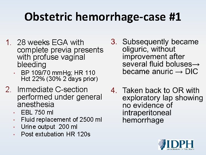
Obstetric hemorrhage-case #1 1. 28 weeks EGA with complete previa presents with profuse vaginal bleeding • BP 109/70 mm. Hg; HR 110 Hct 22% (30% 2 days prior) 2. Immediate C-section performed under general anesthesia • • EBL 750 ml Fluid replacement of 2500 ml Urine output 200 ml Post extubation HR 120 s
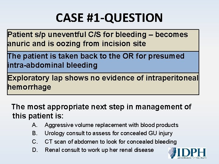
CASE #1 -QUESTION Patient s/p uneventful C/S for bleeding – becomes anuric and is oozing from incision site The patient is taken back to the OR for presumed intra-abdominal bleeding Exploratory lap shows no evidence of intraperitoneal hemorrhage The most appropriate next step in management of this patient is: A. B. C. D. Aggressive volume replacement with blood products Urology consult to assess for concealed GU injury CT scan of abdomen to look for concealed bleeding Renal consult to work up her renal disease
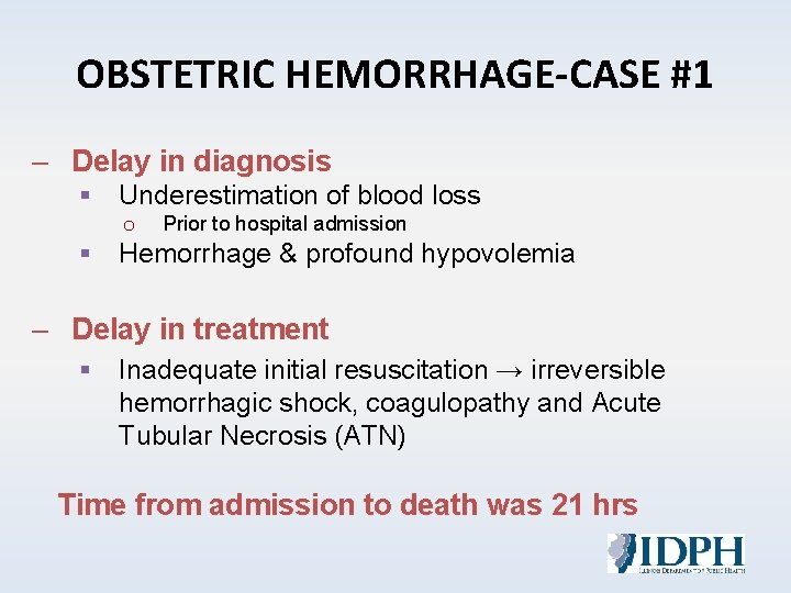
OBSTETRIC HEMORRHAGE-CASE #1 – Delay in diagnosis § Underestimation of blood loss o Prior to hospital admission § Hemorrhage & profound hypovolemia – Delay in treatment § Inadequate initial resuscitation → irreversible hemorrhagic shock, coagulopathy and Acute Tubular Necrosis (ATN) Time from admission to death was 21 hrs
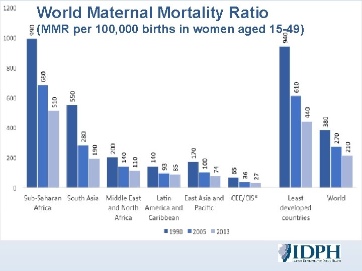
World Maternal Mortality Ratio (MMR per 100, 000 births in women aged 15 -49)
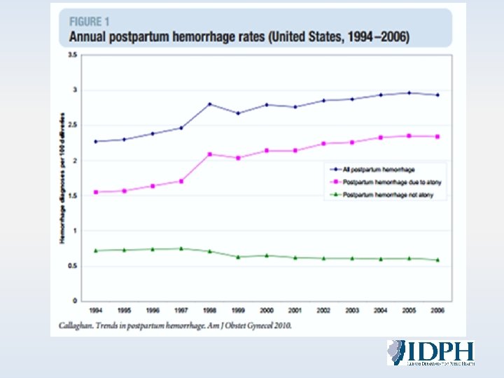
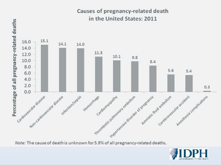
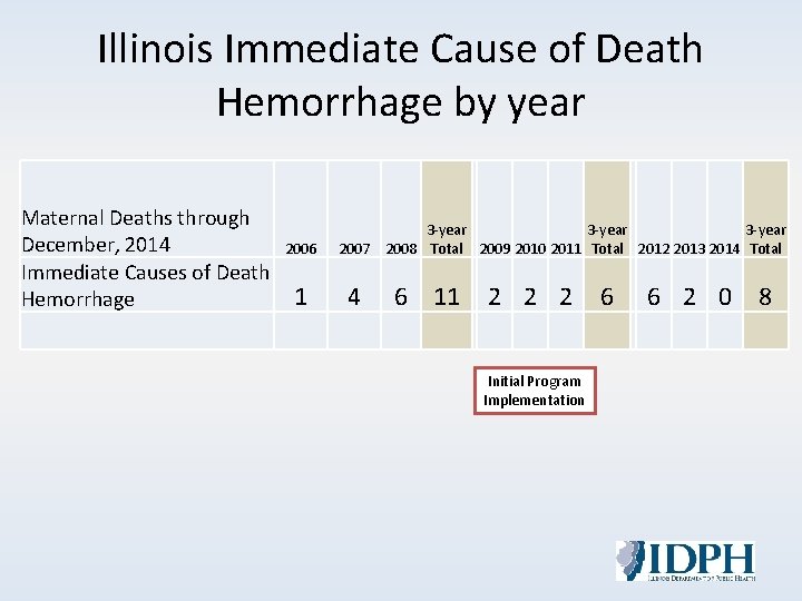
Illinois Immediate Cause of Death Hemorrhage by year Maternal Deaths through December, 2014 Immediate Causes of Death Hemorrhage 2006 1 3 -year 2007 2008 Total 2009 2010 2011 Total 2012 2013 2014 Total 4 6 11 2 2 2 Initial Program Implementation 6 6 2 0 8
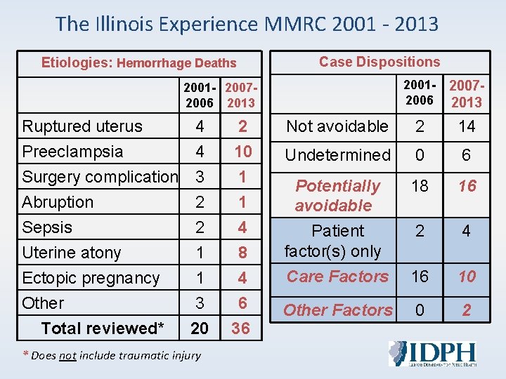
The Illinois Experience MMRC 2001 - 2013 Etiologies: Hemorrhage Deaths Case Dispositions 2001 - 20072006 2013 20012006 20072013 Ruptured uterus 4 2 Not avoidable 2 14 Preeclampsia 4 10 Undetermined 0 6 Surgery complication 3 1 Abruption 2 1 Potentially avoidable 18 16 Sepsis 2 4 2 4 Uterine atony 1 8 Patient factor(s) only Ectopic pregnancy 1 4 Care Factors 16 10 Other 3 6 20 36 Other Factors 0 2 Total reviewed* * Does not include traumatic injury
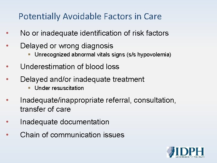
Potentially Avoidable Factors in Care • No or inadequate identification of risk factors • Delayed or wrong diagnosis § Unrecognized abnormal vitals signs (s/s hypovolemia) • Underestimation of blood loss • Delayed and/or inadequate treatment § Under resuscitation • Inadequate/inappropriate referral, consultation, transfer of care • Inadequate documentation • Chain of communication issues
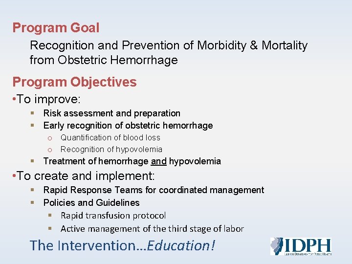
Program Goal Recognition and Prevention of Morbidity & Mortality from Obstetric Hemorrhage Program Objectives • To improve: § Risk assessment and preparation § Early recognition of obstetric hemorrhage o Quantification of blood loss o Recognition of hypovolemia § Treatment of hemorrhage and hypovolemia • To create and implement: § Rapid Response Teams for coordinated management § Policies and Guidelines § Rapid transfusion protocol § Active management of the third stage of labor The Intervention…Education!
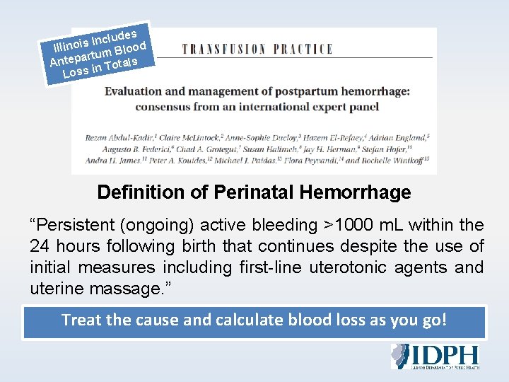
es d u l c n I Illinois m Blood artu Antep n Totals Loss i Definition of Perinatal Hemorrhage “Persistent (ongoing) active bleeding >1000 m. L within the 24 hours following birth that continues despite the use of initial measures including first-line uterotonic agents and uterine massage. ” Treat the cause and calculate blood loss as you go!
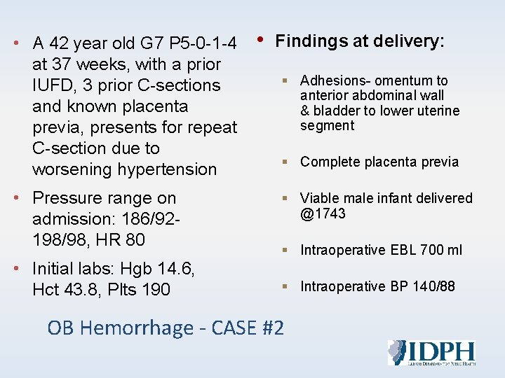
• A 42 year old G 7 P 5 -0 -1 -4 at 37 weeks, with a prior IUFD, 3 prior C-sections and known placenta previa, presents for repeat C-section due to worsening hypertension • Pressure range on admission: 186/92198/98, HR 80 • Initial labs: Hgb 14. 6, Hct 43. 8, Plts 190 • Findings at delivery: § Adhesions- omentum to anterior abdominal wall & bladder to lower uterine segment § Complete placenta previa § Viable male infant delivered @1743 § Intraoperative EBL 700 ml § Intraoperative BP 140/88 OB Hemorrhage - CASE #2
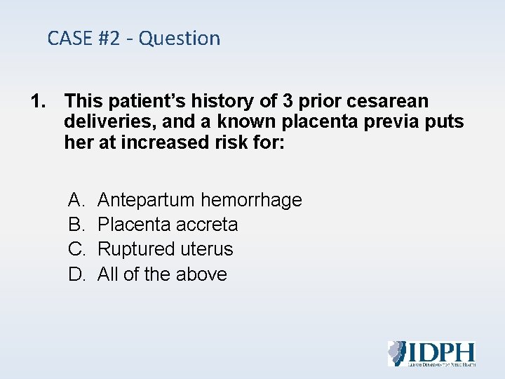
CASE #2 - Question 1. This patient’s history of 3 prior cesarean deliveries, and a known placenta previa puts her at increased risk for: A. B. C. D. Antepartum hemorrhage Placenta accreta Ruptured uterus All of the above
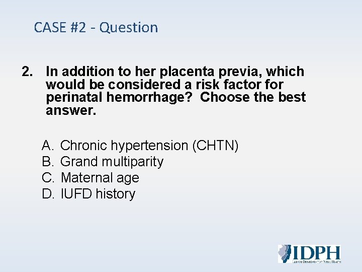
CASE #2 - Question 2. In addition to her placenta previa, which would be considered a risk factor for perinatal hemorrhage? Choose the best answer. A. B. C. D. Chronic hypertension (CHTN) Grand multiparity Maternal age IUFD history
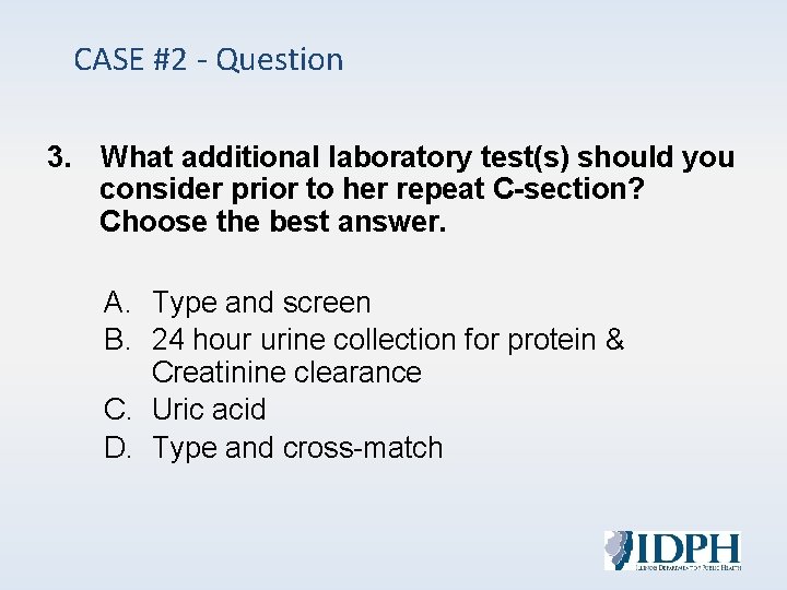
CASE #2 - Question 3. What additional laboratory test(s) should you consider prior to her repeat C-section? Choose the best answer. A. Type and screen B. 24 hour urine collection for protein & Creatinine clearance C. Uric acid D. Type and cross-match
RISK ASSESSMENT AND PREPARATION
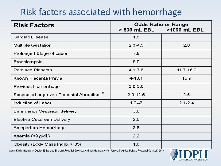
Risk factors associated with hemorrhage *
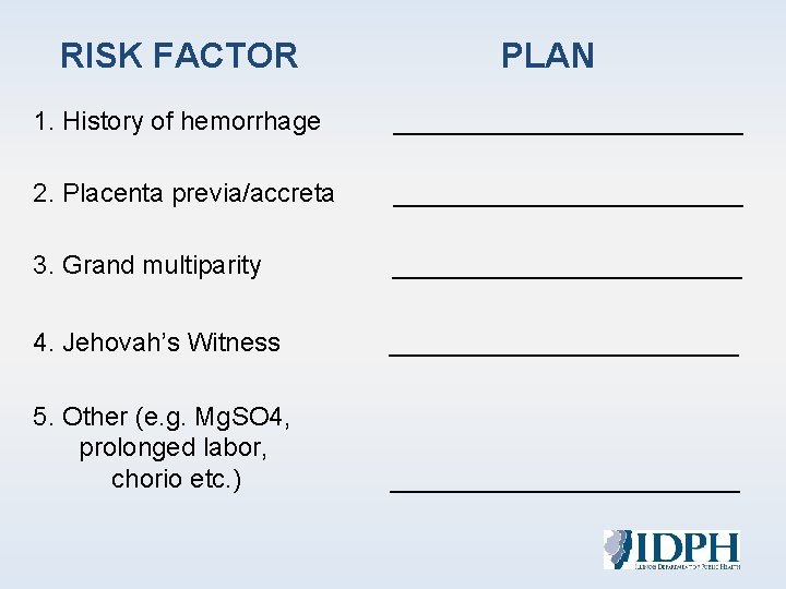
RISK FACTOR PLAN 1. History of hemorrhage ____________ 2. Placenta previa/accreta ____________ 3. Grand multiparity ____________ 4. Jehovah’s Witness ____________ 5. Other (e. g. Mg. SO 4, prolonged labor, chorio etc. ) ____________
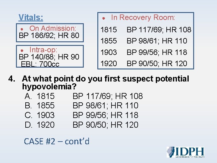
Vitals: ● On Admission: BP 186/92; HR 80 1815 BP 117/69; HR 108 Intra-op: BP 140/88; HR 90 EBL: 700 cc 1903 BP 99/56; HR 118 1920 BP 90/50; HR 120 ● ● In Recovery Room: 1855 BP 98/61; HR 110 4. At what point do you first suspect potential hypovolemia? A. 1815 BP 117/69; HR 108 B. 1855 BP 98/61; HR 110 C. 1903 BP 99/56; HR 118 D. 1920 BP 90/50; HR 120 CASE #2 – cont’d
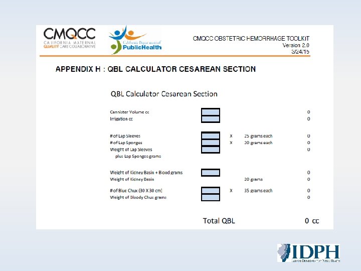
RECOGNITION, VISUALIZATION AND QUANTIFICATION OF BLOOD LOSS (QBL)
The blood loss at a vaginal delivery is given as 350 m. L. To quantify this amount correctly, the blood volume in the collection drape would fill a: A. Standard soda can B. Half gallon of milk C. Pint of milk D. Quart of milk EBL Recognition
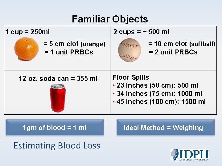
Familiar Objects 1 cup = 250 ml = 5 cm clot (orange) = 1 unit PRBCs 12 oz. soda can = 355 ml 1 gm of blood = 1 ml Estimating Blood Loss 2 cups = ~ 500 ml = 10 cm clot (softball) = 2 unit PRBCs Floor Spills • 23 inches (50 cm): 500 ml • 34 inches (75 cm): 1000 ml • 45 inches (100 cm): 1500 ml Ideal Method = Weighing
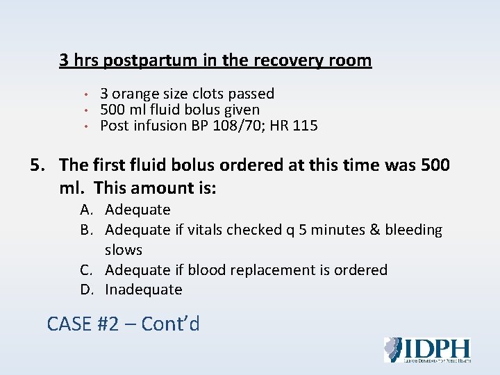
3 hrs postpartum in the recovery room • • • 3 orange size clots passed 500 ml fluid bolus given Post infusion BP 108/70; HR 115 5. The first fluid bolus ordered at this time was 500 ml. This amount is: A. Adequate B. Adequate if vitals checked q 5 minutes & bleeding slows C. Adequate if blood replacement is ordered D. Inadequate CASE #2 – Cont’d
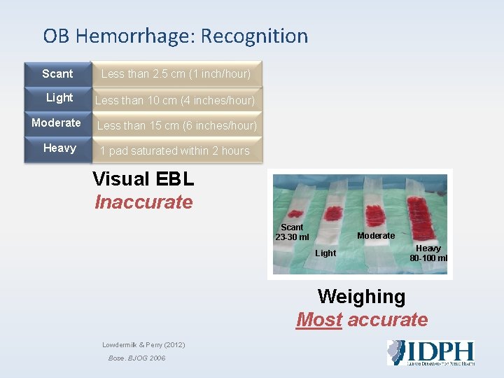
OB Hemorrhage: Recognition Scant Less than 2. 5 cm (1 inch/hour) Light Less than 10 cm (4 inches/hour) Moderate Less than 15 cm (6 inches/hour) Heavy 1 pad saturated within 2 hours Visual EBL Inaccurate Scant 23 -30 ml Moderate Light Heavy 80 -100 ml Weighing Most accurate Lowdermilk & Perry (2012) Bose. BJOG 2006
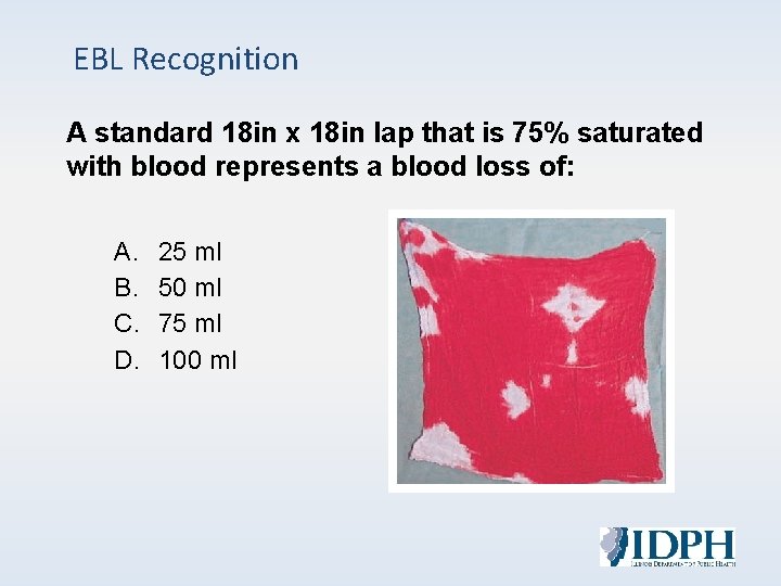
EBL Recognition A standard 18 in x 18 in lap that is 75% saturated with blood represents a blood loss of: A. B. C. D. 25 ml 50 ml 75 ml 100 ml
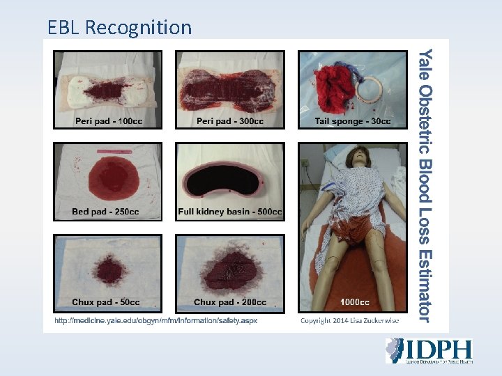
EBL Recognition
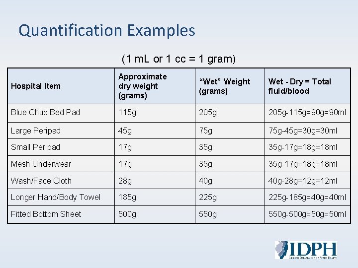
Quantification Examples (1 m. L or 1 cc = 1 gram) Hospital Item Approximate dry weight (grams) “Wet” Weight Wet - Dry = Total (grams) fluid/blood Blue Chux Bed Pad 115 g 205 g-115 g=90 ml Large Peripad 45 g 75 g-45 g=30 ml Small Peripad 17 g 35 g-17 g=18 ml Mesh Underwear 17 g 35 g-17 g=18 ml Wash/Face Cloth 28 g 40 g-28 g=12 ml Longer Hand/Body Towel 185 g 225 g-185 g=40 ml Fitted Bottom Sheet 500 g 550 g-500 g=50 ml
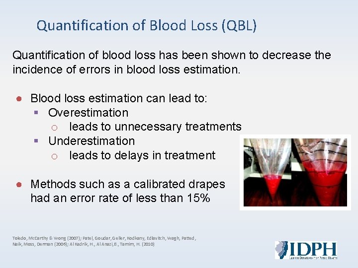
Quantification of Blood Loss (QBL) Quantification of blood loss has been shown to decrease the incidence of errors in blood loss estimation. ● Blood loss estimation can lead to: § Overestimation o leads to unnecessary treatments § Underestimation o leads to delays in treatment ● Methods such as a calibrated drapes had an error rate of less than 15% Toledo, Mc. Carthy & Wong (2007); Patel, Goudar, Geller, Kodkany, Edlavitch, Wagh, Patted, Naik, Moss, Derman (2006); Al Kadrik, H. , Al Anazi, B. , Tamim, H. (2010)
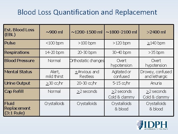
Blood Loss Quantification and Replacement Est. Blood Loss (EBL) ~900 ml Pulse <100 bpm >120 bpm >140 bpm Respirations 14 -20 bpm 20 -30 bpm 30 -40 bpm >35 bpm Blood Pressure Normal Orthostatic changes Overt hypotension Mental Status Alert, mild thirst +Anxious and Restless Agitated or confused Drowsy, confused and lethargic Urine Output >30 cc/hr 20 -30 cc/hr 5 -15 cc/hr Anuria Normal >2 seconds Cold & clammy Crystalloids & blood Cap Refill Fluid Replacement (3: 1 Rule) ~1200 -1500 ml ~1800 -2100 ml >2400 ml
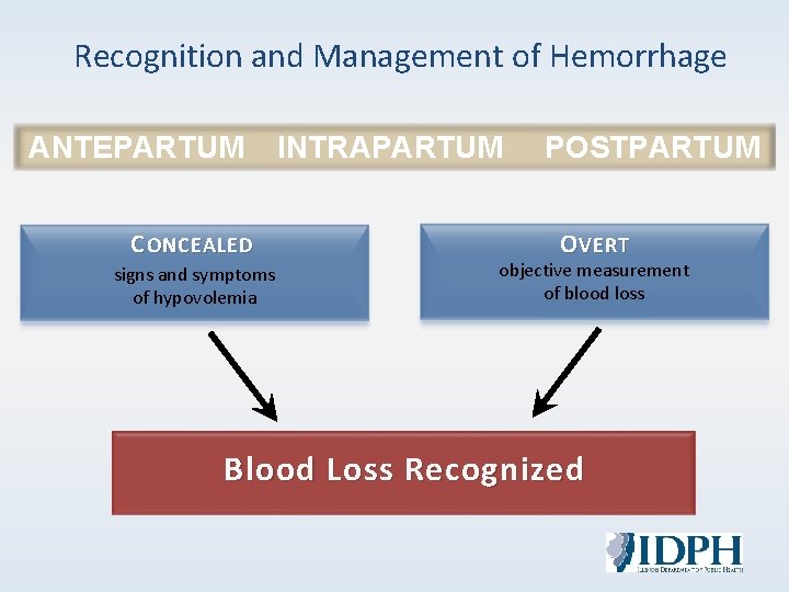
Recognition and Management of Hemorrhage ANTEPARTUM INTRAPARTUM POSTPARTUM C ONCEALED signs and symptoms of hypovolemia O VERT objective measurement of blood loss Blood Loss Recognized
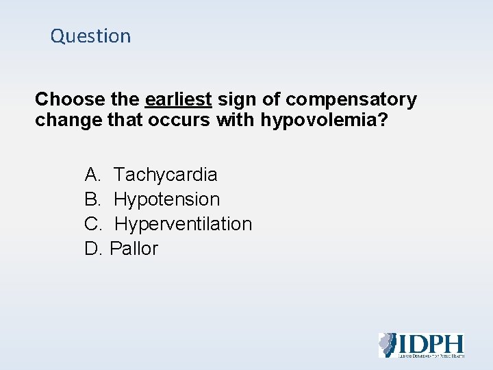
Question Choose the earliest sign of compensatory change that occurs with hypovolemia? A. Tachycardia B. Hypotension C. Hyperventilation D. Pallor
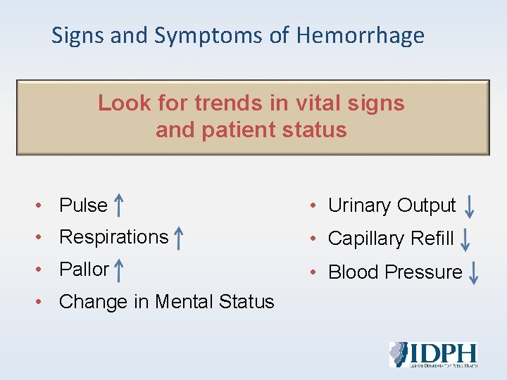
Signs and Symptoms of Hemorrhage Look for trends in vital signs and patient status • Pulse • Urinary Output • Respirations • Capillary Refill • Pallor • Blood Pressure • Change in Mental Status
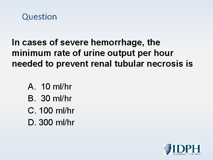
Question In cases of severe hemorrhage, the minimum rate of urine output per hour needed to prevent renal tubular necrosis is A. 10 ml/hr B. 30 ml/hr C. 100 ml/hr D. 300 ml/hr
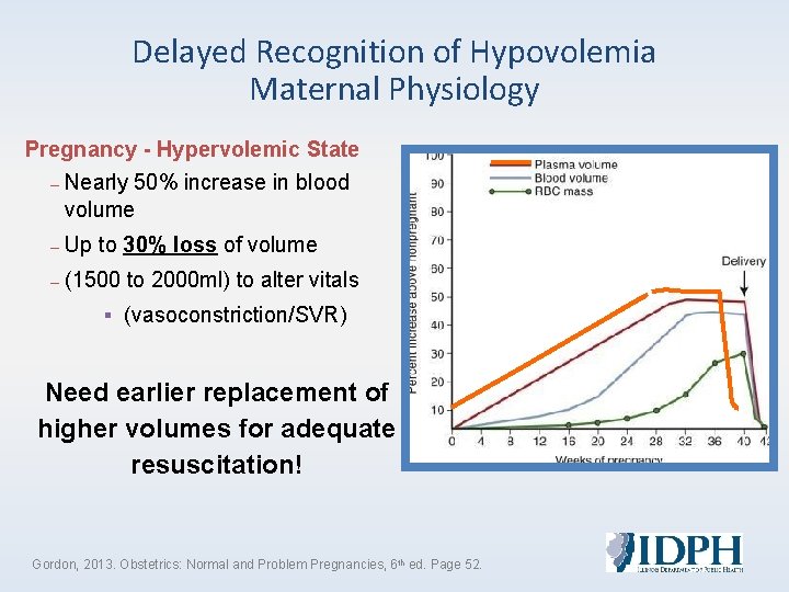
Delayed Recognition of Hypovolemia Maternal Physiology Pregnancy - Hypervolemic State – Nearly 50% increase in blood volume – Up to 30% loss of volume – (1500 to 2000 ml) to alter vitals ▪ (vasoconstriction/SVR) Need earlier replacement of higher volumes for adequate resuscitation! Gordon, 2013. Obstetrics: Normal and Problem Pregnancies, 6 th ed. Page 52.
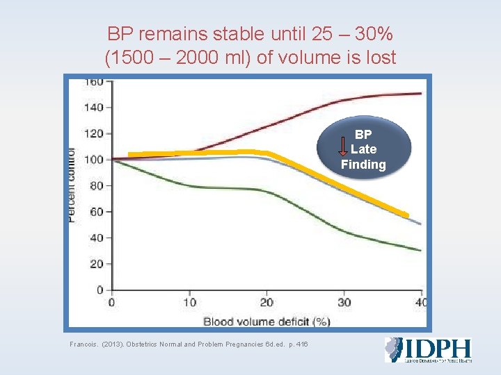
BP remains stable until 25 – 30% (1500 – 2000 ml) of volume is lost BP Late Finding Francois. (2013). Obstetrics Normal and Problem Pregnancies 6 d. ed. p. 416
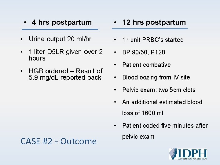
• 4 hrs postpartum • 12 hrs postpartum • Urine output 20 ml/hr • 1 st unit PRBC’s started • 1 liter D 5 LR given over 2 hours • BP 90/50, P 128 • HGB ordered – Result of 5. 9 mg/d. L reported back • Blood oozing from IV site • Patient combative • Pelvic exam: two 5 cm clots • An additional estimated blood loss of 1600 ml • Patient coded five minutes after CASE #2 - Outcome pelvic exam
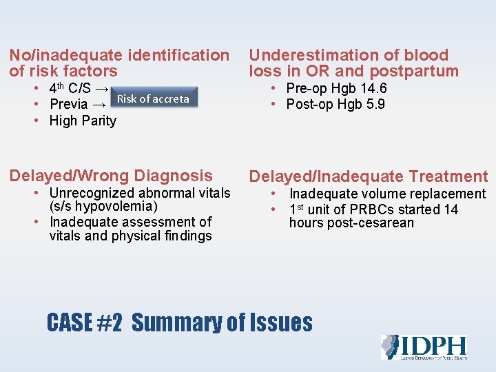
No/inadequate identification Underestimation of blood of risk factors loss in OR and postpartum • 4 th C/S → • Previa → Risk of accreta • High Parity • Pre-op Hgb 14. 6 • Post-op Hgb 5. 9 Delayed/Wrong Diagnosis • Unrecognized abnormal vitals (s/s hypovolemia) • Inadequate assessment of vitals and physical findings Delayed/Inadequate Treatment • Inadequate volume replacement • 1 st unit of PRBCs started 14 hours post-cesarean CASE #2 Summary of Issues
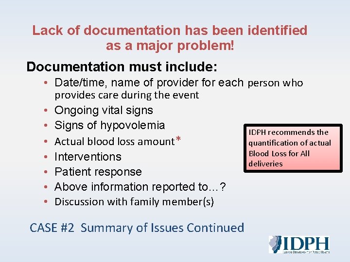
Lack of documentation has been identified as a major problem! Documentation must include: • Date/time, name of provider for each person who provides care during the event • Ongoing vital signs • Signs of hypovolemia IDPH recommends the • Actual blood loss amount* quantification of actual Blood Loss for All • Interventions deliveries • Patient response • Above information reported to…? • Discussion with family member(s) CASE #2 Summary of Issues Continued
Treatment & Product Replacement
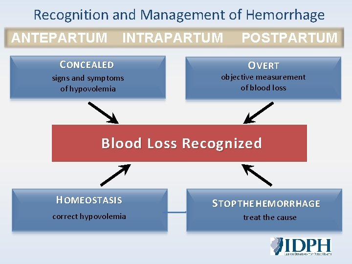
Recognition and Management of Hemorrhage ANTEPARTUM INTRAPARTUM POSTPARTUM C ONCEALED signs and symptoms of hypovolemia O VERT objective measurement of blood loss Blood Loss Recognized H OMEOSTASIS correct hypovolemia S TOP THE HEMORRHAGE treat the cause
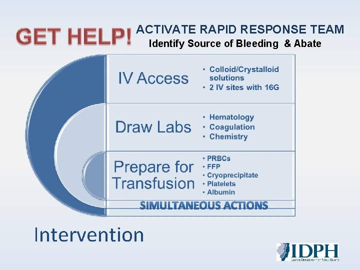
ACTIVATE RAPID RESPONSE TEAM Identify Source of Bleeding & Abate Intervention
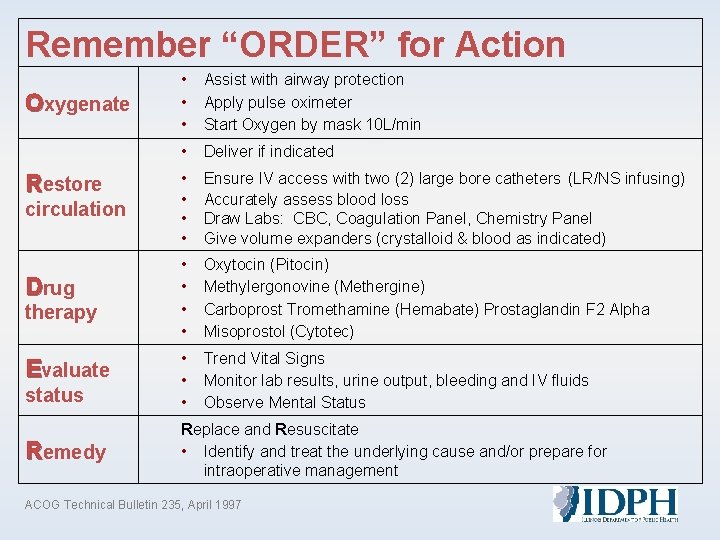
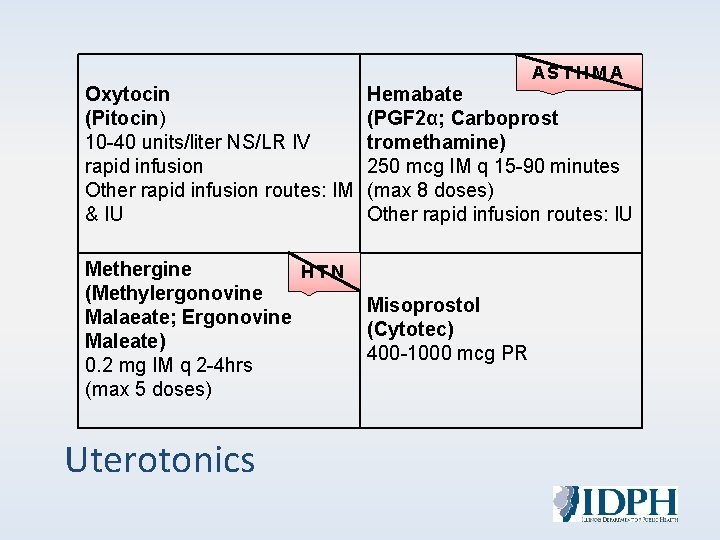
Remember “ORDER” for Action Oxygenate Restore circulation Drug therapy Evaluate status Remedy • • • Assist with airway protection Apply pulse oximeter Start Oxygen by mask 10 L/min • Deliver if indicated • • Ensure IV access with two (2) large bore catheters (LR/NS infusing) Accurately assess blood loss Draw Labs: CBC, Coagulation Panel, Chemistry Panel Give volume expanders (crystalloid & blood as indicated) • • Oxytocin (Pitocin) Methylergonovine (Methergine) Carboprost Tromethamine (Hemabate) Prostaglandin F 2 Alpha Misoprostol (Cytotec) • • • Trend Vital Signs Monitor lab results, urine output, bleeding and IV fluids Observe Mental Status Replace and Resuscitate • Identify and treat the underlying cause and/or prepare for intraoperative management ACOG Technical Bulletin 235, April 1997
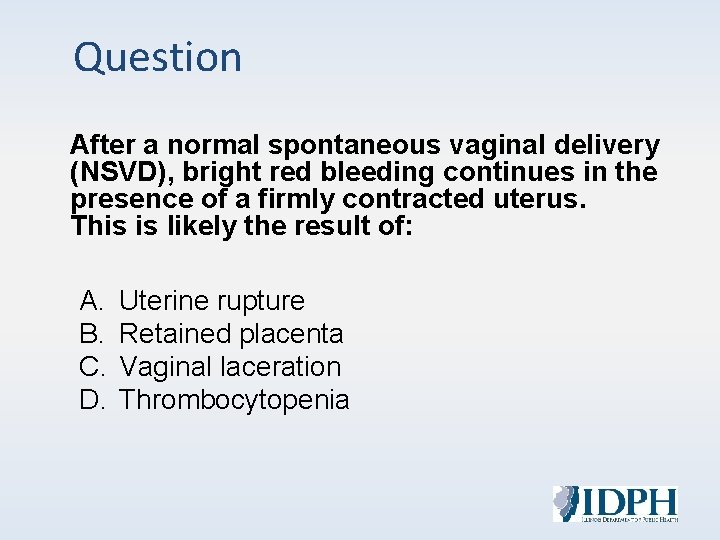
Question After a normal spontaneous vaginal delivery (NSVD), bright red bleeding continues in the presence of a firmly contracted uterus. This is likely the result of: A. B. C. D. Uterine rupture Retained placenta Vaginal laceration Thrombocytopenia
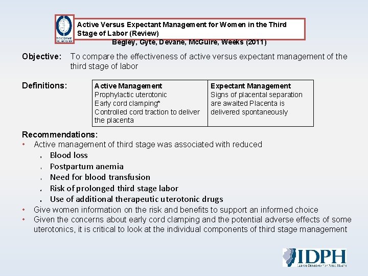
Active Versus Expectant Management for Women in the Third Stage of Labor (Review) Begley, Gyte, Devane, Mc. Guire, Weeks (2011) Objective: To compare the effectiveness of active versus expectant management of the third stage of labor Definitions: Active Management Prophylactic uterotonic Early cord clamping* Controlled cord traction to deliver the placenta Expectant Management Signs of placental separation are awaited Placenta is delivered spontaneously Recommendations: • Active management of third stage was associated with reduced 1. 2. 3. 4. 5. • • Blood loss Postpartum anemia Need for blood transfusion Risk of prolonged third stage labor Use of additional therapeutic uterotonic drugs Give women information on the risk and benefits to support an informed choice Given the concerns about early cord clamping and the potential adverse effects of some uterotonics, it is critical to look at the individual components of third stage management
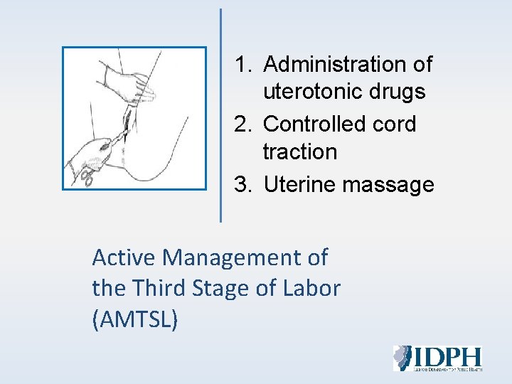
1. Administration of uterotonic drugs 2. Controlled cord traction 3. Uterine massage Active Management of the Third Stage of Labor (AMTSL)
Suggested Third Stage Oxytocin Management Protocol • Standardized, pre-mixed concentration of Oxytocin • Consider a 1 m. U/m. L rate for safe calculation and infusion • Same protocol for vaginal and cesarean deliveries • Safe medication practices for a high-alert drug
Interventions for the Third Stage of Labor • Hemorrhage risk factors • Uterine massage • Utilized uterotonics • Maintain IV site for at-risk sequentially or actual hemorrhage patients • Use of computerized infusion pumps • Assess for signs of hypervolemia and shock • Document dosages (units) – Quantify blood loss of oxytocin administered – Strict intake and output rather than a rate • Empty bladder to maintain uterine tone Burke, 2010
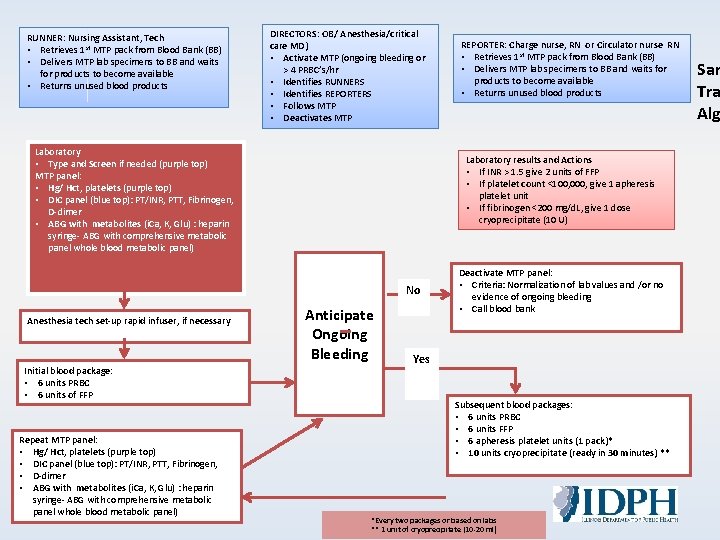
RUNNER: Nursing Assistant, Tech • Retrieves 1 st MTP pack from Blood Bank (BB) • Delivers MTP lab specimens to BB and waits for products to become available • Returns unused blood products DIRECTORS: OB/ Anesthesia/critical care MD) • Activate MTP (ongoing bleeding or > 4 PRBC’s/hr • Identifies RUNNERS • Identifies REPORTERS • Follows MTP • Deactivates MTP Laboratory • Type and Screen if needed (purple top) MTP panel: • Hg/ Hct, platelets (purple top) • DIC panel (blue top): PT/INR, PTT, Fibrinogen, D-dimer • ABG with metabolites (i. Ca, K, Glu) : heparin syringe- ABG with comprehensive metabolic panel whole blood metabolic panel) Laboratory results and Actions • If INR > 1. 5 give 2 units of FFP • If platelet count <100, 000, give 1 apheresis platelet unit • If fibrinogen <200 mg/d. L, give 1 dose cryoprecipitate (10 U) No Anesthesia tech set-up rapid infuser, if necessary Initial blood package: • 6 units PRBC • 6 units of FFP Repeat MTP panel: • Hg/ Hct, platelets (purple top) • DIC panel (blue top): PT/INR, PTT, Fibrinogen, • D-dimer • ABG with metabolites (i. Ca, K, Glu) : heparin syringe- ABG with comprehensive metabolic panel whole blood metabolic panel) REPORTER: Charge nurse, RN or Circulator nurse RN • Retrieves 1 st MTP pack from Blood Bank (BB) • Delivers MTP lab specimens to BB and waits for products to become available • Returns unused blood products Anticipate Ongoing Bleeding Deactivate MTP panel: • Criteria: Normalization of lab values and /or no evidence of ongoing bleeding • Call blood bank Yes Subsequent blood packages: • 6 units PRBC • 6 units FFP • 6 apheresis platelet units (1 pack)* • 10 units cryoprecipitate (ready in 30 minutes) ** *Every two packages or based on labs ** 1 unit of cryoprecipitate (10 -20 ml) Sam Tra Alg
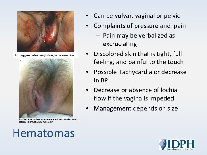
http: //gynaeonline. com/vulvar_hematoma. htm http: //ogscience. org/search. php? where=aview&id=10. 5468/ogs. 2014. 57. 2. 1 68&code=3021 OGS&vmode=PUBREADER Hematomas • Can be vulvar, vaginal or pelvic • Complaints of pressure and pain – Pain may be verbalized as excruciating • Discolored skin that is tight, full feeling, and painful to the touch • Possible tachycardia or decrease in BP • Decrease or absence of lochia flow if the vagina is impeded • Management depends on size
Non-Surgical Techniques for Acute Blood Loss fundal massage bimanual compression uterotonics tamponade devices Recombinant Activated Factor VII uterine packing
Other Options Acute Normovolemic Hemodilution (ANH)* Pre-op Autologous Blood Donation* Cell Saver* Tranexamic Acid (TXA) Rotem http: //www. nordicphotos. is/details/4721109
ASTHMA Oxytocin (Pitocin) 10 -40 units/liter NS/LR IV rapid infusion Other rapid infusion routes: IM & IU Hemabate (PGF 2α; Carboprost tromethamine) 250 mcg IM q 15 -90 minutes (max 8 doses) Other rapid infusion routes: IU Methergine HTN (Methylergonovine Malaeate; Ergonovine Maleate) 0. 2 mg IM q 2 -4 hrs (max 5 doses) Misoprostol (Cytotec) 400 -1000 mcg PR Uterotonics
Method of testing the efficiency of blood coagulation: • Clotting time CT (sec) § • Speed of fibrin formation; influenced by clotting factors, anticoagulants Clot Formation Time CFT (sec) § Kinetics of clot formation • Maximum Clot Firmness, MCF • Percentage of clot firmness loss during measurement Rotational Thromboelastography (TEG)
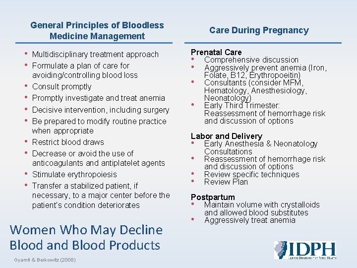
General Principles of Bloodless Medicine Management • • • Multidisciplinary treatment approach Formulate a plan of care for avoiding/controlling blood loss Consult promptly Promptly investigate and treat anemia Decisive intervention, including surgery Be prepared to modify routine practice when appropriate Restrict blood draws Decrease or avoid the use of anticoagulants and antiplatelet agents Stimulate erythropoiesis Transfer a stabilized patient, if necessary, to a major center before the patient’s condition deteriorates Women Who May Decline Blood and Blood Products Gyamfi & Berkowitz (2008) Care During Pregnancy Prenatal Care • Comprehensive discussion • Aggressively prevent anemia (Iron, Folate, B 12, Erythropoeitin) • Consultants (consider MFM, Hematology, Anesthesiology, Neonatology) • Early Third Trimester: Reassessment of hemorrhage risk and discussion of options Labor and Delivery • Early Anesthesia & Neonatology Consultations • Reassessment of hemorrhage risk and discussion of options • Review specific techniques • Review Plan Postpartum • Maintain volume with crystalloids and allowed blood substitutes • Aggressively treat anemia
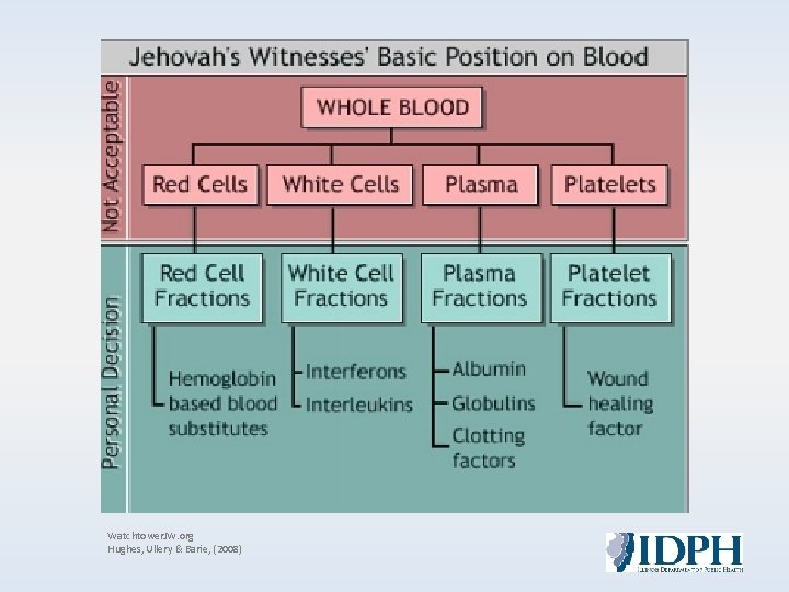
Watchtower. JW. org Hughes, Ullery & Barie, (2008)
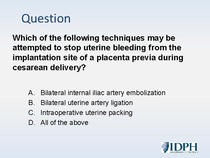
Question Which of the following techniques may be attempted to stop uterine bleeding from the implantation site of a placenta previa during cesarean delivery? A. B. C. D. Bilateral internal iliac artery embolization Bilateral uterine artery ligation Intraoperative uterine packing All of the above
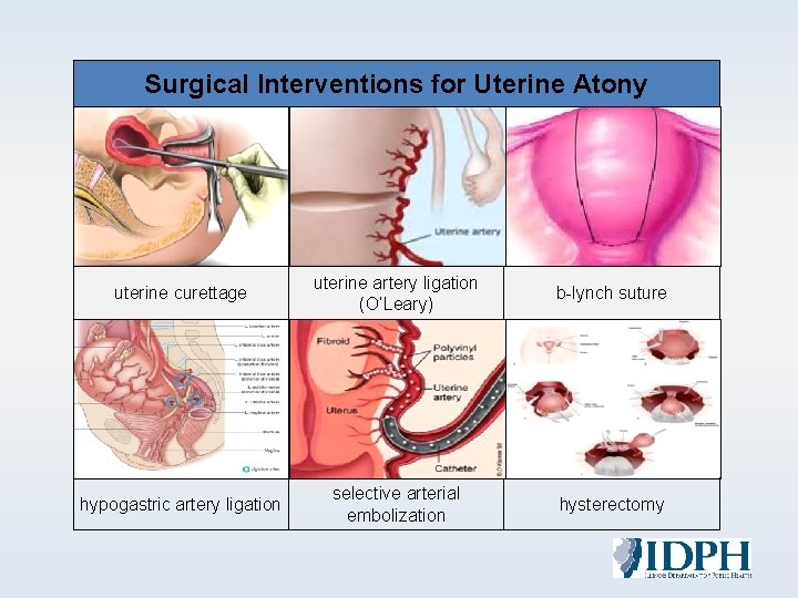
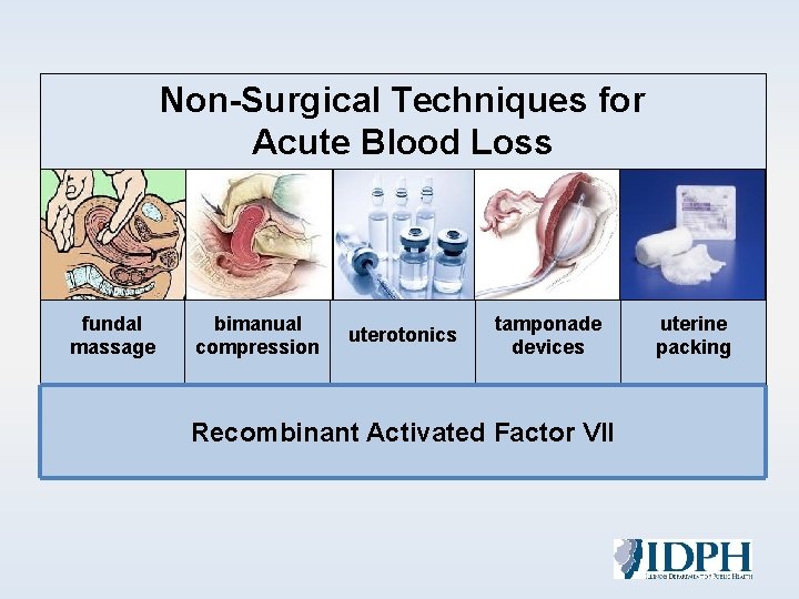
Surgical Interventions for Uterine Atony uterine curettage uterine artery ligation (O’Leary) b-lynch suture selective arterial embolization hysterectomy ? hypogastric artery ligation
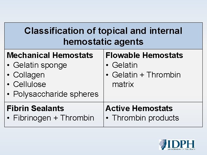
Classification of topical and internal hemostatic agents Mechanical Hemostats Flowable Hemostats • Gelatin sponge • Gelatin • Collagen • Gelatin + Thrombin • Cellulose matrix • Polysaccharide spheres Fibrin Sealants • Fibrinogen + Thrombin Active Hemostats • Thrombin products
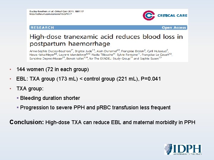
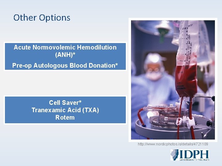
• 144 women (72 in each group) • EBL: TXA group (173 m. L) < control group (221 m. L), P=0. 041 • TXA group: § Bleeding duration shorter § Progression to severe PPH and p. RBC transfusion less frequent Conclusion: High-dose TXA can reduce EBL and maternal morbidity in PPH
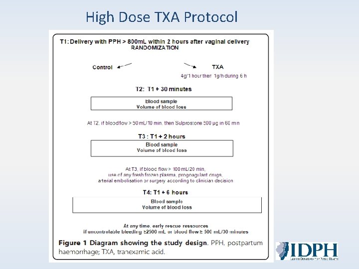
High Dose TXA Protocol
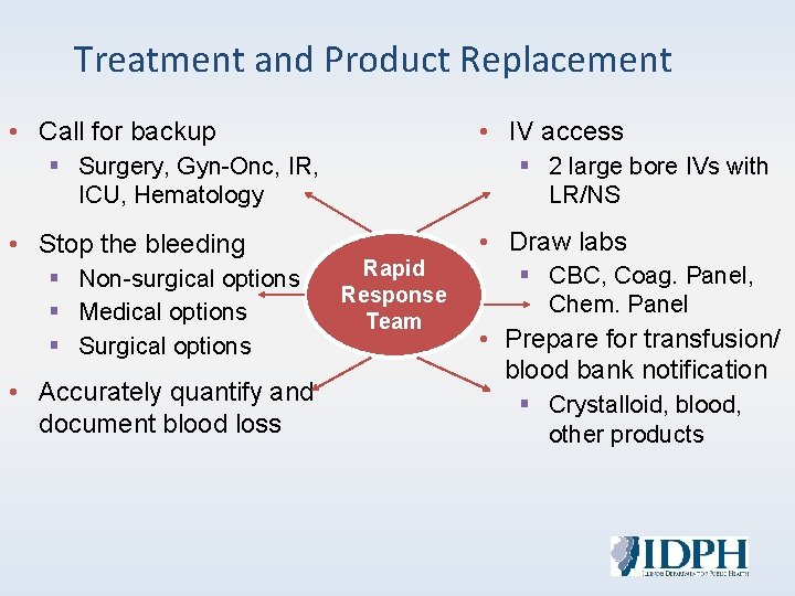
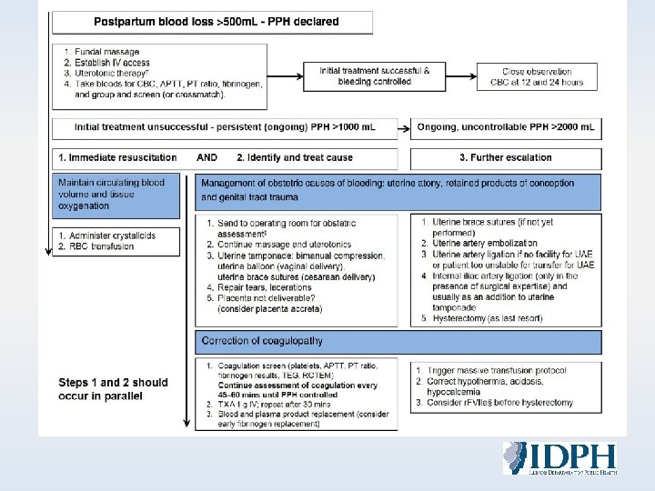
Treatment and Product Replacement • Call for backup • IV access § Surgery, Gyn-Onc, IR, ICU, Hematology • Stop the bleeding § Non-surgical options § Medical options § Surgical options • Accurately quantify and document blood loss § 2 large bore IVs with LR/NS • Draw labs Rapid Response Team § CBC, Coag. Panel, Chem. Panel • Prepare for transfusion/ blood bank notification § Crystalloid, blood, other products
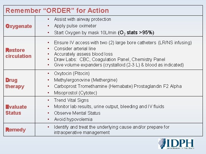
Remember “ORDER” for Action • • • Assist with airway protection Apply pulse oximeter Restore circulation • • • Ensure IV access with two (2) large bore catheters (LR/NS infusing) Consider arterial line Accurately assess blood loss Draw Labs: CBC, Coagulation Panel, Chemistry Panel Give volume expanders (crystalloid (2 -3 L) & blood as indicated) Drug therapy • • Oxytocin (Pitocin) Methylergonovine (Methergine) Carboprost Tromethamine (Hemabate) Prostaglandin F 2 Alpha Misoprostol (Cytotec) Evaluate Status • • Trend Vital Signs Monitor lab results, urine output, bleeding and IV fluids Observe Mental Status Avoid hypovolemia • Identify and treat the underlying cause and/or prepare for intraoperative management Oxygenate Remedy Start Oxygen by mask 10 L/min (O 2 stats >95%)
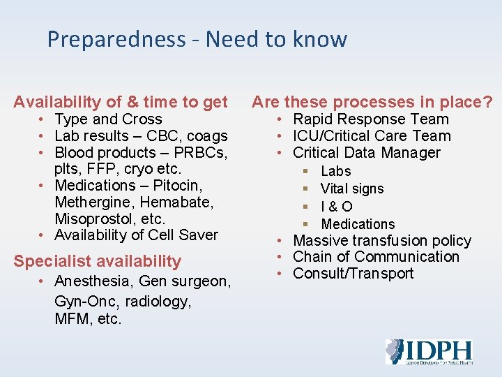
Preparedness - Need to know Availability of & time to get • Type and Cross • Lab results – CBC, coags • Blood products – PRBCs, plts, FFP, cryo etc. • Medications – Pitocin, Methergine, Hemabate, Misoprostol, etc. • Availability of Cell Saver Specialist availability • Anesthesia, Gen surgeon, Gyn-Onc, radiology, MFM, etc. Are these processes in place? • Rapid Response Team • ICU/Critical Care Team • Critical Data Manager § § Labs Vital signs I & O Medications • Massive transfusion policy • Chain of Communication • Consult/Transport
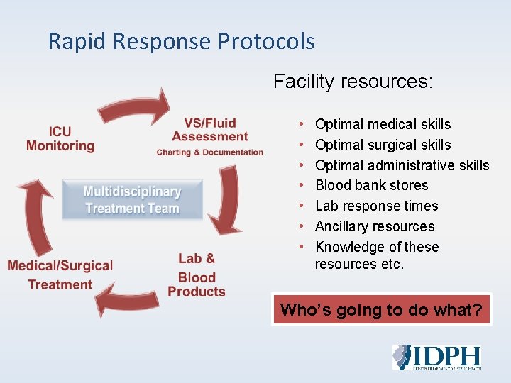
Rapid Response Protocols Facility resources: • • Optimal medical skills Optimal surgical skills Optimal administrative skills Blood bank stores Lab response times Ancillary resources Knowledge of these resources etc. Who’s going to do what?

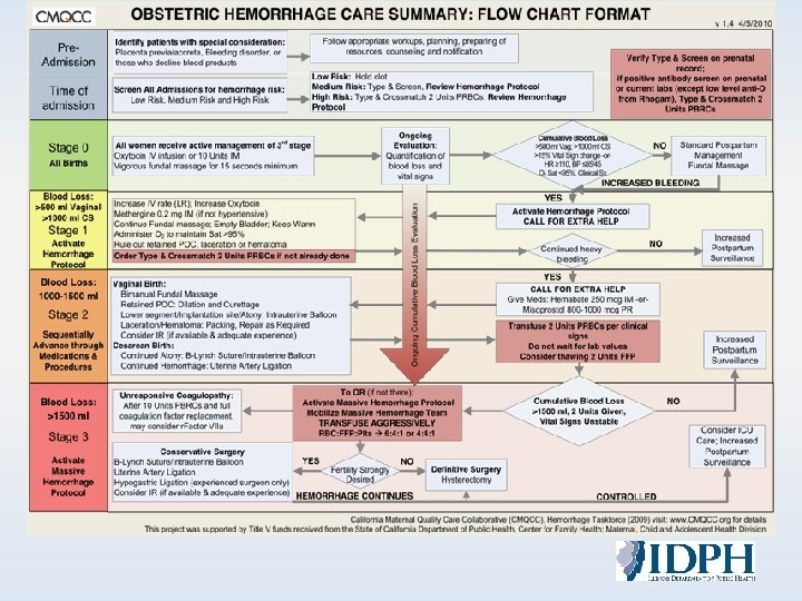
Treatment Algorithms
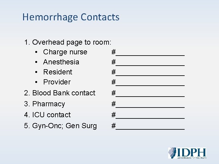
Hemorrhage Contacts 1. Overhead page to room: • Charge nurse #_________ • Anesthesia #_________ • Resident #_________ • Provider #_________ 2. Blood Bank contact #_________ 3. Pharmacy #_________ 4. ICU contact #_________ 5. Gyn-Onc; Gen Surg #_________
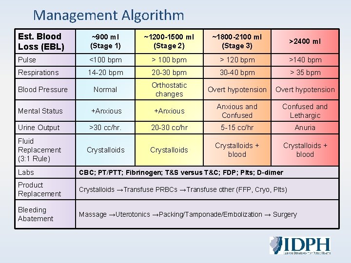
Management Algorithm Est. Blood Loss (EBL) ~900 ml (Stage 1) ~1200 -1500 ml (Stage 2) ~1800 -2100 ml (Stage 3) >2400 ml Pulse <100 bpm > 120 bpm >140 bpm Respirations 14 -20 bpm 20 -30 bpm 30 -40 bpm > 35 bpm Blood Pressure Normal Orthostatic changes Overt hypotension Mental Status +Anxious and Confused and Lethargic Urine Output >30 cc/hr. 20 -30 cc/hr 5 -15 cc/hr Anuria Fluid Replacement (3: 1 Rule) Crystalloids + blood Labs CBC; PT/PTT; Fibrinogen; T&S versus T&C; FDP; Plts; D-dimer Product Replacement Crystalloids →Transfuse PRBCs →Transfuse other (FFP, Cryo, Plts) Bleeding Abatement Massage →Uterotonics →Packing/Tamponade/Embolization → Surgery
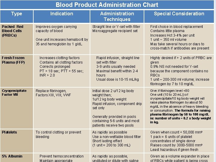
Blood Product Administration Chart Type Packed Red Blood Cells (PRBCs) Indication Improves oxygen carrying capacity of blood Administration Techniques Straight line or Y-set with filter Microaggregate recipient set • • Increases clotting factors Contains all clotting factors Corrects prolonged PT > 18 sec; PTT > 55 sec; INR > 2. 0 • • • Cryoprecipitate Factor VIII • • One unit increases hematocrit by 35 and hemoglobin by 1 g/d. L Fresh Frozen Plasma (FFP) Special Consideration Rapid infusion, straight line set with filter 3 -9 units usually needed Maximal benefit within 2 -4 hours Usual dose is 10 -15 m. L/kg Replace fibrinogen, Initial dose 2 u/12 kg body Factors XIII, VWF weight then; 1 u/12 kg body weight Rapid infusion, component drip set only • • • Generally provided in pools containing 5 -6 units and most patients receive two pools Platelets To control clotting or prevent bleeding As rapidly as possible Use a non-wettable blood filter Short lasting effect (1 unit = 200 to 300 m. L) • • 5% Albumin • • Prevent hemoconcentration Maintain appropriate As rapidly as possible, undiluted or dilute with saline • First choice in blood replacement Contains little plasma Increases Hct 3 -4% per unit 1 unit ~ 350 ml volume May take several hours or days to cross-match if antibodies are present Highly desired if > 2 units of PRBC are given 0. 9% NS not needed for Y-set because the component contains no RBCs 1 unit – 200 -300 ml volume; increase fibrinogen by 7 to 10 mg/d. L Give if fibrinogen level <80 One unit (10 to 20 m. L)) of cryoprecipitate/10 kg body weight will raise plasma fibrinogen by about 50 mg/d. L in the absence of heavy bleeding or consumption. The formula for raising plasma fibrinogen by 50 to 100 mg/d. L is: number of units = 0. 2 x body weight in kg. Given when count < 50, 000 mm 3 1 pack = 6 units of platelet concentrates of single donor Raises count by 3000 -5000 mm 3 Least hazardous if given fresh Given as a volume expander in place of RBCs while patient is being cross
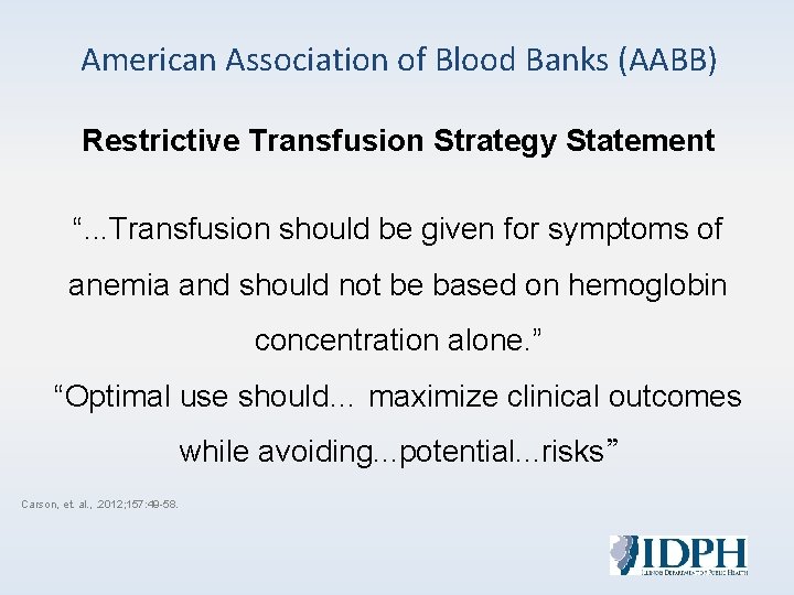
American Association of Blood Banks (AABB) Restrictive Transfusion Strategy Statement “. . . Transfusion should be given for symptoms of anemia and should not be based on hemoglobin concentration alone. ” “Optimal use should… maximize clinical outcomes while avoiding. . . potential. . . risks” Carson, et. al. , . 2012; 157: 49 -58.
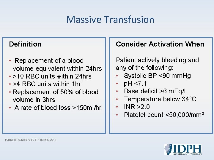
Massive Transfusion Definition Consider Activation When • Replacement of a blood volume equivalent within 24 hrs • >10 RBC units within 24 hrs • >4 RBC units within 1 hr • Replacement of 50% of blood volume in 3 hrs • A rate of blood loss >150 ml/hr Patient actively bleeding and any of the following: • Systolic BP <90 mm. Hg • p. H <7. 1 • Base deficit >6 m. Eq/L • Temperature below 34°C • INR >2. 0 • Platelet count <50, 000/mm³ Pacheco, Saade, Gei, & Hankins, 2011
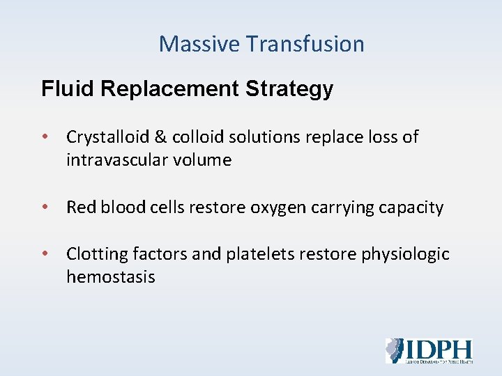
Massive Transfusion Fluid Replacement Strategy • Crystalloid & colloid solutions replace loss of intravascular volume • Red blood cells restore oxygen carrying capacity • Clotting factors and platelets restore physiologic hemostasis
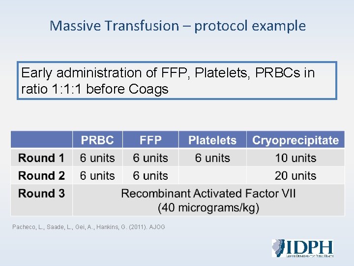
Massive Transfusion – protocol example Early administration of FFP, Platelets, PRBCs in ratio 1: 1: 1 before Coags Pacheco, L. , Saade, L. , Gei, A. , Hankins, G. (2011). AJOG
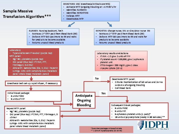
Sample Massive Transfusion Algorithm*** DIRECTORS: OB/ Anesthesia/critical care MD) • Activate MTP (ongoing bleeding or > 4 PRBC’s/hr • Identifies RUNNERS • Identifies REPORTERS • Follows MTP • Deactivates MTP RUNNER: Nursing Assistant, Tech • Retrieves 1 st MTP pack from Blood Bank (BB) • Delivers MTP lab specimens to BB and waits for products to become available • Returns unused blood products REPORTER: Charge nurse, RN or Circulator nurse RN • Retrieves 1 st MTP pack from Blood Bank (BB) • Delivers MTP lab specimens to BB and waits for products to become available • Returns unused blood products Laboratory • Type and Screen if needed (purple top) MTP panel: • Hg/ Hct, platelets (purple top) • DIC panel (blue top): PT/INR, PTT, Fibrinogen, D-dimer • ABG with metabolites (i. Ca, K, Glu) : heparin syringe- ABG with comprehensive metabolic panel whole blood metabolic panel) Laboratory results and Actions • If INR > 1. 5 give 2 units of FFP • If platelet count <100, 000, give 1 apheresis platelet unit • If fibrinogen <200 mg/d. L, give 1 dose cryoprecipitate (10 U) No Deactivate MTP panel: • Criteria: Normalization of lab values and /or no evidence of ongoing bleeding • Call blood bank Anesthesia tech set-up rapid infuser, if necessary Initial blood package: • 6 units PRBC • 6 units of FFP Repeat MTP panel: • Hg/ Hct, platelets (purple top) • DIC panel (blue top): PT/INR, PTT, Fibrinogen, D -dimer • ABG with metabolites (i. Ca, K, Glu) : heparin syringe- ABG with comprehensive metabolic panel whole blood metabolic panel) Anticipate Ongoing Bleeding Yes Subsequent blood packages: • 6 units PRBC • 6 units FFP • 6 apheresis platelet units (1 pack)* • 10 units cryoprecipitate (ready in 30 minutes) ** *Every two packages or based on labs ** 1 unit of cryoprecipitate (10 -20 ml)

Summary
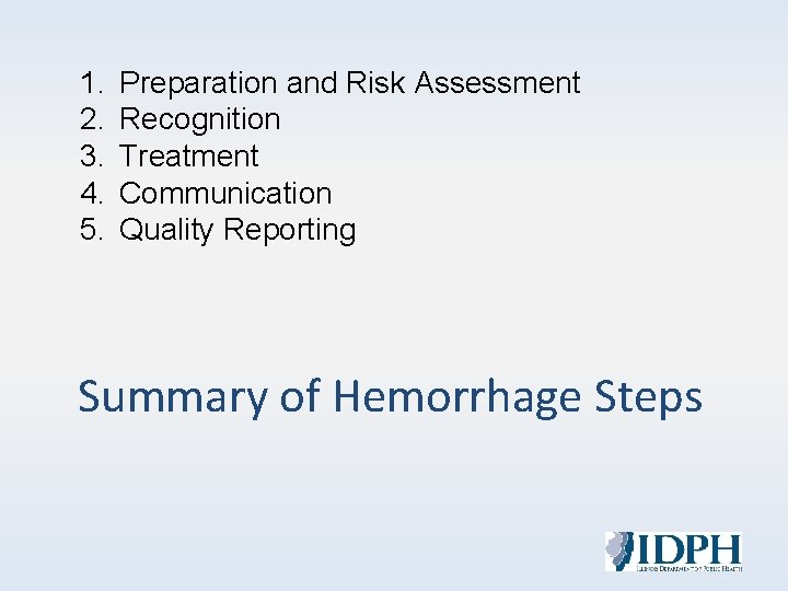
1. 2. 3. 4. 5. Preparation and Risk Assessment Recognition Treatment Communication Quality Reporting Summary of Hemorrhage Steps
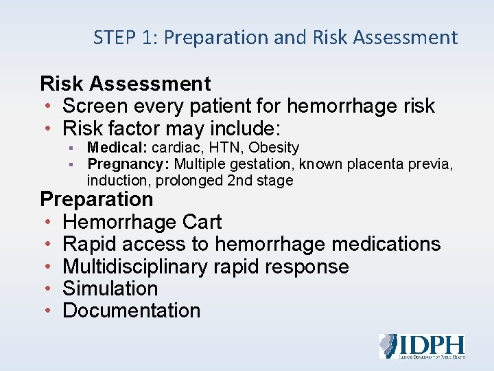
STEP 1: Preparation and Risk Assessment • Screen every patient for hemorrhage risk • Risk factor may include: § § Medical: cardiac, HTN, Obesity Pregnancy: Multiple gestation, known placenta previa, induction, prolonged 2 nd stage Preparation • Hemorrhage Cart • Rapid access to hemorrhage medications • Multidisciplinary rapid response • Simulation • Documentation
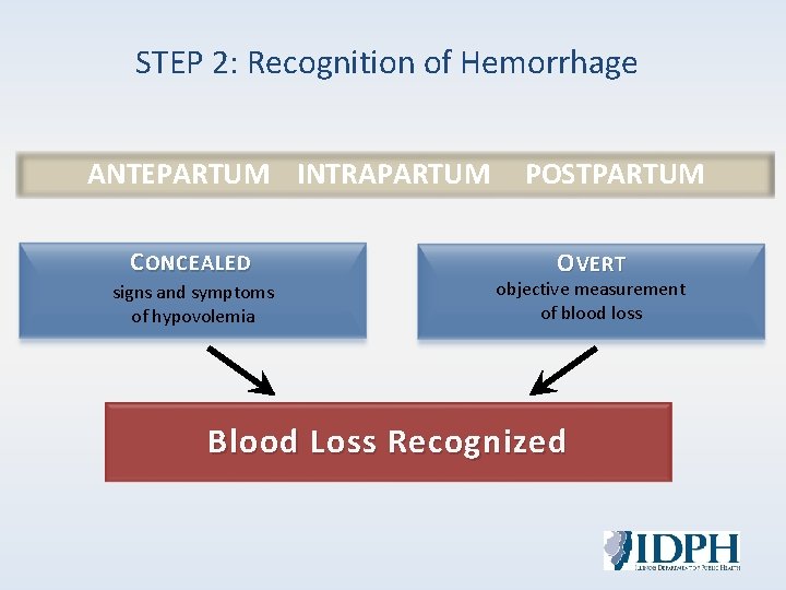
STEP 2: Recognition of Hemorrhage ANTEPARTUM INTRAPARTUM C ONCEALED signs and symptoms of hypovolemia POSTPARTUM O VERT objective measurement of blood loss Blood Loss Recognized
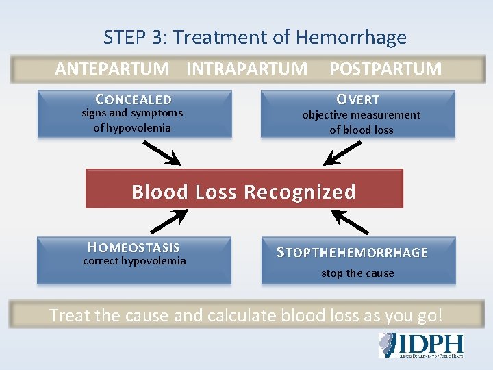
STEP 3: Treatment of Hemorrhage ANTEPARTUM INTRAPARTUM POSTPARTUM C ONCEALED signs and symptoms of hypovolemia O VERT objective measurement of blood loss Blood Loss Recognized H OMEOSTASIS correct hypovolemia S TOP THE HEMORRHAGE stop the cause Treat the cause and calculate blood loss as you go!
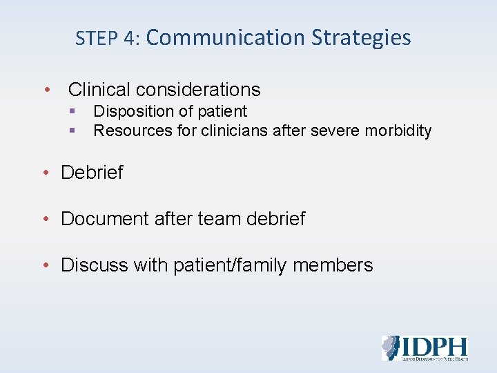
STEP 4: Communication Strategies • Clinical considerations § § Disposition of patient Resources for clinicians after severe morbidity • Debrief • Document after team debrief • Discuss with patient/family members
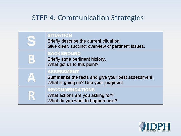
STEP 4: Communication Strategies S B A R SITUATION Briefly describe the current situation. Give clear, succinct overview of pertinent issues. BACKGROUND Briefly state pertinent history. What got us to this point? ASSESSMENT Summarize the facts and give your best assessment. What is going on? Use your judgment. RECOMMENDATIONS What actions are you asking for? What do you want to happen next?
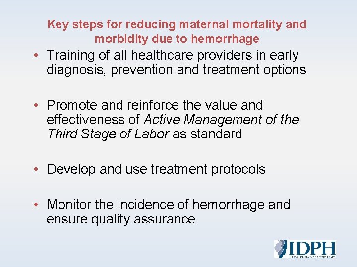
Key steps for reducing maternal mortality and morbidity due to hemorrhage • Training of all healthcare providers in early diagnosis, prevention and treatment options • Promote and reinforce the value and effectiveness of Active Management of the Third Stage of Labor as standard • Develop and use treatment protocols • Monitor the incidence of hemorrhage and ensure quality assurance
STEP 5: Quality Reporting & Systems Learning • • • Establish a culture of huddles for high-risk patients and post-event debriefs Conduct a multidisciplinary review of serious hemorrhages for systems issues Monitor outcomes and processes metrics
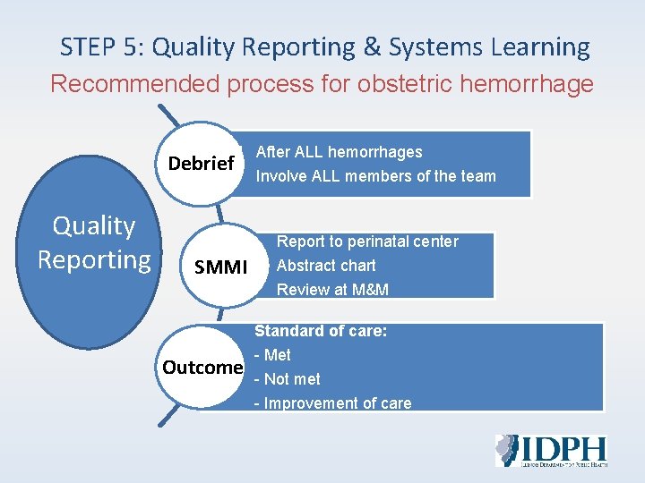
STEP 5: Quality Reporting & Systems Learning Recommended process for obstetric hemorrhage Debrief Quality Reporting After ALL hemorrhages Involve ALL members of the team Report to perinatal center SMMI Abstract chart Review at M&M Standard of care: Outcome - Met - Not met - Improvement of care

THANK YOU
- Slides: 89