OBJECTIVES Review the clinical anatomy and physical exam

OBJECTIVES • Review the clinical anatomy and physical exam of the wrist and hand • Formulate a pathoanatomic diagnosis in the clinical setting • Discuss common clinical conditions that can be elicited from the physical exam

INTRODUCTION: Hand Wrist • Series of complex, delicately balanced joints • Function is integral to every act of daily living • Most active portion of the upper extremity

INTRODUCTION • The least protected joints • Extremely vulnerable to injury • Difficult and complex examination • Diagnosis often vague – If no fracture = “wrist strain or sprain” • Bilateral comparison useful

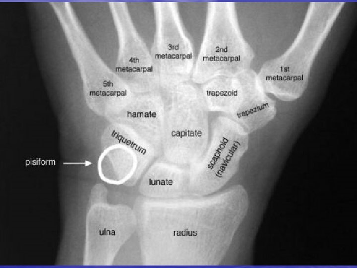
Bony Anatomy • Phalanges: 14 • Sesamoids: 2 • Metacarpals: 5 • Carpals – Proximal row: 4 – Distal row: 4 • Radius and Ulna Lister’s tubercle

ANATOMY • Muscles /Tendons – Volar wrist- 6 – Dorsal wrist- 9 • 6 compartments – Volar hand- 10 – Dorsal hand- dorsal interossei • Nerves - 3 – – – Median Ulnar Radial • Arteries - 2

HISTORY • Age • Handedness • Chief complaint • Occupation • Previous injury • Previous surgery • Sx related to specific • • activities What exacerbates What improves Frequency Duration

HISTORY • 4 principle mechanisms of injury – – Throwing Weight bearing Twisting Impact

PHYSICAL EXAM • Inspection • Palpation • Range of Motion • Neurologic Exam • Special Tests

INSPECTION • Observe upper • • • extremity as patient enters room Examine hand in function Deformities Attitude of the hand

INSPECTION Palmar Surface • Creases • Thenar and • • • Hypothenar Eminence Arched Framework Hills and Valleys Web Spaces

Cascade sign • Assure all fingers point to scaphoid area when flexed at PIPs

INSPECTION of Dorsal Hand Wrist • Hills and Valleys • Height of metacarpal heads • Finger nails – Pale or white=anemia or circulatory – Spoon shaped=fungal infection – Clubbed=respiratory or congenital heart • Deformities

Ganglion • Cystic structure that • • • arises from synovial sheath Discrete mass Dull ache Dorsal or Volar aspect

Boutonniere Deformity • Tear or stretch of • • • the central extensor tendon at PIP Note: unopposed flexion at PIP Extension at DIP Trauma or inflammatory arthritis
 NOTE: Extension at")
Swan Neck Deformity • Contraction of intrinsic • muscles (trauma, RA) NOTE: Extension at PIP

Osteoarthritis • Heberden’s nodes: DIP • Bouchard’s nodes: PIP

Rheumatoid Arthritis • MCP swelling • Swan neck deformities • Ulnar deviation at MCP joints • Nodules along tendon sheaths

Mallet Finger • Hyperflexion injury • Ruptured terminal • • extensor mechanism at DIP Incomplete extension of DIP joint or extensor lag Treatment: – stack splint

Dupuytren’s Contractures • Palmar or digital • • fibromatosis Flexion contracture Painless nodules near palmar crease Male> Female Epilepsy, diabetes, pulmonary dz, alcoholism

RANGE OF MOTION • Active range of motion • Passive range of motion if unable to actively move joint • Bliateral comparison – To determine degrees of restriction

RANGE OF MOTION Wrist • Flexion • Extension • Radial deviation • Ulnar deviation – Ulnar deviation is greater than radial

RANGE OF MOTION Fingers • Flexion/extension at MCP, PIP, DIP – Tight fist and open – Do all fingers work in unison • ABDuction/ADDuction at MCP – Spread fingers apart and then back together

PALPATION of Skin • Warmth? • Dryness? – Anhydrosis= nerve damage • Scars

PALPATION of Wrist Dorsum • Radial Styloid • Scaphoid • 1 st MC/Trapezium jt • Lunate • Lister’s Tubercle • Ulnar Styloid • TFCC • Triquetrum • Pisiform • Hook of Hamate • Guyon’s Tunnel

Radial Styloid palpation Scaphoid Bone palpation Radial styloid

Scaphoid Fracture • Most commonly fractured carpal bone – 70 -80% of all carpal bone injuries – 8% of all sports related fractures – 1 in 100 college football players • Most susceptible to injury – Bridges proximal and distal rows of the carpal bones – Load to the dorsiflexed wrist as in fall onto outstretched hand

Scaphoid Fracture • Painful, swollen wrist after a fall • Tenderness in snuffbox • High frequency of nonunion and avascular necrosis • Initial x-rays often unremarkable

st 1 MC/Trapezium joint palpation

Thumb CMC Joint Arthritis • Painful pinch or • grasp “Grind Test” – Axial pressure to thumb while palpating CMC joint

Lunate Bone palpation

Kienbock’s Disease • Idiopathic osteonecrosis of lunate • Stress or compression fracture of the lunate – Disruption of blood supply with collapse and secondary fragmentation • Pain and stiffness of the wrist in the ABSENCE of TRAUMA

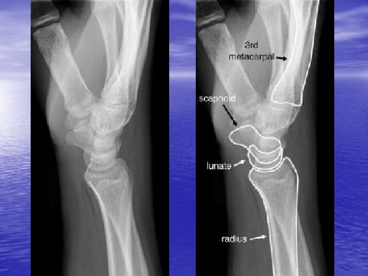
Scapholunate Dissociation • Diagnosis often missed • Pain, swelling, and decreased ROM • Pressure over scaphoid tuberosity elicits pain • Greatest pain over dorsal scapholunate area, accentuated with dorsiflexion • X-ray shows widening of scapholunate joint space by at least 3 mm

Ulnar Styloid palpation Lister’s Tubercle palpation Ulnar styloid
")
Triangular Fibro-Cartilage Complex palpation (TFCC)

Triangular Fibrocartilage Complex Injuries • Thickened pad of connective tissue that functions as a cushion for the ulnar carpus as well as a sling support for the lunate and triquetrum • Injury from compression between lunate and head of ulna – Breaking fall with hand – Rotational forces-racket and throwing sports

Triangular Fibrocartilage Complex Injuries • Ulnar sided wrist pain, • • • swelling, loss of grip strength “Click” with ulnar deviation Point tenderness distal to ulnar styloid TFCC load test

Triquetrum Bone palpation

Triquetrum Fracture • 2 nd most common carpal fracture • Fall onto outstretched hand with wrist in dorsiflexion and ulnar deviation • Swelling and tenderness over the dorsal ulnar aspect of the wrist

PALPATION of HAND Bone • Metacarpals - 5 • Phalanges - 14 • Palpate for swelling, tenderness • Assess for symmetry

PALPATION Soft tissue • 6 Dorsal Compartments – Transport extensor tendons • 2 Palmar Tunnels – Transport nerves, arteries, flexor tendons

1 st Dorsal Compartment • Abductor Pollicis Longus and Extensor Pollicis Brevis • Radial border of Anatomic Snuff Box • Site of stenosing tenosynovitis – De Quervain’s Tenosynovitis – Finkelstein’s Test

De. Quervain’s Tenosynovitis • Inflammation of EXT • • Pollicis Brevis and ABD Pollicis Longus tendons Tenderness 1 st Dorsal Compartment Finkelstein’s Test

2 nd Dorsal Compartment • Extensor Carpi Radialis Longus • and Extensor Carpi Radialis Brevis Make fist—becomes prominent
 • Similar to De. Quervain’s tenosynovitis • Peritendinitis related to")
Intersection Syndrome (Squeaker Wrist) • Similar to De. Quervain’s tenosynovitis • Peritendinitis related to bursal inflammation at the junction of the 1 st and 2 nd dorsal compartments • Overuse of the radial extensor of the wrist
 • Seen in gymnasts, rowers, weightlifters, racket sports • Proximal")
Intersection Syndrome (Squeaker Wrist) • Seen in gymnasts, rowers, weightlifters, racket sports • Proximal to De. Quervain’s- 4 -6 cm from radiocarpal joint • Crepitation or squeaking can be heard with passive or active ROM

3 rd Dorsal Compartment • Extensor Pollicis Longus • Ulnar side of Anatomic Snuff Box • Can rupture secondary to Colles’ Fracture or Rheumatoid Arthritis • Extensor Pollicis Longus Tenosynovitis

4 th Dorsal Compartment • Extensor Digitorum Communis and Extensor Indicis • Palpate from the carpus to the metacarpophalangeal joints • Frequent site of ganglion cysts

5 th Dorsal Compartment • Extensor Digiti Minimi • May become involved in rheumatoid arthritis • May be subject to attrition – friction due to dorsal dislocation of the ulnar head – synovitis

6 th Dorsal Compartment • Extensor Carpi Ulnaris – Tendinitis -repetitive wrist motion or snap of wrist • May dislocate over the styloid process of the ulna – Seen with Colles’ fracture with associated fracture of the distal ulnar styloid – Audible snap

Extensor Carpi Ulnaris Tenosynovitis and Subluxation • 6 th Dorsal Compartment • Second most common site of tenosynovitis (after De. Quervain’s) • Common in racket and rowing sports • Pain and tenderness with ulnar deviation • Suspect subluxation when clicking on ulnar side of forearm

PALPATION Palmar Aspect • Pisiform and Hamate • Tunnel of Guyon • Ulnar Artery • Carpal Tunnel • Flexor Carpi Radialis • Flexor Carpi Ulnaris

Tunnnel of Guyon Pisiform and Hamate palpation

Hamate Hook Fracture • Frequently misdiagnosed as tendonitis or sprain • Pain, swelling, and tenderness over hypothenar eminence • Suspect when patient complains of painful griping and swinging

Tunnel of Guyon • Depression between • • pisiform and hook of hamate Contains ulnar nerve and artery Site of compression injuries – unusually tender if pathology is present

Ulnar Nerve Compression • Tunnel of Guyon • Seen in direct or repetitive trauma, fractures of hamate or pisiform, or sports related – Operating a jackhammer – repetitive power gripping (ex. Cycling) • Sx= pain, weakness, paresthesias in ulnar sensory distribution

Carpal Tunnel • Deep to palmaris • • longus Contains median nerve and finger flexor tendons Most common overuse injury of the wrist

Carpal Tunnel Syndrome • Entrapment of the median nerve – Phalen’s and Tinel’s Test – 2 point discrimination • Symptoms – Aching in hand arm – Nocturnal or AM paresthesias – “Shaking” to obtain relief

Carpal Tunnel Tests • Neurologic exam – Median nerve sensation and motor • Phalen’s Test: • both wrists maximally flexed for 1 minute Tinel’s Test

Flexor carpi ulnaris Palmaris longus Flexor carpi radialis Volar flexor tendons

PALPATION Palm of Hand • Thenar Eminence – 3 muscles of thumb – Atrophy seen in carpal tunnel syndrome • Hypothenar Eminance – 3 muscles of little finger – Atrophy with ulnar nerve compression • Palmar Aponeurosis – Dupuytren’s Contracture

PALPATION of Fingers • Finger Flexor Tendons – Trigger Finger- sudden audible snapping with movement of one of the fingers • Extensor Tendons • Tufts of Fingers – Felon- local infection – Paronychia- hangnail infection

SPECIAL TESTS Long Finger Flexor Test • Flexor Digitorum Superficialis Test – Flex finger at PIP – The only functioning tendon at the PIP • Flexor Digitorum Profundus Test – Flex at DIP • Inability to flex= tendon cut or denervated

Flexor Tendon Injury “Jersey Finger” • Avulsion injury from • rapid passive extension of the clenched fist Loss of flexion at PIP and/or DIP – “+” sublimus or profundus tests

Trigger Finger • Stenosing flexor • • tenosynovitis Painful snap or lock Palpate nodule as digit flexed and extended

Flexor Tenosynovitis • Tendon sheath infection • Usually due to a puncture wound • Bacterial skin flora • Relative surgical emergency

Flexor Tenosynovitis 4 Cardinal Signs of Kanavel • Uniform swelling of • • • the finger Sensitivity along the course of the tendon sheaths Pain upon passive extension Fingers held in flexion

RANGE OF MOTION Thumb • Thumb flexion/extension at MCP and IP – Touch pad at base of little finger • Thumb ABD/ADD at carpometacarpal joint • Opposition – Touch tip of thumb to tip of each finger

Skier’s Thumb Gamekeeper’s Thumb • Ulnar Collateral • • Ligament rupture of the thumb MCP joint Instability, weak and ineffective pinch Radially directed stress at MCP jointstable if opens <35 degrees

NEUROLOGIC EXAM • Muscular assessment using grading system • Sensation testing • Bilateral comparison

NEUROLOGIC EXAM Muscle Testing • WRIST – EXT C 6 – FLEX C 7 • FINGERS – EXT C 7 – FLEX C 8 – ABD T 1 – ADD T 1

Sensation Testing Dorsal hand Radial hand

NEUROLOGIC EXAM Sensation Testing • Neurological Level – Dermatomes- 3 neurologic levels – C 6, C 7, C 8

RADIOLOGIC STUDIES • AP and Lateral of • hand wrist Consider Obliques and special views if fracture suspected but not seen on AP and Lateral

EXAMINATION OF RELATED AREAS • Referred pain can be due to: – Herniated cervical discs – Osteoarthritis – Brachial plexus outlet syndrome – Elbow and shoulder entrapment syndrome

Sites of Pain and Common Pathology • Dorsal pain – Ganglion (#1 cause of dorsal pain) – Extensor tendonitis (overuse) – Kienbach’s Disease • Volar Pain – Ganglion – Flexor tendinitis – Carpal tunnel syndrome – Thumb CMC joint arthritis

Site of Pain and Common Pathology • Radial pain – Thumb CMC DJD – De. Quervain’s tendinitis – Scaphoid fracture • Ulnar pain – EXT carpi ulnaris tendinitis – Synovitis – Triangular fibrocartilage complex tear
- Slides: 78