Objectives Present the pathology clinical presentation and management

Objectives • Present the pathology, clinical presentation and management of chronic infection of the middle ear cleft • Outline the indications of the common middle ear surgical procedures

CHRONIC OTITIS MEDIA

Definition • Chronic inflammation of the middle ear cleft

Classification of Chronic Otitis Media • Chronic Non Suppurative Otitis Media – Otitis media with effusion “OME” – Adhesive otitis media • Chronic Suppurative Otitis Media “CSOM” – Tubo-tympanic (Safe) – Attico-antral (Unsafe)

Chronic Non Suppurative Otitis Media • Otitis media with effusion “OME” • Adhesive otitis media
")
OTITIS MEDIA WITH EFFUSION (OME)

Definition Presence of non-purulent fluid within the middle ear cleft

Pathology of OME • Eustachian tube dysfunction • Chronic non suppurative inflammatory changes in the mucosa • Formation of fluid in the middle ear – Transudate – Excudate – Secretion
 • Secretory otitis media • Serous otitis")
Synonyms of otitis media with Effusion (OME) • Secretory otitis media • Serous otitis media • Excudative (Catarrhal) otitis media • Sero-mucinous otitis media • Glue ear

Etiology • Eustachian tube dysfunction – Poor muscular function

Etiology • Eustachian tube dysfunction – Poor muscular function – Adenoids

Etiology • Eustachian tube dysfunction – Poor muscular function – Adenoids – Barotrauma

Otitic Barotrauma • Due to failure of the ET to equalize the middle ear pressure during descent from high altitudes in aircraft or during diving

ETIOLOGY • Eustachian tube dysfunction – Poor muscular function – Adenoids – Barotrauma – Others (e. g. nasopharyngeal carcinoma)

Important Nasopharygeal carcinoma should be suspected in adults with OME especially if it is unilateral

Etiology • Eustachian tube dysfunction – Poor muscular function – Adenoids – Barotrauma – Others (e. g. nasopharyngeal carcinoma) • Infections – Unresolved AOM – Adenoiditis and other URTIs

Etiology • Eustachian tube dysfunction – Poor muscular function – Adenoids – Barotrauma – Others (e. g. nasopharyngeal carcinoma) • Infections – Unresolved AOM – Adenoiditis and other URTIs

Prevalence of OME • Between 20% and 50% of children do have OME at some time between 2 and 10 years of age

Symptoms of OME • Hearing impairment – Is usually the only symptom • ± Mild Otalgia • Other symptoms (Fluid sensation, tinnitus)

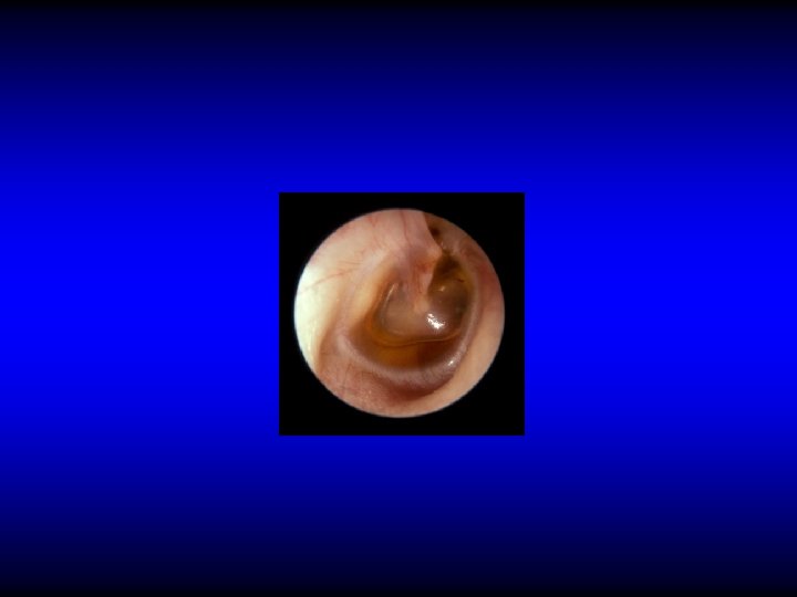
Examination • Otoscopy • Tuning fork tests

Otoscopy NORMAL

Otoscopy

Tuning fork tests • Rinne’s & Weber’s
 • Tympanometry")
Investigations • Pure tone audiogram (PTA) • Tympanometry

PTA

Tympanometry
 Type B (OME) Type C (ET dysfunction ? OME)")
Type A (Normal) Type B (OME) Type C (ET dysfunction ? OME)

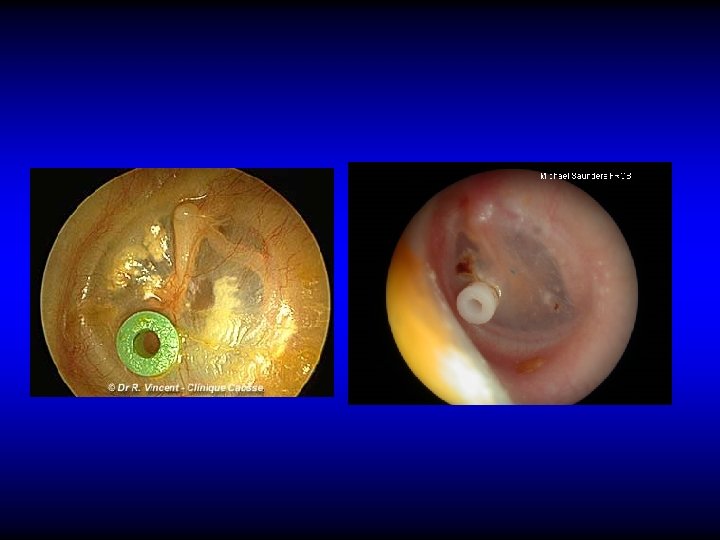
Diagnostic Myringotomy

Other indications of myringotomy • AOM with bulging TM – Relieve pain –C&S – To produce a clean cut incision which is more likely to heal spontaneously

Sequelae of OME • Spontaneous resolution – 50% resolve within 3 months. Only 5% persists for more than 12 months • Tympanosclerosis • Scarring • Cholesteatoma

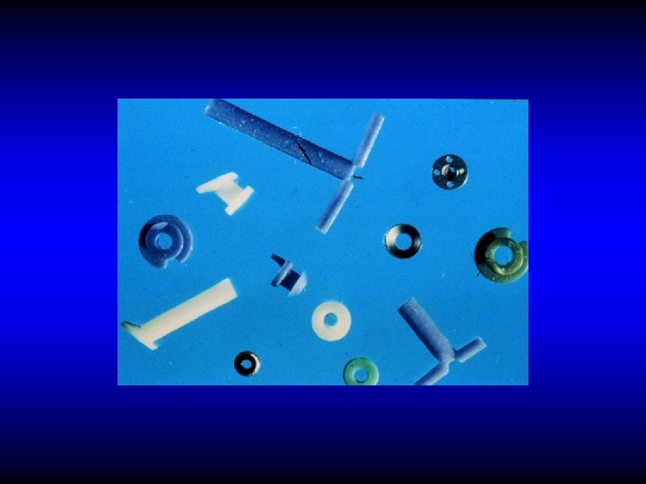
Treatment • Treatment of the cause if feasible • Observation • ? Medical treatment – Antibiotics – Decongestants, ? Auto-inflation • Surgical – Ventilation tubes (grommets) insertion

Ventilation tubes insertion

Other indication for VT insertion • Repeated attacks of AOM

Complications of Ventilation Tubes • Infection

Complications of Ventilation Tubes • Infection • Blockage

Complications of Ventilation Tubes • Infection • Blockage • Early extrusion

Complications of Ventilation Tubes • Infection • Blockage • Early extrusion • Tympanosclerosis

Complications of Ventilation Tubes • Infection • Blockage • Early extrusion • Tympanosclerosis • Persistent perforation

Factors affecting treatment choise • • • Age Duration Unilateral or bilateral Degree of hearing impairment Previous treatment Tympanic membrane changes

Conclusion • OME is very common in children • Etiology is associated with ET dysfunction and or chronic infection • In adults: nasopharyngeal pathology should be considered • Most cases resolve spontaneously • Conservative treatment is of doubtful value • VT insertion restores hearing in the selected cases

Classification of Chronic Otitis Media • Chronic Non Suppurative Otitis Media – Otitis media with effusion “OME” – Adhesive otitis media • Chronic Suppurative Otitis Media “CSOM” – Tubo-tympanic (Safe) – Attico-antral (Unsafe)

Chronic Adhesive Otitis Media • Formation of adhesion in the middle ear following CSOM or OME

Clinical Features • History of CSOM or OME • Deafness is usually the only symptoms • TM shows various structural changes

Treatment • Observation • Surgical treatment • Hearing aid

Classification of Chronic Otitis Media • Chronic Non Suppurative Otitis Media – Otitis media with effusion “OME” – Adhesive otitis media • Chronic Suppurative Otitis Media “CSOM” – Tubo-tympanic (Safe) – Attico-antral (Unsafe)

CHRONIC SUPPURATIVE OTITIS MEDIA
 Attico-antral (Unsafe)")
Clinico-pathological types Tubo-tympanic (Safe) Attico-antral (Unsafe)

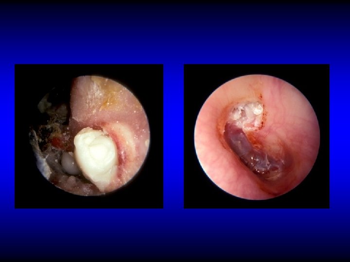
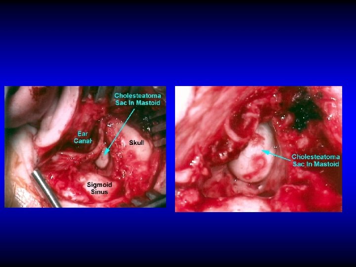
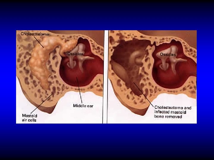
Pathology • Purulent or muco purulent discharge • TM perforation • Granulation tissue or polyps • Fibrosis & tympanosclerosis • Cholesteatoma (attico-antral type)

CHOLESTEATOMA
 in the middle")
Definition • The presence of a desquamating stratified squamous epithelium (skin) in the middle ear
 • Metaplasia • Epithelial migration")
Pathogenesis of cholesteatoma • Implantation (congenital or acquired) • Metaplasia • Epithelial migration

Effects of Cholesteatoma • Persistence of the infection • Bone erosion – Pressure necrosis – Enzymes

Clinical Features of CSOM

Symptoms of CSOM • Otorrhea – Intermittent, profuse & odorless in TT type – Persistent, scanty & malodorous in AA type • Deafness • Tinnitus N. B. Any other symptom means complication

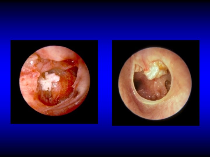
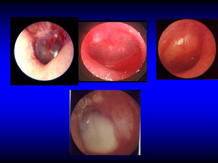
Otoscopic examination • Discharge – Present in TT type if active but may be absent – Usually is present in AA type • Perforation – Central: in TT type – Marginal or attic in AA type with cholesteatoma

Perforation in TT CSOM
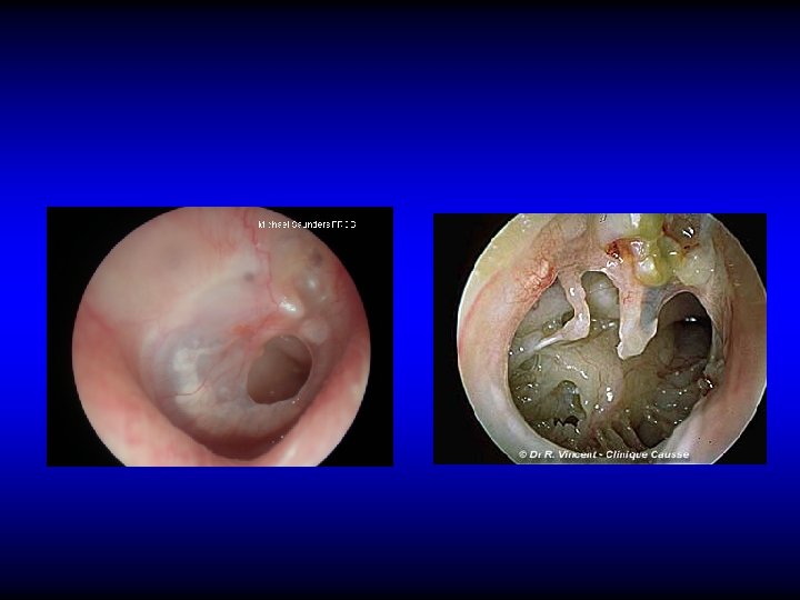

Perforation in AA CSOM

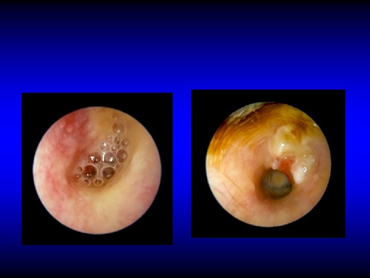
Otoscopic examination • Discharge – Present in TT type if active but may be absent – Usually is present in AA type • Perforation – Central: in TT type – Marginal or attic in AA type with cholesteatoma • Polyps, granulation tissue, tympanosclerosis
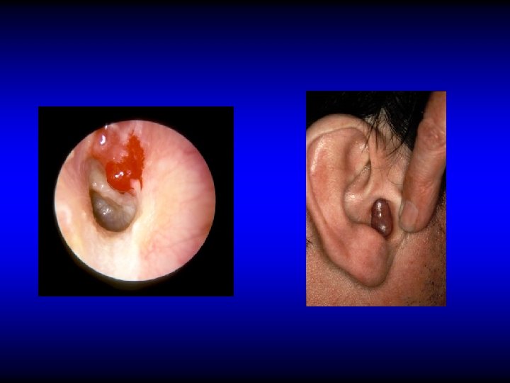

Tympanosclerosis
")
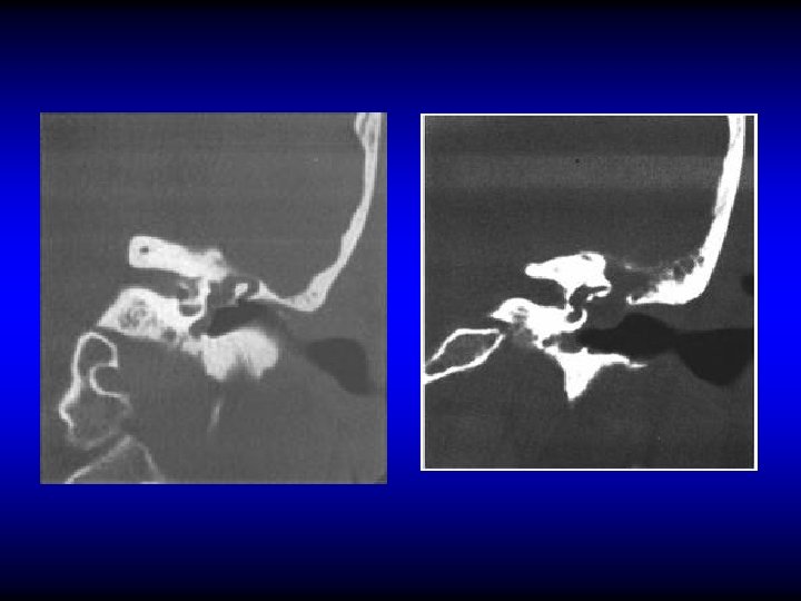
Investigations • Audiometry • Bacteriology • CT (mainly in the AA type)

Treatment of CSOM

Active TT type Inactive TT type Attico-antral type

Conservative treatment Active TT type Inactive TT type Conservative Treatment • Treat any predisposing factor • Ear toilet MYRINGOPLASTY • Antibiotics OR • Keep the ear dry • Removal of polyps and granulations TYMPANOPLASTY

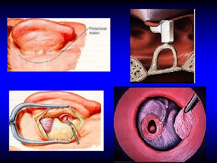
Myringoplasty Tympanoplasty • An operation performed to repair the tympanic membrane cavity (TM and/or the ossicles)

Aims of Tympanoplasty and Myringoplasty • To prevent re-infection • To improve hearing

Treatment of Attico-antral CSOM Removal of cholesteatoma by radical or modified radical mastoidectomy

Radical & Modified Radical Mastoidectomy Radical An operation in which the mastoid antrum and middle ear and the external canal are converted into common cavity. The tympanic membrane, malleus and incus are removed leaving only the stapes in situ. Modified Radical An operation in which the mastoid antrum and middle ear and the external canal are converted into common cavity. The tympanic membrane and ossicles remnants are retained

Aims of radical & modified radical mastoidectomy • Remove cholesteatoma to provide – Safety – Dry ear

Conclusion • In TT type the discharge is usually copious, intermittent and odorless. The perforation is central. Treatment is by elective tympanoplasty to prevent re-infection and improve hearing. • In the AA type the discharge is usually scanty, persistent and of bad odor. The perforation is attic or marginal with cholesteatoma. Treatment is by mastoidectomy to provide safety and dry ear


COMPLICATIONS OF SUPPURATIVE OTITIS MEDIA

Definition • The spread of infection beyond the boundaries of the middle ear cleft

Routes of spread • Direct extension – Usually due to bone erosion by cholesteatoma • Thrombophlebitis • Normal anatomical pathways – e. g. oval window, round window etc
 complications • Intracranial")
Complications of suppurative otitis media • Extracranial complications • Cranial (intra-temporal) complications • Intracranial complications

Extracranial complications • Otitis externa • Retropharyngeal abscess • Septicemia
 complications • Acute mastoiditis & mastoid abscess • Petrositis • Facial nerve")
Cranial (intratemporal) complications • Acute mastoiditis & mastoid abscess • Petrositis • Facial nerve paralysis • Labyrinthine fistula and labyrinthitis

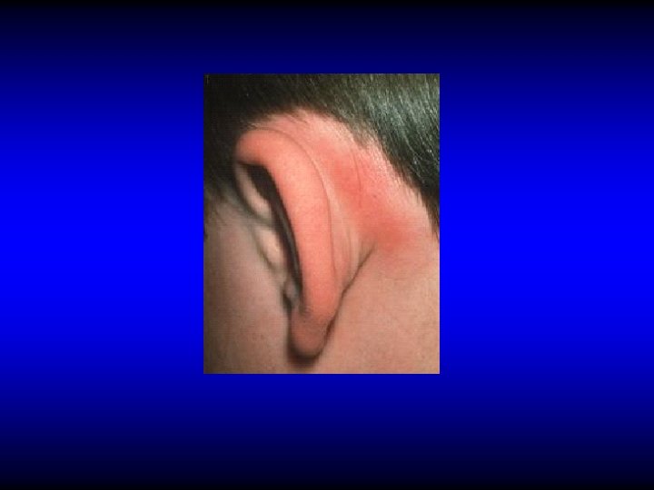
Acute mastoiditis & mastoid abscess

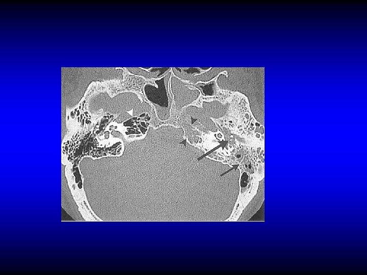
Pathology of acute mastoiditis Involvement of the bone of the mastoid air cells by acute suppurative inflammation

NORMAL ACUTE MASTOIDITIS

Pathology of mastoid abscess • Formation of pus subperiosteally

Diagnosis of acute mastoiditis • General constitutional manifestations • Tympanic membrane changes

Diagnosis of acute mastoiditis • General constitutional manifestations • Tympanic membrane changes • Otorrhea and reservoir sign • Retroauricular tenderness and redness

Diagnosis of mastoid abscess


DIAGNOSIS OF ACUTE MASTOIDITIS & MASTOID ABSCESS • General constitutional manifestations • Tympanic membrane changes • Otorrhea and reservoir sign • Retroauricular tender ness and red ness • Subperiosteal and Bezold’s abscess • Imaging

NORMAL ACUTE MASTOIDITIS

MASTOID ABSCESS

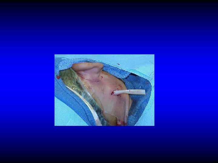
Treatment of acute mastoiditis • IV antibiotics • Cortical mastoidectomy if medical treatment fails or if there are signs of abscess formation

Cortical mastoidectomy An operation performed to covert the mastoid antrum and air cells into one cavity, without disturbing the existing middle ear content

Aim • Drainage

Indications of cortical mastoidectomy • Acute mastoiditis not responding to medical treatment • Mastoid abscess

Technique of Cortical Mastoidectomy
 complications • Acute mastoiditis • Petrositis (apical petrositis) • Facial nerve paralysis")
Cranial (intratemporal) complications • Acute mastoiditis • Petrositis (apical petrositis) • Facial nerve paralysis • Labyrinthine fistula and labyrinthitis
 An extension of infection from the middle ear pneumatized petrous apex.")
Petrositis (petrous apicitis) An extension of infection from the middle ear pneumatized petrous apex. into a
 – Retro-orbital pain –")
Diagnosis of petrositis • Gradenigo’s syndrome – Otitis media (otorrhea) – Retro-orbital pain – Squint (VI cranial nerve palsy) • Imaging

Treatment of petrositis • Antibiotics and myringotomy • Surgical drainage if medical treatment fails
 complications • Acute mastoiditis • Petrositis • Facial nerve paralysis • Labyrinthine")
Cranial (intratemporal) complications • Acute mastoiditis • Petrositis • Facial nerve paralysis • Labyrinthine fistula and labyrinthitis

Facial paralysis in AOM • Mostly due to pressure on a dehiscent nerve by inflammatory products • Usually is partial and sudden in onset • Treatment myringotomy is by antibiotics and

Facial paralysis in CSOM • Usually is due to cholesteatoma • Insidious in onset • May be partial or complete • Treatment is by immediate surgical exploration (mastoidectomy and repair)
 complications • Acute mastoiditis • Petrositis (apical apicitis) • Facial nerve paralysis")
Cranial (intratemporal) complications • Acute mastoiditis • Petrositis (apical apicitis) • Facial nerve paralysis • Labyrinthine fistula and labyrinthitis

Pathology of labyrinthitis • Labyrinthine fistula – Erosion of the bony labyrinth usually due to cholesteatoma – Mostly in the lateral SCC • Acute labyrinthitis

Diagnosis of labyrinthine fistula • May be asymptomatic • Vertigo • SNHL • Fistula test • CT scan

Treatment of labyrinthine fistula • Mastoidectomy and surgical repair

Acute Labyrinthitis • Vertigo • SNHL • Nystagmus • Treatment is by antibiotic and myringotomy if due to AOM or by antibiotics and mastoid surgery if due to CSOM

Intracranial complications • Extradural abscess • Lateral sinus thrombophlebitis • Subdural empyema • Meningitis • Brain abscess • Otitic hydrocephalus

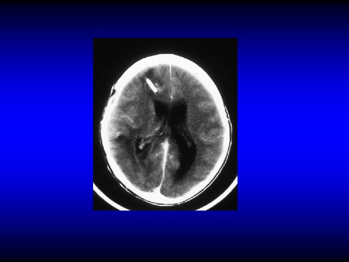
Extradural abscess • Accumulation of pus between dura and bone • Causes headache but may be silent • Diagnosis is confirmed by CT or MRI • Treatment is by drainage
 • Suppuration of the subdural space • Sever headache, fever, irritative")
Subdural abscess (empyema) • Suppuration of the subdural space • Sever headache, fever, irritative and paralytic focal neurological symptoms • CT and MRI
 • Suppuration of the subdural space • May be localized, multiple")
Subdural abscess (empyema) • Suppuration of the subdural space • May be localized, multiple or diffuse • Sever headache, fever, irritative and paralytic focal neurological symptoms • CT and MRI • Treatment is by neurosurgical drainage

Lateral sinus thrombophlebitis Pathology

Lateral sinus thrombophlebitis Diagnosis • Fever, rigor, and sweating • Headache and neck pain • Tenderness and edema in the neck • Blood culture • CT, MRI

Treatment of sinus thrombophlebitis • IV antibiotics and myringotomy if due to AOM • Mastoid surgery if no improvement or if due to CSOM

Intracranial complications • Extradural abscess • Lateral sinus thrombophlebitis • Subdural empyema • Meningitis • Brain abscess • Otitic hydrocephalus

Otogenic meningitis • Infection of the subarachnoid space • The most common intracranial complication • Fever, headache, neck phonophobia, restlessness etc • Kernig’s & Brudziniski signs stiffness,
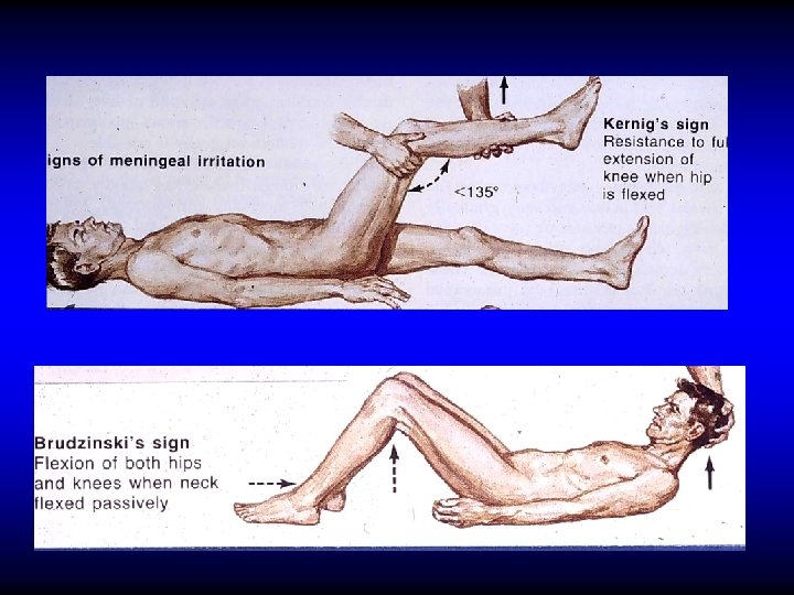

Otogenic meningitis • Infection of the subarachnoid space • The most common intracranial complication • Fever, headache, neck phonophobia, restlessness etc • Kernig’s & Brudziniski signs • Lumber puncture stiffness,

Treatment • IV antibiotics and myringotomy if due to AOM • Mastoid surgery if secondary to CSOM

Intracranial complications • Extradural abscess • Lateral sinus thrombophlebitis • Subdural empyema • Meningitis • Brain abscess • Otitic hydrocephalus

Otogenic brain abscess • 25% of children's and 50% of adult’s brain abscesses are otogenic • Mostly in temporal lobe or cerebellum (2: 1)

Otogenic brain abscess Clinical manifestations • General manifestations: fever, lethargy, headache. • Manifestation of raised IC pressure • Focal manifestations – Temporal: Aphasia, hemianopia, paralysis – Cerebellar: ataxia, vertigo, nystagmus, muscle incoordination

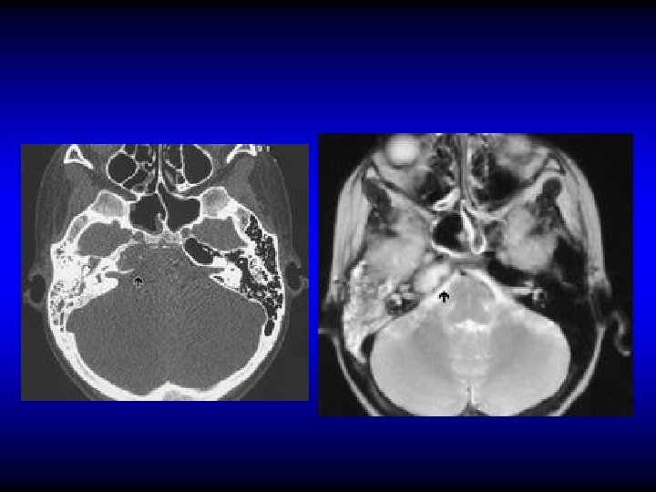
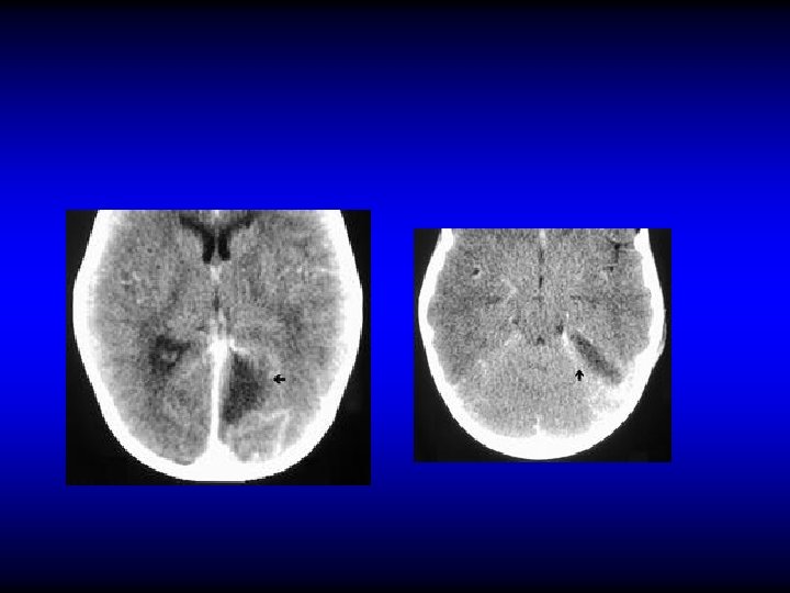
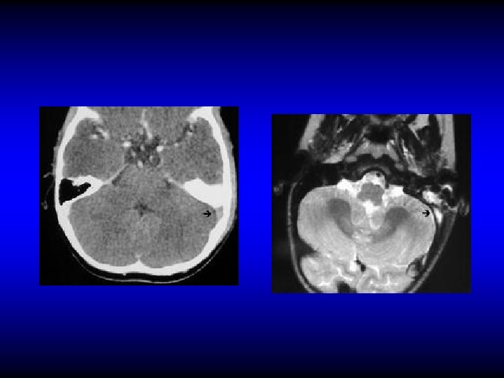
Otogenic brain abscess Diagnosis • CT • MRI

CT


MRI

Otogenic brain abscess Treatment • Repeated aspiration or Excision • IV antibiotics and myringotomy if due to AOM • Mastoid surgery if due to CSOM

Intracranial complications • Extradural abscess • Lateral sinus thrombophlebitis • Subdural empyema • Meningitis • Brain abscess • Otitic hydrocephalus

Otitic hydrocephalus • Very rare • Intracranial hypertension associated with ear disease. • Most often it follows lateral sinus thrombophlebitis • Clinically: Manifestations of increased IC pressure • CT

Otitic hydrocephalus • Very rare • Intracranial hypertension associated with ear disease. It most often follows lateral sinus thrombophlebitis • Clinically: Manifestations of increased IC pressure • CT • Treatment: steroids, diuretics, hyperosmolar dehydrating agents, repeated LP

General principles of management of the complications • • High index of suspicion May be multiple CT and MRI are the mainstay investigations Treatment is by parental antibiotics & surgery for the complication if applicable • Treatment of the ear lesion – Myringotomy in AOM – Mastoidectomy in CSOM

OFFICE HOURS Prof. YOUSRY EL-SAYED • Flat 407 Building 5 King Abdel-Aziz University Hospital • Mondays from 11 am to 1 pm • Thursdays from 11 am to 1 pm

THANK YOU
- Slides: 152