Objectives Physiological anatomy of the breast Physiological changes
 Physiological changes")
Objectives Physiological anatomy of the breast Physiological changes during breast development (Mamogenesis) Physiological changes during lactation (Lactogenesis) and their physiological action Phases of lactogenesis. Physiological changes during Galactopoeisis. Endocrine and autocrine control. Involution (the termination of milk production).

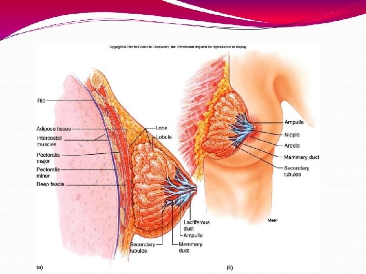
Small group activity What is the structure of human breast?
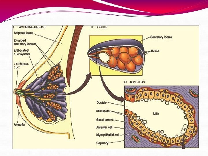

Small group activity Where does milk come from?

Theories Uterine milk theory: Vessel connecting the uterus to the breast (diversion of menstrual blood to the breast) Chyle theory: Milk is derived directly from chyle (milky fluid of emulsified fat absorbed from the intestinal tract into lymphatic system). Synthesis theory: Milk is formed from substrates carried to the gland in the blood

Where does milk come from?
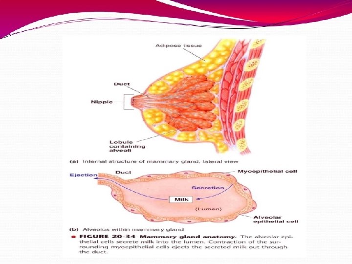


Fig. 1. Mammary gland development. The postnatal development of female mammary tissue occurs in several steps regulated by hormones. At birth, the mammary epithelium consists of limited ducts. At puberty, high levels of circulating hormones stimulate both the proliferation of the mammary epithelial cells (MECs) and the enlargement of the surrounding fat pad. At the onset of pregnancy, epithelial ducts elongate, branch and alveoli develop. During lactation, the mammary epithelium reach its maximal development containing numerous alveoli, which produce huge amounts of milk. Upon weaning, milk production ceases, the mammary alveoli regress (involution) and the mammary epithelium returns to a non-pregnant state.
 During puberty Estrogen stimulate proliferation of ducts and deposition of fat")
Breast development (mamogenesis) During puberty Estrogen stimulate proliferation of ducts and deposition of fat Progesterone stimulate development of lobules
 During pregnancy Complete development of glandular tissue")
Breast development (mamogenesis) During pregnancy Complete development of glandular tissue
 Endocrine system plays a major role in synchronizing development (mamogenesis) and")
Breast development (mamogenesis) Endocrine system plays a major role in synchronizing development (mamogenesis) and function (lactogenesis) of mammary gland with reproduction Three categories of hormones: Reproductive hormones (endocrine) Estrogen, progesterone, prolactin, oxytocin and h. PL Metabolic hormones (endocrine) GH, corticosteroids, thyroxin, PTH and insulin Mammary hormones (autocrine) GH, prolactin, parathyroid hormone-related protein (PTHr. P) and leptin

Size of the breast & lactation
 Reproductive hormones (direct effect) Estrogen (placenta) Growth & branching of ductal")
reast development (mammogenesis) Reproductive hormones (direct effect) Estrogen (placenta) Growth & branching of ductal system (with GH) Fat deposition in the stroma Progesterone (placenta) Growth of lobule-alveolar system(budding of alveoli and secretory changes in epithelial cells ) Although estrogen and progesterone are essential for physical development of the breasts, they inhibit actual secretion of milk
 Prolactin (anterior pituitary) Its level increases during pregnancy (10 -20 times)")
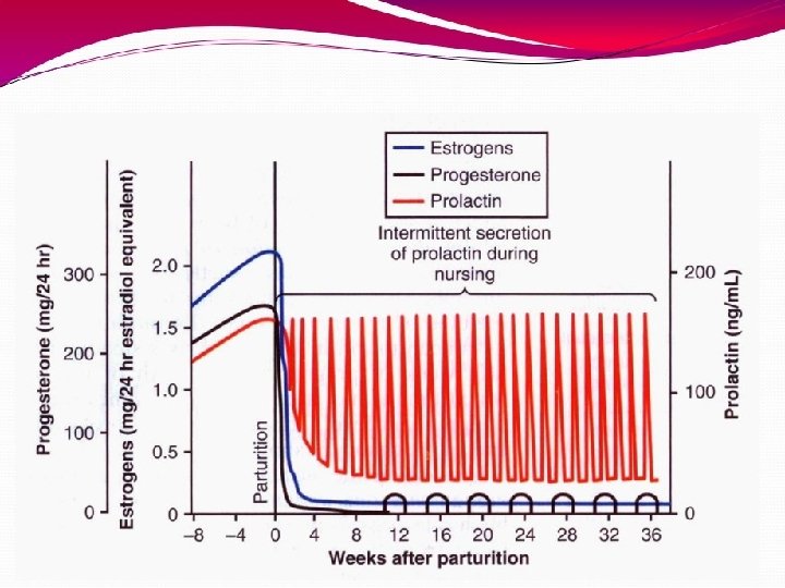
Breast development (mamogenesis) Prolactin (anterior pituitary) Its level increases during pregnancy (10 -20 times) Its main function is milk production Sudden drop in E & P after delivery allows milk production It is controlled mainly by hypothalamic hormone PIH (Dopamine) Human placental lactogen (placenta) Facilitate mammogenesis Delay milk production

ctogenesis Lactogenesis: cellular changes by which mammary epithelial cells are converted from a non secretory state to a secretory state: 2 stages Lactogenesis 1: (Cytologic and enzymatic differentiation of alveolar epithelial cells). Starts in mid pregnancy and characterized by expression of many genes involved in synthesis of milk components (increase in the uptake transport systems for amino acids, glucose, and calcium required for milk synthesis). Hormones involved: 1. 2. 3. 4. Progesterone (suppresses milk secretion) Prolactin and/or placental lactogen Growth hormone Glucocorticoids (Cortisol)
. Around parturition withdrawal of progesterone +")
Lactogenesis 2: (Copious secretion of all milk components). Around parturition withdrawal of progesterone + high level of prolactin leads to: Further increase in expression of milk protein genes Glands absorb increased quantities of metabolic substrates from the blood. Movement of cytoplasmic lipid droplets and casein into alveolar Lumina Transfer of immunoglobulins Secretion of colostrum followed by milk

Lactogenesis 2: Suckling stimulates further increase in expression of genes involved in milk secretion with expansion of alveolar epithelium Lactation is maintained by removal of milk 2 hormones involved Prolactin (milk production) Oxytocin (milk let-down)


Stages of lactogenesis Mamogenesis Lactogenesis Galctopoesis

 Metabolic hormones (direct effect) GH Its secretion is stimulated")
ormonal regulation of lactogenesis 1) Metabolic hormones (direct effect) GH Its secretion is stimulated by progesterone Increases production of IGF-1 by the liver and locally. Mediate cell survival and ductal growth Corticosteroids Increases during pregnancy (five fold) Involved in breast development (permissive action on milk protein synthesis)

rmonal regulation of lactogenesis Thyroxin Essential for milk production Thyroxin & TSH level decreases during lactation TRH increases leading to stimulation of PRL (nasal administration to treat inadequate lactation ) Insulin Low during lactation Shunt of nutrients from storage depots to milk synthesis
 Mammary hormones GH Progesterone stimulates its secretion Leptin Increases")
rmonal regulation of lactogenesis 2) Mammary hormones GH Progesterone stimulates its secretion Leptin Increases during pregnancy (increase adipose tissue) Decreases with lactation PTHr. P Increases during lactation Mobilizes bone calcium Increase in alkaline phosphatase
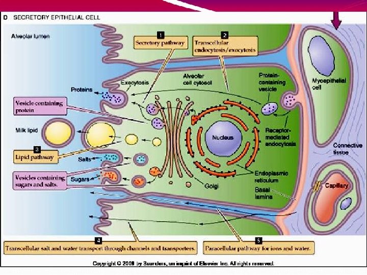

The pathways for milk secretion and synthesis by the mammary epithelial cell. I: Exocytosis of milk protein, lactose, and other components of the aqueous phase in Golgi-derived secretory vesicles. II: Milk fat secretion via the milk fat globule. III: Direct movement of mono valent ions, water, and glucose across the apical membrane of the cell. IV: Transcytosis of components of the interstitial space. V: The paracellular pathway for plasma components and leukocytes. Pathway V is open only during pregnancy, involution, and in inflammatory states such as mastitis. SV = Secretory vesicle; RER = Rough endoplasmic reticulum; BM = Basement membrane; MFG = Milk fat globule; CLD = Cytoplasmic lipid droplet; N = Nucleus; PC = Plasma cell; FDA = Fat-depleted adipocyte; TJ = Tight junction; GJ = Gap junction; D = Desmosome; ME = Myoepithelial cell.

Galactopoeisis

lactopoeisis Definition: Galactopoeisis is defined as the maintenance of lactation once lactation has been established. Role of Hormones Prolactin: milking-induced surge is a direct link between the act of nursing (or milk removal) and the galactopoeitic hormones involved in maintaining lactation. Growth Hormone: support increase in synthesis of lactose, protein, and fat in the mammary gland Glucocorticoids: galactopoeitic in physiological doses

Thyroid Hormones: galactopoeitic Ovarian Hormones : Estrogen in very low doses is galactopoietic Progesterone alone has no effect on galactopoeisis because there are no progesterone receptors in the mammary gland during lactation

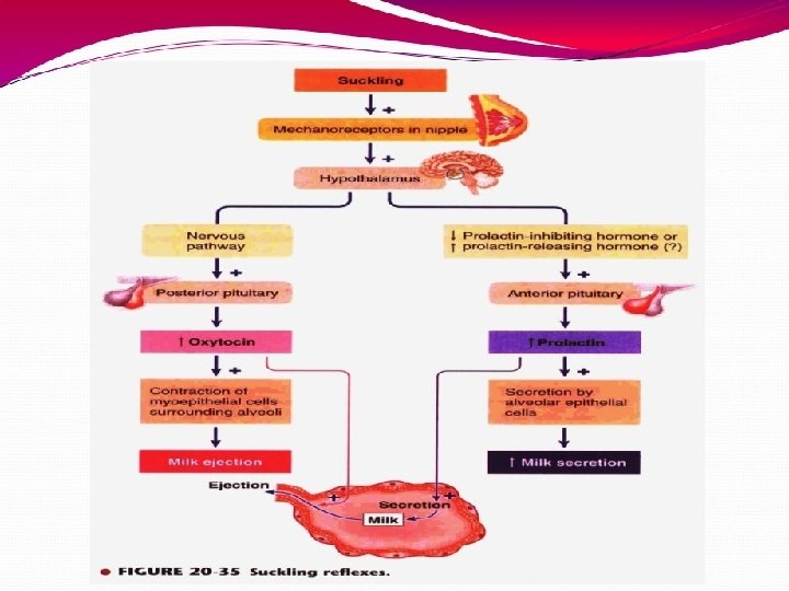
Suckling reflex What about---?

Suckling reflex

Milk production is a "use it or lose it" process. The more often and effectively the baby nurses, the more milk will be produced Milk production <100 ml/day in day 1 postpartum Milk production by day 3 reaches 500 ml/day Milk composition changes dramatically(↓Na+2& Cl- ) due to closure of tight junctions that block paracellular pathway

AAP Recommendations Exclusive breast feeding for the first six months of life Continued breast feeding for at least one year, ‘As long as is desired by mother and child’. American Academy of Pediatrics (2005). "Breastfeeding and the Use of Human Milk. " Pediatrics 115(2): 496 -506.

Failure of breast feeding

- Slides: 41