Objectives Name the four major tissue types and

Objectives: • Name the four major tissue types and their subcategories • Explain how each type of tissue is classified • Relate each types structure to the function it performs in the body • What are the common characteristics for each tissue type
 • Kings:")
Jigsaw activity • Aces: Explain classification of Epithelial tissue (page 78 -79) • Kings: Characteristics of Epithelial tissue • Queens: Simple Epithelial (subcategories, function and location) • Jacks: Stratified Epithelial (subcategories, function and location) • Tens: Glandular Epithelium (subcategories, function and location)

Body Tissues · Cells are specialized for particular functions · Tissues · Groups of cells with similar structure and function · Four primary types · Epithelium · Connective tissue · Nervous tissue · Muscle Copyright © 2003 Pearson Education, Inc. publishing as Benjamin Cummings Slide 3. 41

Epithelial Tissues · Found in different areas · Body coverings · Body linings · Glandular tissue · Functions · Protection · Absorption · Filtration · Secretion Copyright © 2003 Pearson Education, Inc. publishing as Benjamin Cummings Slide 3. 42
 · Tissue layer always has")
Epithelium Characteristics · Cells fit closely together (Sheet Like) · Tissue layer always has one free surface · The lower surface is bound by a basement membrane · Avascular (have no blood supply) · Regenerate easily if well nourished Copyright © 2003 Pearson Education, Inc. publishing as Benjamin Cummings Slide 3. 43

Classification of Epithelium · Number of cell layers · Simple – one layer · Stratified – more than one layer Figure 3. 16 a Copyright © 2003 Pearson Education, Inc. publishing as Benjamin Cummings Slide 3. 44 a

Classification of Epithelium · Shape of cells · Squamous – flattened · Cuboidal – cubeshaped · Columnar – column-like Figure 3. 16 b Copyright © 2003 Pearson Education, Inc. publishing as Benjamin Cummings Slide 3. 44 b

Simple Epithelium · Simple squamous · Single layer of flat cells · Usually forms membranes · Lines body cavities · Lines lungs and capillaries Copyright © 2003 Pearson Education, Inc. publishing as Benjamin Cummings Figure 3. 17 a Slide 3. 45

Squamous • The epithelium is a single layer of flat, or squamous, cells. • The peripheral cytoplasm of each cell is so attenuated that it is usually not resolvable; the nucleus is usually the only cellular structure visible. • The nucleus usually has a flattened appearance (sometimes spherical), and, due to the width of the cells, nuclei of adjacent cells are usually separated by a distance of many nuclear diameters. • The cells' surfaces are void of specialized surface structures, such as cilia or microvilli.

Basement membrane

Apical surface Basement membrane

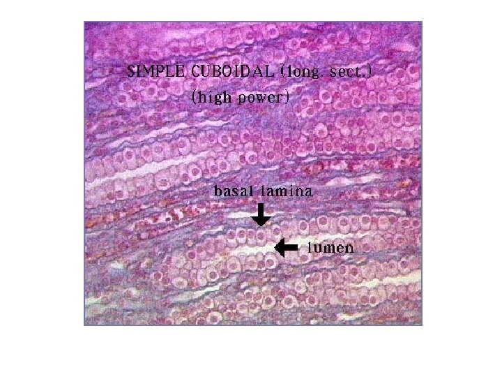
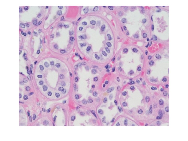
Simple Epithelium · Simple cuboidal · Single layer of cube-like cells · Common in glands and their ducts · Forms walls of kidney tubules · Covers the ovaries Copyright © 2003 Pearson Education, Inc. publishing as Benjamin Cummings Figure 3. 17 b Slide 3. 46

Cuboidal • This epithelium consists of a single layer of cuboidal cells resting on an undetectable basement membrane. • The height of each epithelial cell is nearly equal to the width of the cell. • The cells are tightly packed, forming a continuous sheet of cells that effectively forms the wall of a kidney tubule. • The nucleus is spherical and sits at the near-center of the cell. • The nuclei of adjacent cells are relatively close together, certainly closer than in a simple squamous epithelium, and are aligned in a single row.

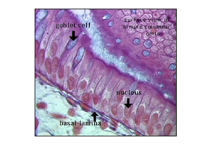
Simple Epithelium · Simple columnar · Single layer of tall cells · Often includes goblet cells, which produce mucus · Lines digestive tract Figure 3. 17 c Copyright © 2003 Pearson Education, Inc. publishing as Benjamin Cummings Slide 3. 47

Cuboidal • The epithelium consists of a single layer of columnar cells (the cells' height is significantly greater then their width) resting on an undetectable basement membrane. • The cells are tightly packed, forming a continuous sheet of cells that effectively forms the protective lining of the stomach. • The nucleus is generally ovoid and sits at the lower to mid-portion of the cell. • Because the width of the cell is relatively narrow, the nuclei of adjacent cells are relatively close together and are aligned in a single row.
. Lu, stomach lumen Lu. S, lumenal surface.")
Fig. 2. Mammalian stomach (H & E). Lu, stomach lumen Lu. S, lumenal surface. Epi, simple secretory columnar epithelium

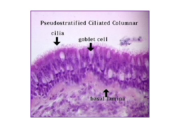
Simple Epithelium · Pseudostratified · Single layer, but some cells are shorter than others · Often looks like a double cell layer · Sometimes ciliated, such as in the respiratory tract · May function in absorption or secretion Copyright © 2003 Pearson Education, Inc. publishing as Benjamin Cummings Figure 3. 17 d Slide 3. 48

Pseudostratified • The nuclei staggered, while not obvious, all the epithelial cells in this membrane rest on the basement membrane, but they vary in height - all do not reach the apical surface. This contributes to the staggered locations of the nuclei. • Note the ciliated border of the columnar cells. Cilia are relatively flexible, motile hair-like surface projections. The cilia of the respiratory epithelium beat constantly toward the pharynx and nasal passages, creating a continuous flow of mucus in that direction. • Note the presence of goblet cells. While individual goblet cells are seldomly distinct, a mucus cup can occasiionally be seen. Tracheal goblet cells secret mucus over the surface of the epithelium to prevent dessication and to trap air-borne microscopic particulates, thus cleansing air as it passes to the lung alveoli.

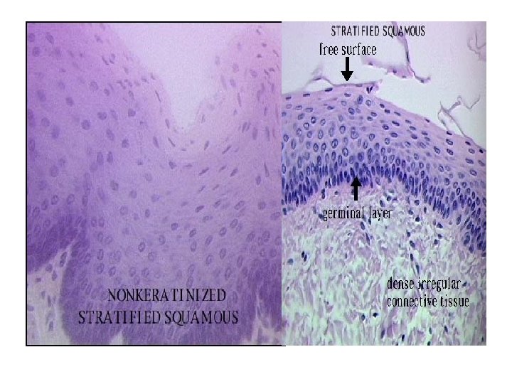
Stratified Epithelium · Stratified squamous · Cells at the free edge are flattened · Found as a protective covering where friction is common · Locations · Skin · Mouth · Esophagus Copyright © 2003 Pearson Education, Inc. publishing as Benjamin Cummings Figure 3. 17 e Slide 3. 49
 a) When found, you see it as")
Stratified Epithelium · Stratified cuboidal · (RARE) a) When found, you see it as two layers of cuboidal cells b) Ducts (sweat glands, mammary glands). · Stratified columnar (very limited in distribution) · *Present in Large ducts of some glands* Pharynx, male urethra, and lining some glandular ducts. Copyright © 2003 Pearson Education, Inc. publishing as Benjamin Cummings Slide 3. 50

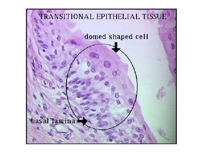
Stratified Epithelium · Transitional epithelium · Shape of cells depends upon the amount of stretching · Lines organs of the urinary system (Ureters, urinary bladder, part of the urethra) Copyright © 2003 Pearson Education, Inc. publishing as Benjamin Cummings Figure 3. 17 f Slide 3. 51

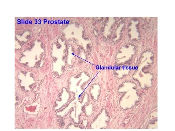
Glandular Epithelium · Gland – one or more cells that secretes a particular product · Two major gland types · Endocrine gland · Ductless · Secretions are hormones · Exocrine gland · Empty through ducts to the epithelial surface · Include sweat and oil glands Copyright © 2003 Pearson Education, Inc. publishing as Benjamin Cummings Slide 3. 52
- Slides: 29