Objectives 1 Describe the anatomical structure of skin

Objectives: 1. Describe the anatomical structure of skin and soft tissues. 2. Differentiate the various types of skin and soft tissue infections and there clinical presentation. 3. Name bacteria commonly involved in skin and soft tissue infections 4. Describe the pathogenesis of various types of skin and soft tissue infections 5. Recognize specimens that are acceptable and unacceptable for different types of skin and soft tissue infections 6. Describe the microscopic and colony morphology and the results of differentiating bacteria isolates in addition to other non-microbiological investigation 7. Discuss antimicrobial susceptibility testing of anaerobes including methods and antimicrobial agents to be tested. 8. Describe the major approaches to treat of skin and soft tissue infections , either medical or surgical.

* *This picture summarize the lecture return to it when you finish. *Only in males slides

key to developing an adequate differential diagnosis requires: • History: patient’s immune status, the geographical locale, travel history, recent trauma or surgery, previous antimicrobial therapy, lifestyle, and animal exposure or bites • Physical examination: severity of infection • Investigation: -CBCs, Chemistry -Swab, biopsy or aspiration* -Radiographic procedures* -Level of infection and the presence of gas or abscess. • Diagnostic and therapeutic: • Surgical* exploration or debridement • Antibiotics treatment *Surgical : diagnostic (very important to determine if the infection and whither it is deep or superficial ) and therapeutic (if it's severe it might need amputating) *Radiology ( CT or X-Ray) : primary to determine if there is bone infection or if there is gas. *Aspirate : For pus or exudate

Introduction: *Zoom in the image if not clear -Soft tissue definition: anything other than bones. • Soft tissue infections : It is a Common disease. • Can be mild to moderate or sever muscle or bone and lungs or heart valves infection. • Most common Cause are: Staphylococcus aureus and streptococcus. • Emerging antibiotic resistance among: -Staphylococcus aureus (methicillin resistance). -Methicillin drug is the laboratory name for cloxicillin drug -Streptococcus pyogenes (erythromycin resistance). Important to know the: 1 - Name of the syndrome. 2 - Organisms causing it. 3 - Clinical presentation. 4 - Antibiotics. -You should differentiate between epidermis, subcutaneous infections and is it invading other tissues like muscles or bones to choose the right management. -The more the organism has powerful toxins and enzyme the more it spread deeply, and he may devolve gas gangrene. For example : clostridium perfringase
: Definition / causes / characteristics / treatment • It’s a")
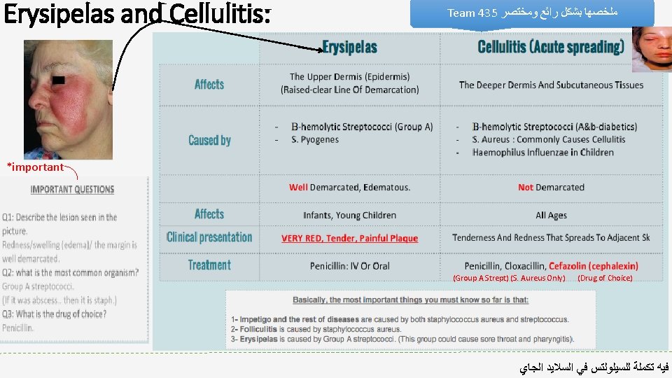
Definition: Impetigo - (pyoderma*): Definition / causes / characteristics / treatment • It’s a common skin infection, in Children 2– 5 years in tropical or subtropical regions. Very superficial in epidermis Causes: • Always caused by β-hemolytic streptococci (Nonbullous) and/or S. aureus (Bullous*) Characteristics: • • • Systemic symptoms are usually absent. Consists of discrete purulent* lesions (Blister* if it rupture, it’s produce fluid (honey crust)) Exposed areas of the body( face and extremities) Skin colonization- Inoculation by abrasions, minor trauma, or insect bites Poststreptococcal glomerulonephritis*. (anti–DNAse B) : (antideoxyribonuclease B) ; if there is a lot of Anti. DNAse in the test then it means he have impetigo. Treatment: Cefazolin: covers Both Staphylococcus and streptococcus. • Cloxacillin: covers Staphylococcus only • Erythromycin: covers streptococcus only • Mupirocin It may lead to immune complication in some cases. • Pyoderma*: any skin disease that is pyogenic (has pus)) Purulent*: ﺻﺪﻳﺪﻱ If patient has cellulitis, 1 st drug you think of = Cefazolin Blister*: ﺍﻟﺤﺒﺔ ﺃﻮ ﺍﻟﺒﺜﺮﺓ *After infection of impetigo by "group A"(beta hemolytic) Streptococcus , the patient might have post streptococcal glomerulonephritis: it is immunological response which affects kidney, it is primarily antigen-antibody complex reaction. This antibody is produced against cell wall of "A streptococcus "and accumulate in the glomerulus and cause acute glomerulonephritis and shows reversible renal failure Bulbous* =pus

Cutaneous abscesses: Definition / causes / characteristics / treatment Definition: • Collections of pus within the dermis and deeper skin tissues. Causes : • • Typically polymicrobial, S. aureus alone in ∼ 25 % Do Gram stain, culture, and systemic antibiotics Characteristics: • • • Painful, tender, and fluctuant Multiple lesions: cutaneous gangrene. severely impaired host defenses. extensive surrounding cellulitis. high fever. Treatments: • Incision and evacuation* of the pus. ( )ﻓﺘﺢ ﺍﻟﺨﺮﺍﺝ ﻭﺍﺳﺘﺨﺮﺍﺝ ﺍﻟﺼﺪﻳﺪ ﻣﻨﻪ • Antibody: Cloxacillin (S. Aureus only) *Evacuation =drainage

Furuncles and carbuncles: Definition / causes / characteristics / treatment Definition: • Furuncle or bolis: infection of the hair follicle (folliculitis) , small and affect one hair follicle • Carbuncle: infection of many hair follicles, big and with coalescent inflammatory mass Causes: • usually caused by S. aureus Characteristics • extends through the dermis into the subcutaneous tissue (so it is Deeper than Cutaneous abscesses. ) • Carbuncle present in the back of neck especially diabetes patients Treatment: • Larger furuncles and all carbuncles require incision and drainage. • Systemic antibiotics are usually unnecessary Outbreaks* of Furunculosis: • • • Caused by MSSA*, and MRSA* Families, prisons , sports teams Inadequate personal hygiene Repeated attacks of furunculosis Presence of S. aureus in the anterior nares- 20 -40% Mupirocin ointment( )ﺍﺳﻢ ﻣﺮﻫﻢ -to eradicate staphylococcal carriage nasal colonization Outbreaks*: ﺗﻔﺸﻲ MSSA*=methicillin sensitive staph-aureus MRSA*= methicillin resistance staph-aureus -S. aureus it is the normal flora in anterior nares ﺇﺫﺍ ﻓﻴﻪ ﺇﺻﺎﺑﺔ ﻓﻲ ﺍﻟﻌﺎﺋﻠﺔ ﺭﺍﺡ ﺗﺎﺧﺬﻫﻢ ﻛﻠﻬﻢ ﻭﺗﺎﺧﺬ ﻣﻨﻬﻢ Nose swap ﻭﺗﺸﻮﻑ ﻣﻴﻦ ﻳﺤﻤﻞ ﺍﻟﺒﻜﺘﻴﺮﻳﺎ ﻭﺗﺴﻮﻱ ﻟﻪ Decolonization ﻟﻼﺳﺘﺤﻤﺎﻡ ﻣﺜﻼ ، ﻭﺗﻌﻄﻴﻪ ﻣﻌﻘﻤﺎﺕ + topical antibiotics ﺍﻟﻜﺎﺭﺑﻨﻜﻞ ﻋﺒﺎﺭﺓ ﻋﻦ ﺃﻜﺜﺮ ﻣﻦ ﻓﺮﻧﻜﻞ
, preexisting skin infections-ulceration,")
Cont-Cellulitis: • Risk factors: • Obesity, venous insufficiency, lymphatic obstruction (operations), preexisting skin infections-ulceration, or eczema, • CA-MRSA ( Community Association MRSA) ü Carry Panton-Valentine leukocidin gene ü More sensitive to antibiotics ü Can lead to sever skin and soft tissue infection or septic shock • Diagnosis and Treatment • Clinical diagnosis Symptoms and Signs • High WBCs, blood culture rarely needed • Aspiration and biopsy might be needed in diabetes mellitus, malignancy, animal bites, neutropenia (Pseudomonas aeruginosa ), immunodeficiency, obesity and renal failure • Observe for progression to sever infection(increased in size with systemic manifestation ie. fever, leukocytosis • Treatment: ü Vancomycin or linazolid in case of MRSA ü Clindamycin, TMP-SMZ for Ca. MRSA

Necrotizing Fasciitis: • • ** Also known as flesh eating disease. It is a rare deep skin and subcutaneous tissues infection. The Most serious infection in Microbiology can kill patient. Deep inflammation of fascia, systemic Usually present in diabetic patient Sometimes present with as cellulitis Most common in arms, legs, and abdominal wall and is fatal in 30%-40% of cases. • Two types: Type 1: is Polymicrobial Type 2 : which is Monomicrobial. (caused by one microorganism) o Caused by: ü ü ü ü Fournier’s gangrene (testicular), ( if localized in the testis or scrotum, or perineal region ), Necrotizing cellulitis ( another name for it ). Group A streptococcus ( streptococcus pyogens ) Staphylococcus Aureus or CA-MRSA ( community acquired – methicillin resistant staph aureus ) Clostridium Perfringens ( gas in tissues ) Bacteroides Fragillis ( other causative microorganism ) Vibrio Vulnificus ( liver function ) Gram negative bacteria ( synergy ) ﻳﻌﻨﻲ ﻣﺘﻌﺎﻭﻧﺔ ﻣﻊ ﺑﻜﺘﻴﺮﻳﺎ ﺛﺎﻧﻴﺔ : E. Coli , klebseilla, Pseudomonas. -Fungi ** important (from 435 team)

Doctors Notes - Most important sign is SEVERE PAIN. It is considered a clinical emergency. Type 1 can be clostridium perfringens and group A streptococcus Type 2 is group A alone It is confirmed by biopsy. Also, MRI is used before that. We do a Gram stain for it to determine the type ( 1 or 2 ) and choose the correct antibiotic. - We can give 2 antibiotics ( Penicillin and Clindamycin ) to treat it because it is an emergency, BUT surgery is always the best treatment. - you don’t have to memorize the risk factors for Necrotizing Fasciitis , but read it
")
Necrotizing Fasciitis: Super-antigens : are super inducer for immune system)

Necrotizing Fasciitis: v Signs and symptoms: • • • Rapid progression of sever pain with fever , chills (typical) Swelling , redness, hotness, blister, gas formation, gangrene and necrosis Blisters with subsequent necrosis , necrotic eschars Diarrhea and vomiting (very ill) Shock organ failure Mortality as high as 73 % if untreated v Diagnosis: • A delay in diagnosis is associated with a grave prognosis and increased mortality • Clinical-high index of suspicion ü Blood tests : • • CBC-WBC , differential , ESR BUN (blood urea nitrogen)- to check for renal failure. ü Surgery debridement : amputation ü Radiographic studies: • X-rays : subcutaneous gases • Doppler CT or MRI - to evaluate the spread. ü Microbiology: • Culture &Gram's stain • ( blood, tissue, pus aspirate) Susceptibility tests

Necrotizing Fasciitis: Treatment: • • If clinically suspected patient needs to be hospitalized OR require admission to ICU(intensive care unite). Start intravenous antibiotics immediately. Antibiotic selection based on bacteria suspected. broad spectrum antibiotic combinations against: • MRSA: methicillin-resistant Staphylococcus aureus (MRSA) • Anaerobic bacteria • Gram-negative and gram-positive Bacilli Surgeon consultation : ** • Extensive Debridement of necrotic tissue and collection of tissue samples • Can reduce morbidity and mortality Antibiotics combinations : • Penicillin/clindamycin/gentamicin. (Given together, and they’re the most important) • Ampicillin/sulbactam • Cefazolin plus metronidazol • Piperacillin/tazobactam • Clostridium perfringens - penicillin G • Hyperbaric oxygen therapy (HBO) treatment ** important (team 435) https: //www. youtube. com/watch? v=tu. RCq 4 nyn 2 Y

Pyomyositis: Definition: Acute bacterial infection of skeletal muscle, usually caused by Staph. Aureus. - No predisposing penetrating wound, vascular insufficiency or contiguous infection. - Most cases occur in the tropics ( )ﺍﻟﺪﻭﻝ ﺍﻻﺳﺘﻮﺍﺋﻴﺔ. - 60% of cases outside of tropics have predisposing risk factors (RF): Diabetes mellitus, ethyl alcohol, liver disease, steroid prescriptions, HIV, hematologic malignancy. History: - Blunt trauma or vigorous exercise (50%), then period of swelling without pain. - 10 -21 days later : pain, tenderness, swelling and fever. - Pus can be aspirated from muscle. - 3 rd stage: sepsis, later metastatic abscesses if untreated. Diagnosis: X-ray, US, MRI or CT. Treatment: Surgical drainage and antibiotics. You can read this slide

Doctors Notes • • • - staph arues is resistant to penicillin -S. aureus it is the normal flora in anterior nares - cafazolin is the drug of choice in treatment of impetigo -we usually treat pus by draining it - erysipelas is well defined - erysipelas is most common in legs - cellulitis is poorly-defined - necrotizing fasciitis is treated with surgery and antibiotics If the abscess deep> anaerobic+ gram +ve If it superficial > staph
 Therapy Cat/Dog Bites Pasturella multocida; Capnocytophaga Amox/clav")
Other Specific Skin Infections Epidemiology Common Pathgen(s) Therapy Cat/Dog Bites Pasturella multocida; Capnocytophaga Amox/clav (Doxy; FQ or SXT + Clinda) Human bites Mixed flora eikenella corrodens Hand Surgeon; ATB as above Fresh water injury Aeromonas FQ; Broad Spectrum Betalactam Salt water injury (warm) Vibrio vulnificus FQ; Ceftazidime Thorn , Moss sporothrix schenckii Meat-packing Erysipelothrix Penicillin Cotton sorters Anthrax Penicillin Cat scratch Bartonella Azithromycin Potassium iodine

TAKE HOME POINTS • Most commonly caused by Staphylococcus aureus and Streptococcus pyogenes • Risk factors for developing SSTIs include breakdown of the epidermis, surgical procedures , crowding, comorbidities, venous stasis, lymphedema • Most of the infection are mild and can be managed on an outpatient basis In case • Most SSTIs can be managed on an outpatient basis, although patients with evidence of rapidly progressive infection, high fevers, or other signs of systemic inflammatory response should be monitored in the hospital setting. • Superficial SSTIs typically do not require systemic antibiotic treatment and can be managed with topical antibiotic agents, heat packs, or incision and drainage. • Systemic antibiotic agents that provide coverage for both Staphylococcus aureus and Streptococcus pyogenes are most commonly used as empiric therapy for both uncomplicated and complicated deeper infections.

Summary: Impetigo Coetaneous abscesses Furuncles /Carbuncles Part affected epidermis Dermis and deeper skin tissue dermis and subcutaneous tissue Etiology Caused by Beta Hemolytic streptococci / Staph. Aureus Characteristics Treatment Staph Aureus or polymicrobial Staph Aureus Erysipelas Cellulitis Necrotizing fasciitis Upper dermis Deep dermis and subcutaneous tissue Deep skin and subcutaneous tissue Beta hemolytic streptococci group A Beta hemolytic streptococci (diabetics) Staph. Aureus (trauma) Homophiles influenza (children) Streptococcus A and Clostridium perfrenges Systemic symptom usually absent Painful, tender and fluctuant Hair follicle infection (folliculitis) Red , tender , painful plaque Risk factors : obesity , venous insufficiency , lymphatic obstruction, preexisting skin infection , ulceration , eczema Cefazolen Incision and evacuation of pus Systemic antibiotics are usually unnecessary Penicillin, cloxacillin Cefazolen Did you study well ? https: //www. onlineexambuilder. com/micro-1/exam-121025 The most serious infection in Microbiology. Associated with gangrene and shock organ failure Penicillin , Clindamycin and Surgery

SAQ: 42 year old male with Diabetes Mellitus came with sever pain, fever and his right leg was swelling, redness and blisters with subsequent necrosis. Q 1: What is the most likely Diagnose in this case ? Necrotizing fasciitis Q 2: How can we differentiate between this infection in this case and cellulitis, Both of them, the patient come with pain and mild redness ? In Necrotizing fasciitis, the patient has SEVER PAIN and feels heaviness in the leg infected. Q 3: Can radiologist confirm our diagnose ? Yes , by X-rays to see if there is any subcutaneous gases or not. by MRI & CT for soft tissue. Q 4: If we say that this case is Type(I) infection. What does it mean ? This infection cause by polymicrobial agent ( mixed organisms / more than one ). Q 5: List some organism can cause this infection ? Group A streptococcus / Clostridium perfringens / Bacteroides fragilis / Fungi. Q 6: The surgeon play an important role in this case by removing the necrotic tissue and sending a sample to Microbiology lab. What is the purpose of sending that sample after removing the necrotic tissue ? To Know what are the causative organisms and therefore determine the suitable Antibiotics for treatment. Q 7: Why in this case we recommend to use combinations of Penicillin-clindamycin ? The Clindamycin is more penetrate into soft tissue. The Clindamycin works on the ribosome which lead to inhibit the synthesis of proteins which are in this case the toxins. Q 8: Streptococcus pyogenes are destroying the skin and muscle by releasing exotoxins which sometimes known as (Superantigen). What does it mean ? They are a class of antigen that cause non-specific activation of T-cells, therefore overproduction of cytokines which may lead to Severe systemic illness such as Toxic shock syndrome. The answers are below the questions , ZOOM to see it clear =)
 Shrooq Alsomali (leader) Ibrahim Fetyani Rawan Alqahtani")
GOOD LUCK! MICROBIOLOGY TEAM: Waleed Aljamal (leader) Shrooq Alsomali (leader) Ibrahim Fetyani Rawan Alqahtani Meshal Eiaidi Hanin Bashaikh Khalid Alhusainan Jawaher Alkhayyal Hussam Alkhathlan Reem Alshathri Faisal Alqumaizi Ohoud Abdullah Abdulaziz Alangari Lama Al-musallam Khalid Alshehri Wateen Alhamoud Nasir Aldosarie Ruba Barnawi Mohammad Al-Kahil Shooq Albugami The Editing File We are waiting for your feedback @microbio 436 microbioteam@gmail. com
- Slides: 22