
O M20 adm 06 2 1 Chief complaint
- Slides: 16
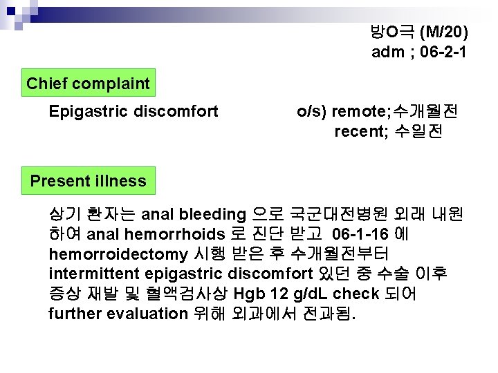
방O극 (M/20) adm ; 06 -2 -1 Chief complaint Epigastric discomfort o/s) remote; 수개월전 recent; 수일전 Present illness 상기 환자는 anal bleeding 으로 국군대전병원 외래 내원 하여 anal hemorrhoids 로 진단 받고 06 -1 -16 에 hemorroidectomy 시행 받은 후 수개월전부터 intermittent epigastric discomfort 있던 중 수술 이후 증상 재발 및 혈액검사상 Hgb 12 g/d. L check 되어 further evaluation 위해 외과에서 전과됨.
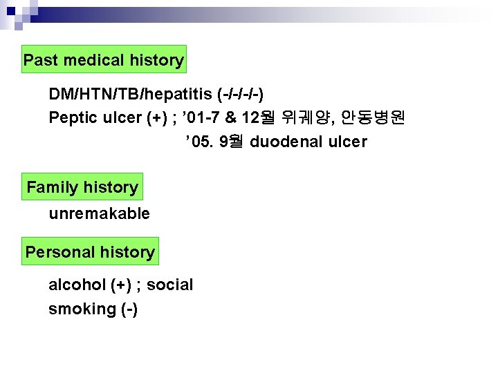
Past medical history DM/HTN/TB/hepatitis (-/-/-/-) Peptic ulcer (+) ; ’ 01 -7 & 12월 위궤양, 안동병원 ’ 05. 9월 duodenal ulcer Family history unremakable Personal history alcohol (+) ; social smoking (-)
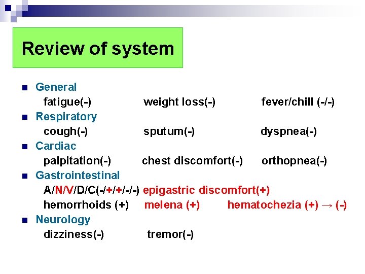
Review of system n n n General fatigue(-) weight loss(-) fever/chill (-/-) Respiratory cough(-) sputum(-) dyspnea(-) Cardiac palpitation(-) chest discomfort(-) orthopnea(-) Gastrointestinal A/N/V/D/C(-/+/+/-/-) epigastric discomfort(+) hemorrhoids (+) melena (+) hematochezia (+) → (-) Neurology dizziness(-) tremor(-)
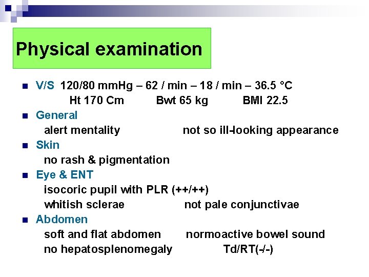
Physical examination n n V/S 120/80 mm. Hg – 62 / min – 18 / min – 36. 5 °C Ht 170 Cm Bwt 65 kg BMI 22. 5 General alert mentality not so ill-looking appearance Skin no rash & pigmentation Eye & ENT isocoric pupil with PLR (++/++) whitish sclerae not pale conjunctivae Abdomen soft and flat abdomen normoactive bowel sound no hepatosplenomegaly Td/RT(-/-)
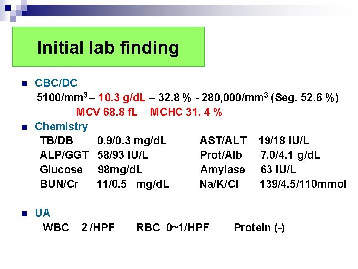
Initial lab finding n n n CBC/DC 5100/mm 3 – 10. 3 g/d. L – 32. 8 % - 280, 000/mm 3 (Seg. 52. 6 %) MCV 68. 8 f. L MCHC 31. 4 % Chemistry TB/DB 0. 9/0. 3 mg/d. L AST/ALT 19/18 IU/L ALP/GGT 58/93 IU/L Prot/Alb 7. 0/4. 1 g/d. L Glucose 98 mg/d. L Amylase 63 IU/L BUN/Cr 11/0. 5 mg/d. L Na/K/Cl 139/4. 5/110 mmol UA WBC 2 /HPF RBC 0~1/HPF Protein (-)
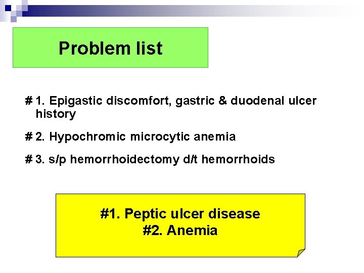
Problem list # 1. Epigastic discomfort, gastric & duodenal ulcer history # 2. Hypochromic microcytic anemia # 3. s/p hemorrhoidectomy d/t hemorrhoids #1. Peptic ulcer disease #2. Anemia
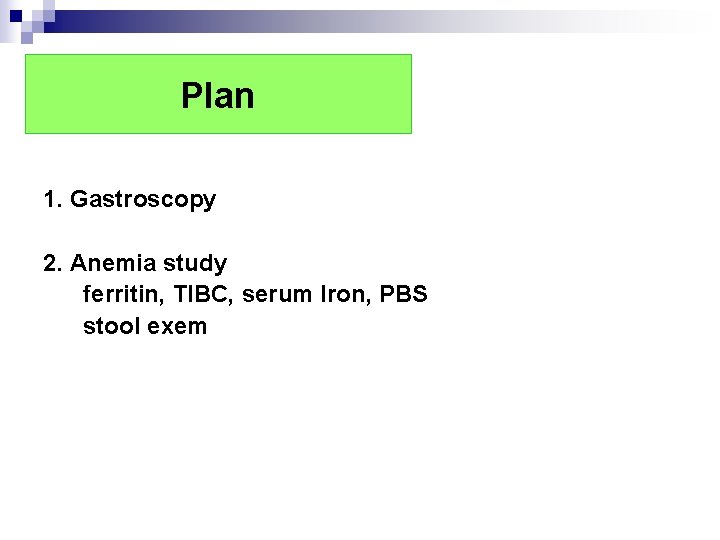
Plan 1. Gastroscopy 2. Anemia study ferritin, TIBC, serum Iron, PBS stool exem
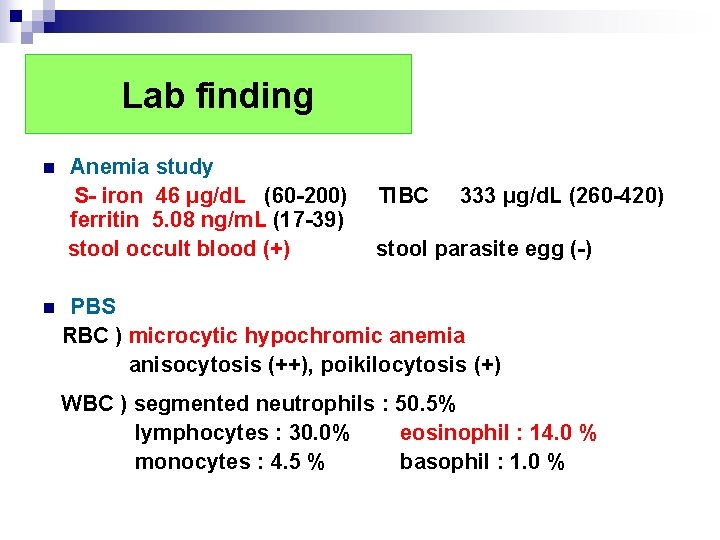
Lab finding n n Anemia study S- iron 46 μg/d. L (60 -200) ferritin 5. 08 ng/m. L (17 -39) stool occult blood (+) TIBC 333 μg/d. L (260 -420) stool parasite egg (-) PBS RBC ) microcytic hypochromic anemia anisocytosis (++), poikilocytosis (+) WBC ) segmented neutrophils : 50. 5% lymphocytes : 30. 0% eosinophil : 14. 0 % monocytes : 4. 5 % basophil : 1. 0 %
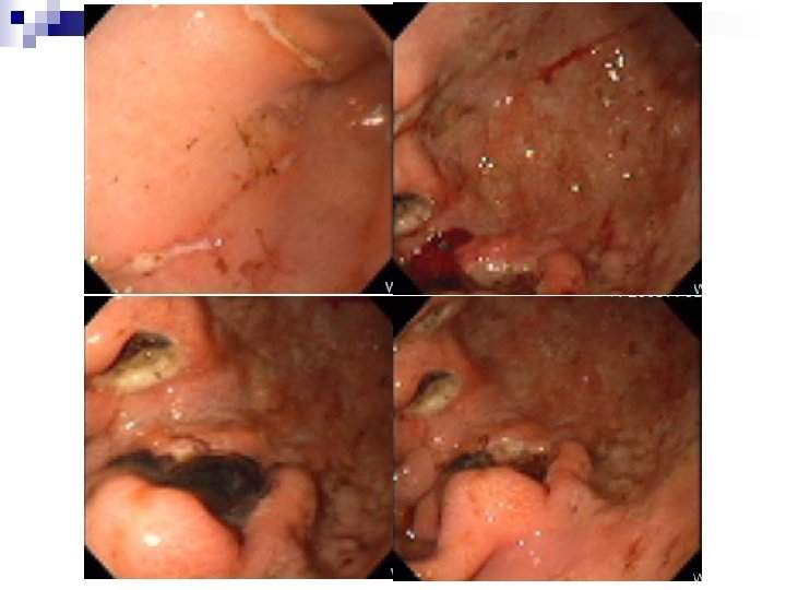
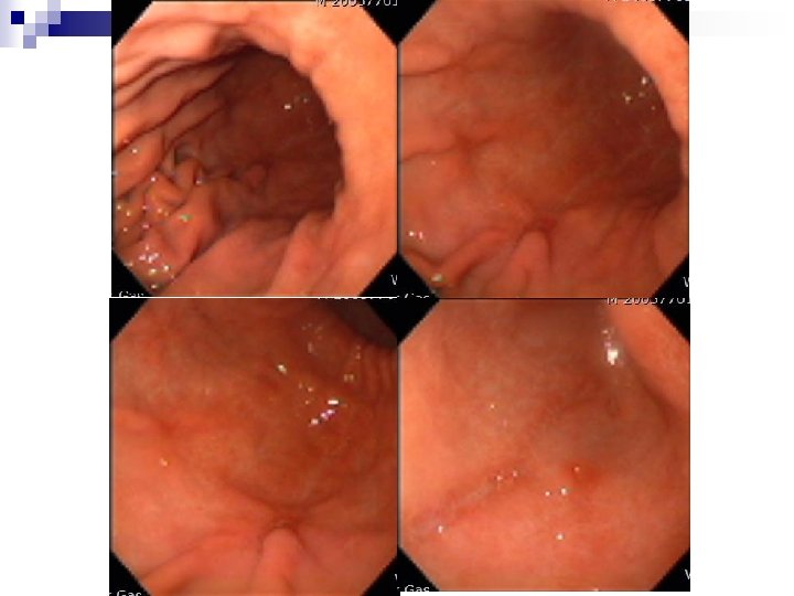
Initial gastroscopy
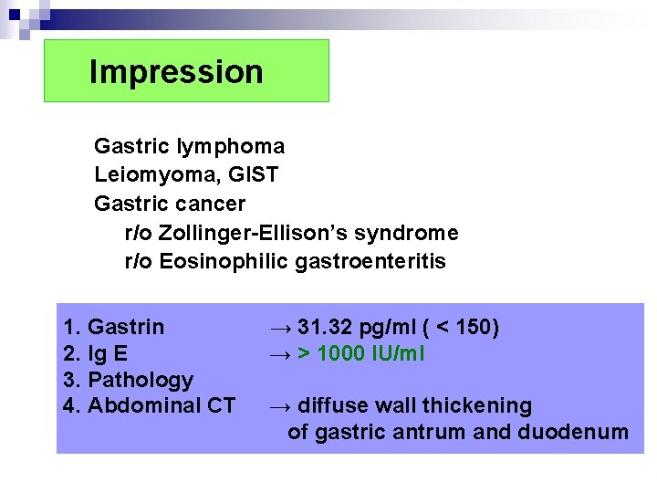
Impression Gastric lymphoma Leiomyoma, GIST Gastric cancer r/o Zollinger-Ellison’s syndrome r/o Eosinophilic gastroenteritis 1. Gastrin 2. Ig E 3. Pathology 4. Abdominal CT → 31. 32 pg/ml ( < 150) → > 1000 IU/ml → diffuse wall thickening of gastric antrum and duodenum
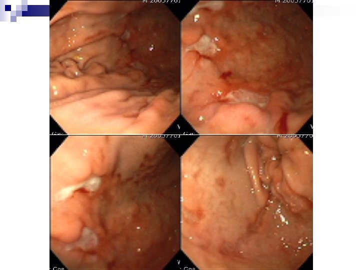
F/U gastroscopy (5일 후)
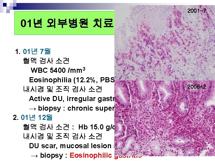
2001 -7 01년 외부병원 치료 기록 1. 01년 7월 혈액 검사 소견 WBC 5400 /mm 3 Hb 7. 4 g/d. L Eosinophilia (12. 2%, PBS) 2006 -2 내시경 및 조직 검사 소견 Active DU, irregular gastric mucosal lesion → biopsy : chronic superficial gastritis 2. 01년 12월 혈액 검사 소견 : Hb 15. 0 g/d. L 내시경 및 조직 검사 소견 DU scar, mucosal lesion at G-C side of low body → biopsy : Eosinophilic gastritis
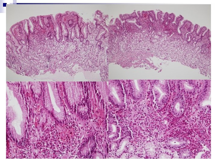
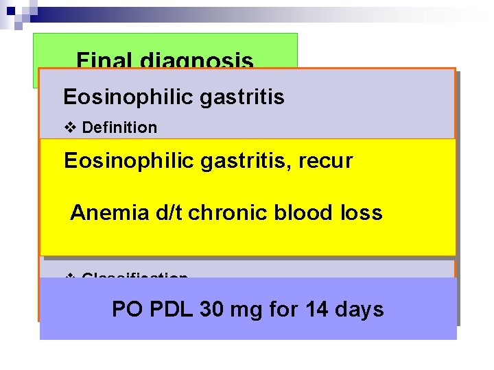
Final diagnosis Eosinophilic gastritis v Definition 1. Presence of abnormal GI symptoms Eosinophilic gastritis, 2. Eosinophilic infiltration in 1 orrecur more areas of the GI tract, defined as 20↑ eosinophils/HPF 3. Anemia Absence of cause of eosinophilia d/tidentified chronic blood loss 4. Exclusion of eosinophilic involvement in organs other than the gut v Classification Mucosal / Muscularis/ Serosal form PO PDL 30 mg for 14 days
F/U gastroscopy (Post PDL 14 days )