o Hemoglobin is a protein in red blood

o Hemoglobin is a protein in red blood cells that carries oxygen. o Each Hb molecule has a complex quaternary shape. o It has two alpha chains and two beta chains of polypeptides. o Each individual chain is a globular protein subunit. o Each subunit contains a single molecule of heme a ring surrounding a single ion of iron.
 Hemoglobin A 2 (Hb. A 2) Hemoglobin F")
Type of Hemoglobin A (Hb. A) Hemoglobin A 2 (Hb. A 2) Hemoglobin F (Hb. F) Polypeptide Chains Notes 2 alpha, 2 beta 97% of normal adult haemoglobin 2 alpha , 2 delta 2. 5% of normal adult haemoglobin 2 alpha , 2 gamma Normal fetal haemoglobin

This type of hemoglobin is present in sickle 1. cell disease. This type of hemoglobin does not carry 2. oxygen well. 3. 4. 5. This type reduced synthesis of β chain may cause β thalassemia. This type of hemoglobin is present in a sickle cell disorder. This type of hemoglobin may be present in certain types of thalassemia.

� Hemoglobin S is responsible for most types of sickle cell disease, a condition in which the red blood cells have a crescent or sickle shape. � It is caused by a point mutation in the β -globin chain of haemoglobin, causing the hydrophilic amino acid glutamic acid to be replaced with the hydrophobic amino acid valine at the sixth position.
 gives up its oxygen to the tissues, Hb.")
� When sickle haemoglobin (Hb. S) gives up its oxygen to the tissues, Hb. S sticks together. ◦ Forms long rods form inside RBC ◦ RBC become rigid, inflexible, and sickle-shaped ◦ Unable to squeeze through small blood vessels, instead blocks small blood vessels ◦ Less oxygen to tissues of body
 § Hb. S –Recessive")
§ 2 copies of the gene for Hb (each parent) § Hb. S –Recessive • S=Sickle • A=Normal
 + Sickle haemoglobin (Hb. S) - Sickle haemoglobin (Hb.")
- Sickle haemoglobin (Hb. S) + Sickle haemoglobin (Hb. S) - Sickle haemoglobin (Hb. S) + (Hb. C) - Sickle haemoglobin (Hb. S) + reduced Hb. A

q 1. 2. 3. 4. 5. 6. Most people with sickle cell disease have at least mild symptoms of chronic anemia, including: Weakness and fatigue. A pale appearance. Yellowing of the skin and the whites of the eyes (jaundice). Shortness of breath. Enlargement of spleen Liver cirrhosis

Hemoglobin S solubility test and Sodium Metabisulfite Test: This test detects the presence of hemoglobin S but does not distinguish between sickle cell disease and trait. o Hemoglobinopathy (Hb) evaluation. : - Hemoglobin electrophoresis the presence of various hemoglobins. - Hemoglobin fractionation by HPLC - Isoelectric focusing. o o o Complete blood count (CBC) Blood smear Reticulocytes Iron test

� In thalassemia the genetic defect, which could be either mutation , results in reduced rate of synthesis or no synthesis of one of the globin chains that make up hemoglobin. � Severe forms of thalassemia are usually diagnosed in early childhood and are lifelong conditions. � 1. 2. Alpha Thalassemia. Beta Thalassemia

� Four genes are involved in making the alpha globin part of hemoglobin two from each parent. Alpha thalassemia occurs when one or more of these genes is variant or missing. 1. 2. 3. One gene affected Silent Alpha Thalassemia Two genes affected Alpha Thalassemia Trait (minor) Three genes affected Hemoglobin H Disease

� Two genes are involved in making the beta globin part of hemoglobin one from each parent. Beta thalassemia occurs when one or both of the two genes are variant. 1. One gene is affected Beta Thalassemia Trait (minor) Both genes are variant Beta Thalassemia Intermedia Beta Thalassemia Major (Not Common) 2.

1. 2. 3. 4. 5. 6. 7. 8. 9. 10. Ø Fatigue Weakness Shortness of breath Pale appearance Yellow discoloration of skin (jaundice) Irritability Facial bone deformities Slow growth Abdominal swelling Dark urine The signs and symptoms you experience depend on the type and severity of thalassemia you have.

- WBC - Nucleated RBC - Microcytic Hypochromic - Anisocytosis ( Schistocyte / Target cells ) - Reticulocytes - Iron test

� It is a test that measures the different types of the oxygen carrying protein (hemoglobin) in the blood.

� Electrophoresis is a technique used to separate and sometimes purify macromolecules especially proteins and nucleic acids that differ in size, charge or conformation � When charged molecules are placed in an electric field, they migrate toward either the positive or negative pole according to their charge. In contrast to proteins, which can have either a net positive ( NH 3+) or net negative charge ( COO- )

1. 2. 3. 4. 5. 6. Strength of electric field Size of molecules Viscosity Temperature Ionic strength Net charge

� The blood sample must be drawn on the tube containing EDTA anti clotting. � Wash is part of the sample (approximately from 500 to 700 microns) several times by saline solution (Normal Saline) and in order to eliminate impurities and proteins found in the sample. � Placed 200 -400 microns of the sample in a special tube then add the solution after crushing (haemoliyzed), which works to break down red blood cells where hemoglobin is liberated from.

� Processing the paper cellulose acetate and soaked it in the buffer solution gradually from the top paper to bottom and vice. � Making sure the completely immersing of the paper cellulose acetate in a solution of buffer. � Then, 10 microns of sample processed are added to the pleats in the paper of cellulose acetate by pipette. � Finally, the apparatus is hooked up to a power source under appropriate running conditions to separate the protein bands.
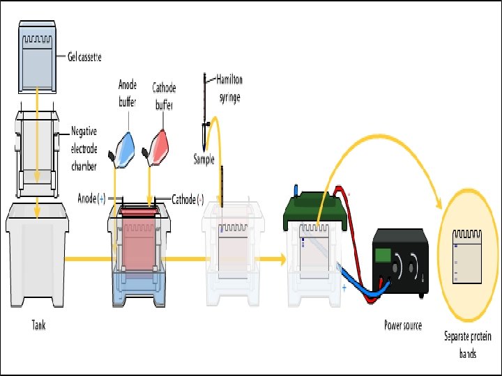

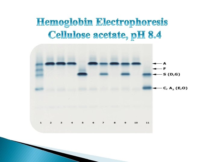
� The gel be stained by ponceaus to allowing visualization of the separated proteins. � After staining, different proteins will appear as distinct bands within the gel. � It is common to run molecular weight size markers of known molecular weight in a separate lane in the gel, in order to calibrate the gel and determine the weight of unknown proteins by comparing the distance traveled relative to the marker.

A 2/C Normal S F A+

A 2/C Normal Hb SS S F A+

A 2/C Normal Hb SS Hb AS S F A+

A 2/C Normal Hb SS Hb AS Hb SC S F A+

A 2/C Normal Hb SS Hb AS Hb SC Hb CC S F A+

A 2/C Normal Hb SS Hb AS Hb SC Hb CC HB AD S F A+

Normal Hgb A 1 Hgb A 2 95 -98% Hgb F 0. 8 -2. 0% Hgb S Hgb C Hgb H Sickle cell 2 -3% 0% 0% 0% Thalasemia Hgb C disease HPFH 4 -5. 8% 15% 70 -98% Hgb H disease under 2% ( minor ) 2 -5% 5 -35% ( minor ) Heterozygous or or 10 -90% 100% (Homozygous) ( major ) 90 -98%

� http: //labtestsonline. org/understanding/analytes /hemoglobin-var/tab/glance � http: //www. nlm. nih. gov/medlineplus/ency/articl e/003639. htm � http: //sickle. bwh. harvard. edu/thalover. html � http: //www. as. miami. edu/chemistry/2086/chap 19/newchapter%2019 -part 1. htm � http: //www. sicklecelldisease. org/index. phtml � http: //en. wikipedia. org/wiki/Sickle-cell_disease � ﻣﻘﺪﻣﺔ ﻓﻲ ﻋﻠﻢ ﻭﺍﻣﺮﺍﺽ ﺍﻟﺪﻡ ﻭﻃﺮﻕ ﺍﻟﻜﺸﻒ ﻋﻨﻬﺎ ﻓﻲ ﺍﻟﻤﺨﺘﺒﺮ )ﻋﺒﺪﺍﻟﻐﻨﻲ ( ﻋﻴﻀﺔ ﺍﻟﺜﺒﻴﺘﻲ

- Slides: 32