nucleus Nucleus blue and Microtubules green Nucleus and
 and Microtubules (green).")
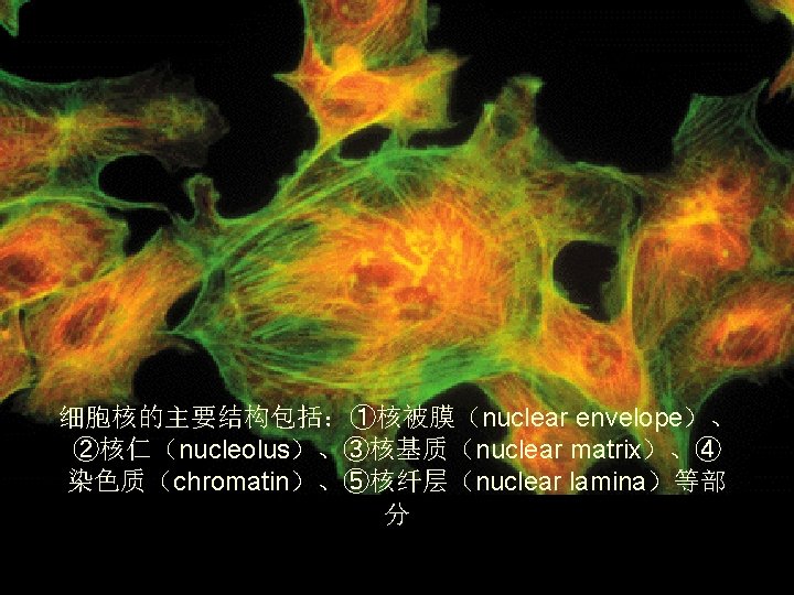
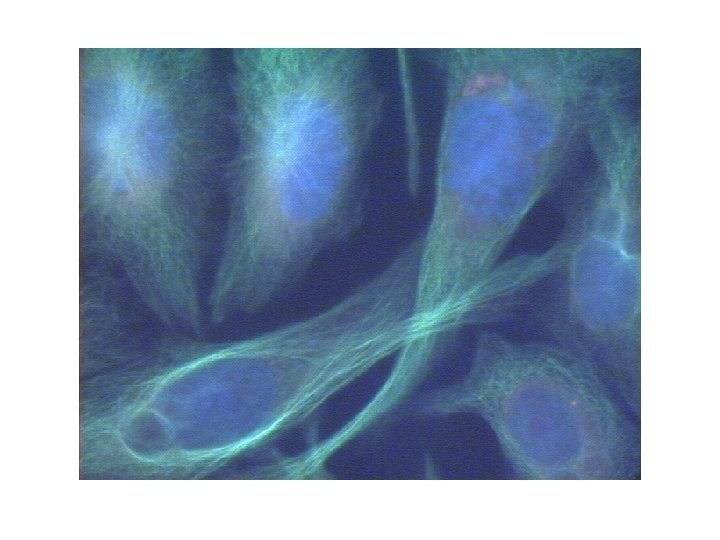
第八章 细胞核nucleus 与染色体 Nucleus (blue) and Microtubules (green).
 labeled")
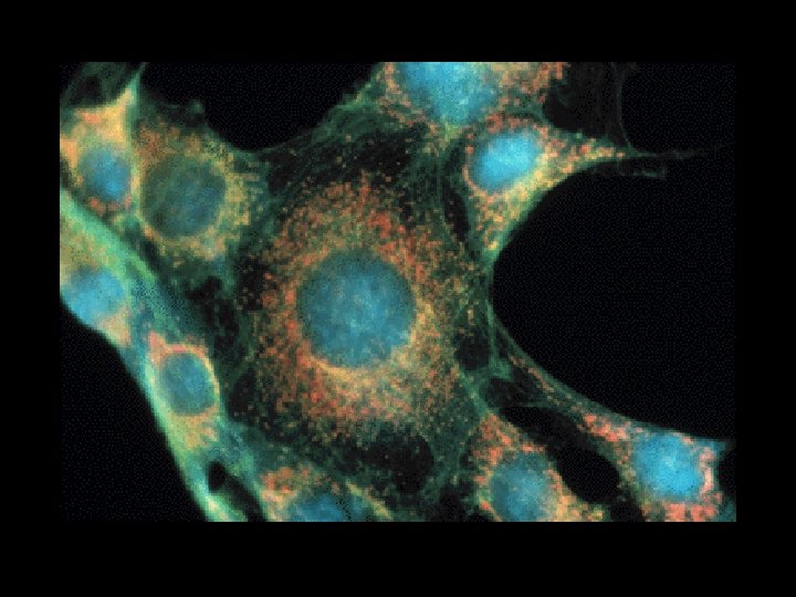
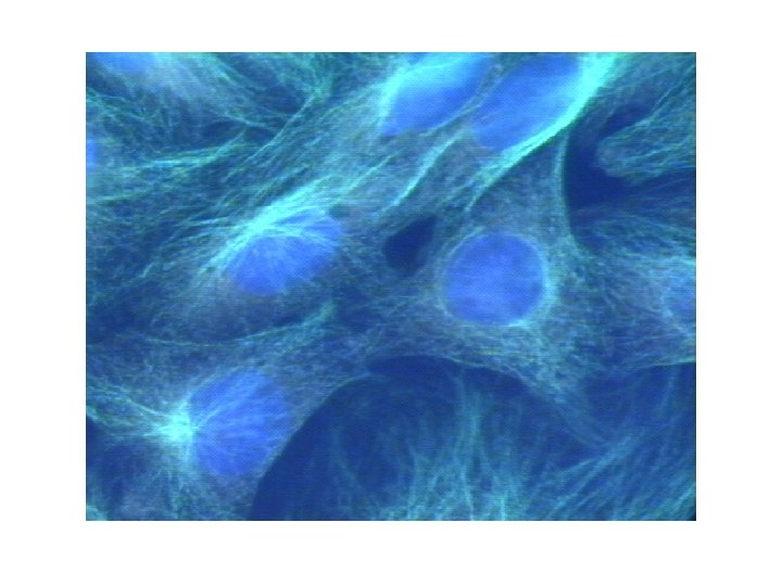
• Nucleus and Internal Membranes • Chinese hamster ovary cells (CHO cells) labeled with Hoechst 33342 (Molecular Probes # H-1399) and Di. OC 6 (Molecular Probes # D-273). The Hoechst stains the nuclear DNA blue, and the Di. OC 6 stains the internal organelles green. (Omega Optical Multi-dye Filter Set # XF 50). Photographs by Victoria Osborne.


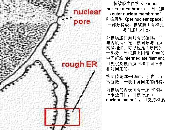
• The space between the outer and inner membranes is also continuous with rough endoplasmic reticulum space. It can fill with newly synthesized proteins just as the rough endoplasmic reticulum does. The nuclear envelope is enmeshed in a network of filaments for stability.

• Nucleus and Microtubules • Chinese hamster ovary cells labeled with Hoechst 33342 (Molecular Probes # H 1399) to stain the nuclear DNA blue and by immunofluorescence with an antitubulin antibody and a FITC-conjugated secondary antibody (green). (Omega Optical Multi-dye Filter Set # XF 64).
 labeled")
• Nucleus and Filamentous Actin • Chinese hamster ovary cells (CHO cells) labeled with Hoechst 33342 (Molecular Probes # H-1399) and BODIPY TR-X phallacidin (Molecular Probes # B-7464). The Hoechst stains the nuclear DNA blue, and BOPIPY TR-X phallacidin stains filamentous actin red. (Omega Optical Multi -dye Filter Set # XF 56).


与核孔 复合体(Nuclear Pore Complex ) 一、核被膜(nuclear membrane) 外核膜outer nuclear membrane 内核膜 Inner")
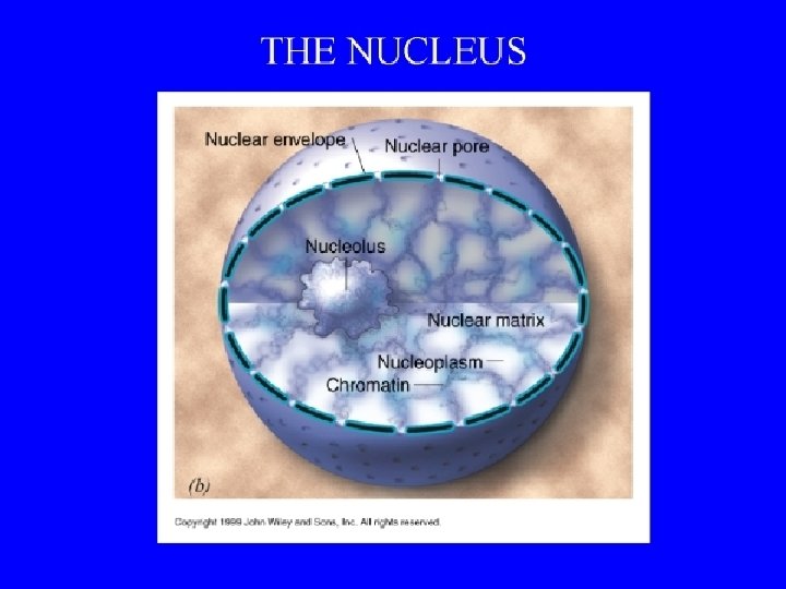
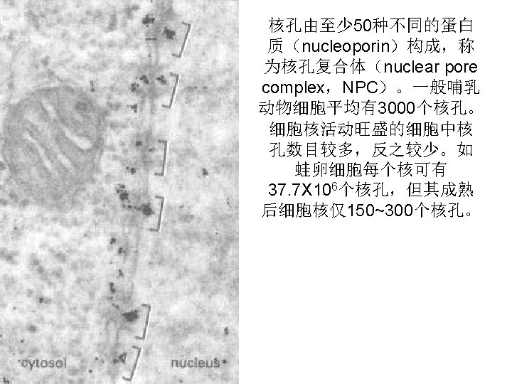
第一节 核被膜(nuclear membrane)与核孔 复合体(Nuclear Pore Complex ) 一、核被膜(nuclear membrane) 外核膜outer nuclear membrane 内核膜 Inner nuclear membrane 核膜孔nuclear pore 核周隙 perinuclear space
,又 称核被膜(nuclear envelope) 核周间隙(perinuclear space) 核孔(nuclear pore) 核纤层(nuclear lamina) 核骨架(nuclear")
• • (nuclear membrane),又 称核被膜(nuclear envelope) 核周间隙(perinuclear space) 核孔(nuclear pore) 核纤层(nuclear lamina) 核骨架(nuclear skeleton) 染色质(chromatin)
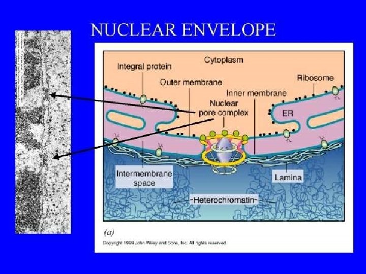
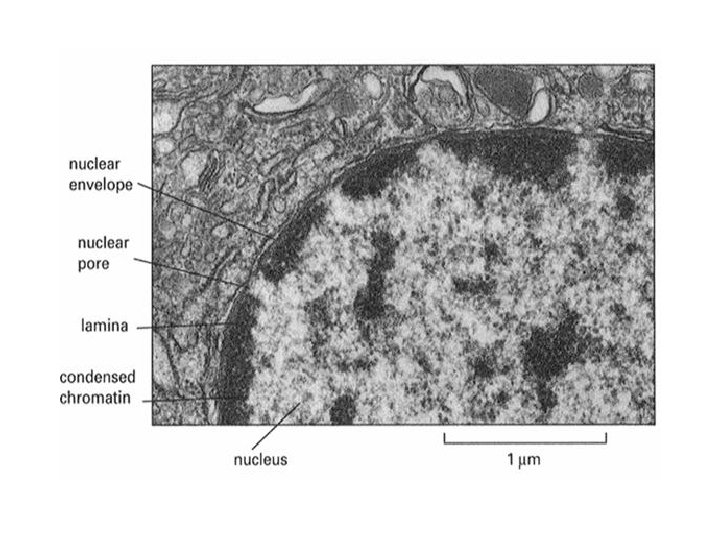

• Nuclear Envelope • The nuclear envelope has two membranes, each with the typical unit membrane structure. They enclose a flattened sac and are connected at the nuclear pore sites. The outermost membrane is continuous with the rough endoplasmic reticulum (ER) and has ribosomes attached (see figure to the left).

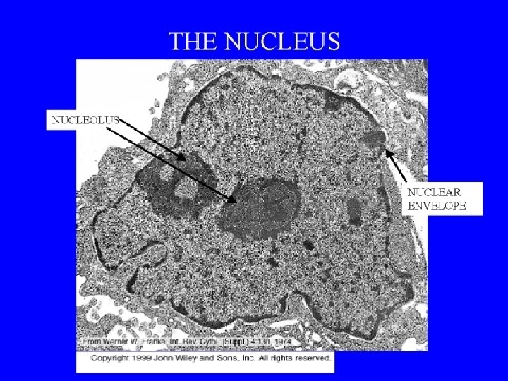
• The nuclear envelope is shown in an electron micrograph in the figure to the right. The filaments outside the envelope are not visualized with these protocols. Also, the nuclear lamina just inside the nuclear envelope is not shown well (see paragraph below for description).

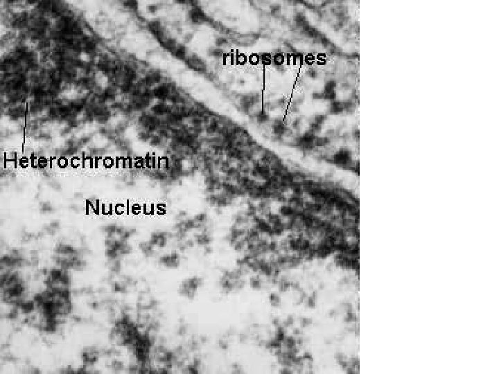
• However, one can see ribosomes on the outer membrane and the sac enclosed by the two membranes. Dense patches of Heterochromatin are seen just inside the inner membrane.

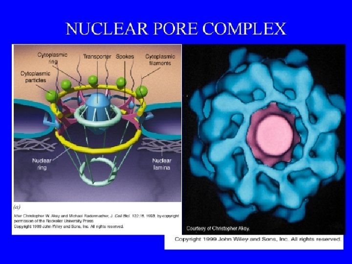
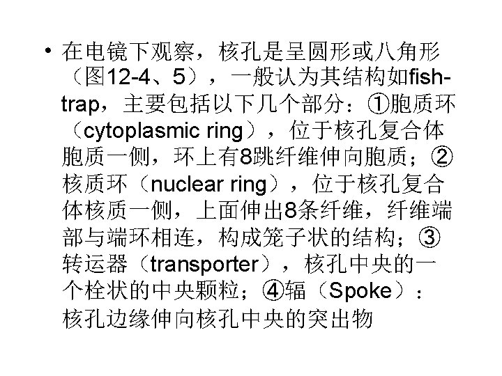
• Nuclear Pore Complex: Structure •
. The cytoplasm also contains numerous ribosomes")
Nucleus with Nuclear Pores (TEM x 73, 200). The cytoplasm also contains numerous ribosomes
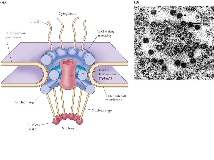
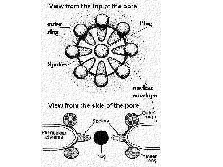

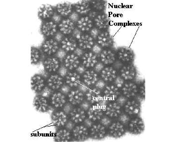
• The above figure shows a view of the nuclear pore from the top. It contains 8 subunits that "clamp" over region of the inner and outer membrane where they join. Actually, they form a ring of subunits 15 -20 nm in diameter. Each subunit projects a spoke-like unit into the center so that the pore looks like a wheel with 8 spokes from the top. Inside is a central "plug". •
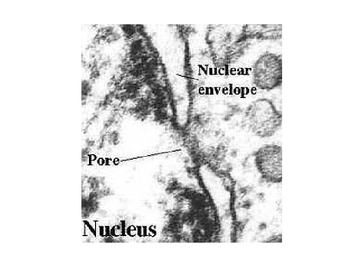

• This electron micrograph shown in the figure to the right depicts a nuclear pore complex seen with the transmission electron microscope. As is obvious, little detail can be seen. The inner and outer membranes of the nuclear envelope are joined and there appears to be a diaphragm-like structure in the center.

• However, the intricate detail pictured in the foregoing figure cannot be appreciated. One needs to use different preparative techniques to see the subunits and their organization. These will be discussed and illustrated in the following sections.
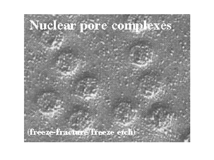

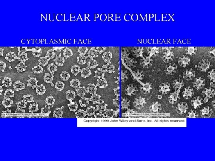
• Freeze-fracture/freeze-etch
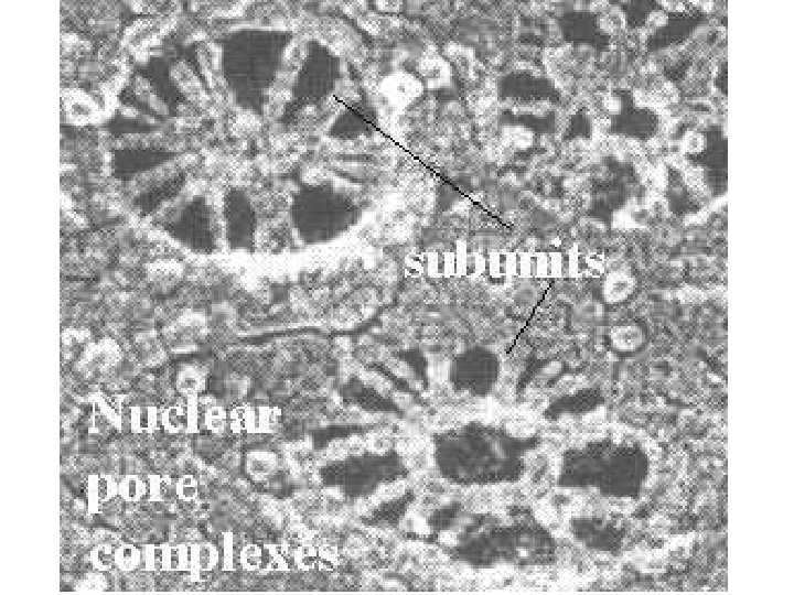

• Another preparation shows more details of the structure of the nuclear pore complex. Here we see the subunits forming the rings and their spokes. Note that one of the pores appears to be open in the center, forming a channel. Figure modified from Bloom and Fawcett, A Textbook of Histology, Chapter 1, Figure 1 -9, Chapman and Hall, Publishers, 1994

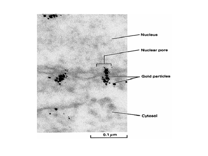
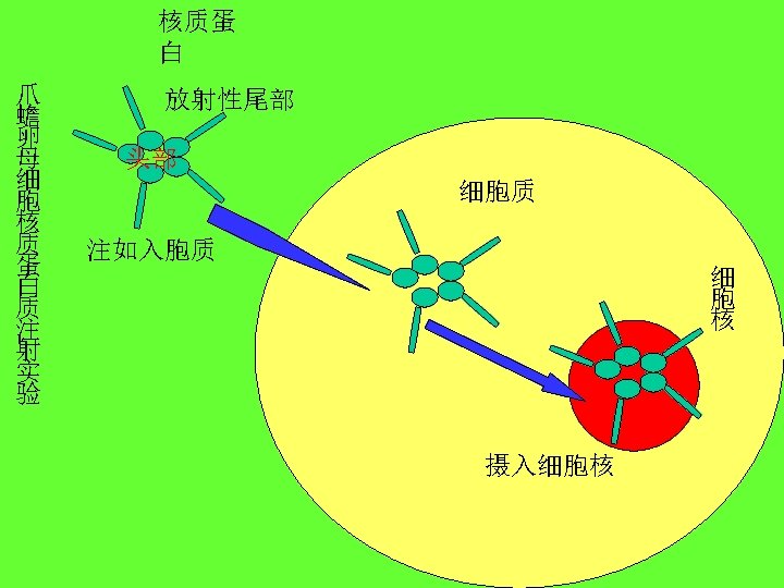
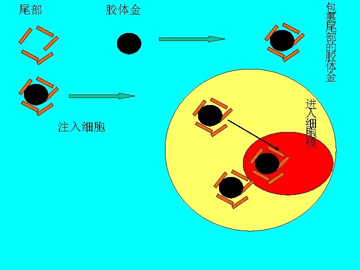
• A peptide sequence, called nucleoplasmin, was isolated and linked to colloidal gold. It was then injected into an oocyte and traced with electron microscopy. As shown in the figure to the left, the gold particles mark the site of transport of the nucleoplasmin and the studies showed that it was transported into the nucleus. Small gold markers are evident inside the nucleus. Figure was taken from Alberts et al. , Molecular Biology of the Cell, Garland Pub. , N. Y. 1994, Fig 12 -15. •

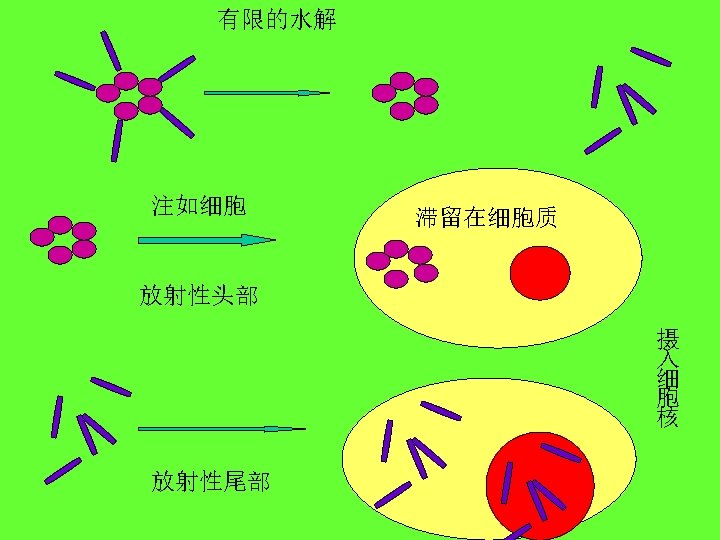
• In another test, workers took advantage of the fact that nucleoplasmin has a head region which is not transported into the nucleus and a tail region which is transported. When the heads and tails were separated and each linked to gold, only the gold-labeled tail region was transported. Gold-labeled head regions remained in the cytoplasm.
- Slides: 59