Nucleus 5 m in diameter The nucleus is


 • Separates the enclosed nuclear compartment from cytoplasm • Maintains the")


")

 or karyoplasm • Analogy with cytoplasm, that part of the nuclear")

 In general terms, there are three levels of")


 DNA is a nucleic acid that contains the genetic instructions used")
 RNA “bases” –")
 is a nonmembrane bound structure composed of proteins")
- Slides: 26
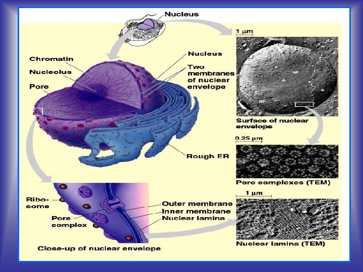
Nucleus ~ 5 µm in diameter The nucleus is the headquarters (controls all cell activity) of the cell. It is the most obvious organelle The Nucleus is a membrane-enclosed organelle which house most of the genetic information and regulatory machinery responsible for providing the cell with its unique characteristics Functions • It stores the cell's hereditary material (DNA) • Site of DNA replication • Site of DNA transcription to m. RNA • Ribosomal formation • Nucleolus: RNA & protein required for ribosomal synthesis • It coordinates the cell's activities, which include growth, intermediary metabolism, protein synthesis, and reproduction (cell division) by regulating gene expression.
STRUCTURE 1. Nuclear envelope, double membrane and nuclear pores 2. Nucleoplasm 3. Chromatin, Chromosome, DNA and RNA 4. Nucleolus (concentrated area of chromatin, RNA and proteins)
Nuclear envelope (Nucleolemm) • Separates the enclosed nuclear compartment from cytoplasm • Maintains the shape of nucleus • Controls exchanges between nucleus and cytoplasm • Important role in organization of nucleus content Structure q. External (outer) nuclear membrane q. Internal (inner) nuclear membrane q Perinuclear space q. Lamina densa (nuclear lamina) q. Nuclear pores
External nuclear membrane • Fluid mosaic (lipid bilayer of ~ 7 nm thick with 70% proteins) • Visible only by electron microscopy • Ribosome attached on external face • It continues with RER membrane The perinuclear space • 10 40 nm • It communicates with the RER internal space • Contains the same molecules as RER • Contains Ca 2+ Internal nuclear membrane • Fluid mosaic (lipid bilayer) • Visible only by electron microscopy; • The inner surface of the nuclear envelop is bound to a thin filamentous network (lamins polypeptides) called the nuclear lamina.
Nuclear lamina a network of intermediate filaments composed of various lamins The lamina acts as a site of attachment for chromosomes and provides structural stability to the nucleus. The lamins have been associated with various genetic disorders collectively termed laminopathies (e. g. a rare form of muscular dystrophy).
THE NUCLEAR PORE • Openings in the nuclear envelope, diameter about 10 nm • Area where the nuclear envelope is interrupted • Regulates exchanges between nucleus and cytoplasm • Ensures the selective transport for big molecules • There are 3000 4000 nuclear pores (10 pores/μm 2) • Dynamic structures – their number grows if it’s necessary
• The nuclear pores are the gateways across which movement of RNAs and proteins takes place between the nucleus and cytoplasm in both direction. • Proteins synthesized in the cytoplasm cross the nuclear envelop to initiate replication and transcription of genetic material. Similarly, m. RNA, t. RNA and ribosomal subunits built in the nucleus cross through the nuclear pores to the cytoplasm The Nuclear Pore Complex Cytoplasmic face Nuclear face
• Octagonal symmetry • 8 fold repetition of glycoprotein subunits (Nucleoporins or Nups) • Nucleoporins: symmetrical on cytoplasmic and nuclear sides Nucleoplasm Nuclear Basket Nuclear Ring Filaments Nuclear Membrane Central Transporter Spoke Ring (inner & outer) Cytoplasmic Ring Cytoplasmic Filaments
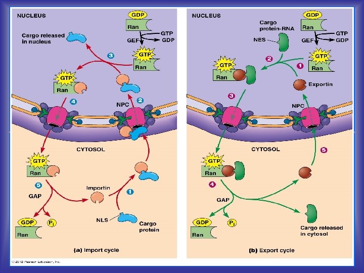
Nuclear transport The entry and exit of large molecules from the cell nucleus is tightly controlled by the nuclear pore complexes (NPCs). Although small molecules can enter the nucleus without regulation. Macromolecules such as RNA and proteins require association with importins proteins to enter the nucleus and exportins to exit Nuclear Import To get materials into the nucleus there is amino acid tag (zip code) called nuclear localization signals (NLS) added onto molecules and are assisted by proteins called importins Once inside the nucleus, interaction with Ran GTP causes a conformational change in the importin that causes it to dissociate from its cargo {RANs are hydrolase enzymes that can bind and hydrolyze guanosine triphosphate (GTP)} Ran (RAs-related Nuclear protein) Nuclear Export Nuclear export roughly reverses the import process; in the nucleus, the exportin binds the cargo and Ran-GTP and diffuses through the pore to the cytoplasm carrying nuclear export signals (NES) or the zip code bound by exportins. The complex can diffuse to the cytoplasm where GTP is hydrolyzed and the NES protein is released Hence, whereas importins depend on Ran GTP to dissociate from their cargo, exportins require Ran GTP in order to bind to their cargo.
Nucleoplasm (nucleus sap) or karyoplasm • Analogy with cytoplasm, that part of the nuclear contents other than the nucleolus. • Highly viscous liquid that surrounds the chromosomes and nucleolus • Many substances such as nucleotides and enzymes are dissolved in the nucleoplasm • A network of fibers known as the nuclear matrix can also found in the nucleoplasm
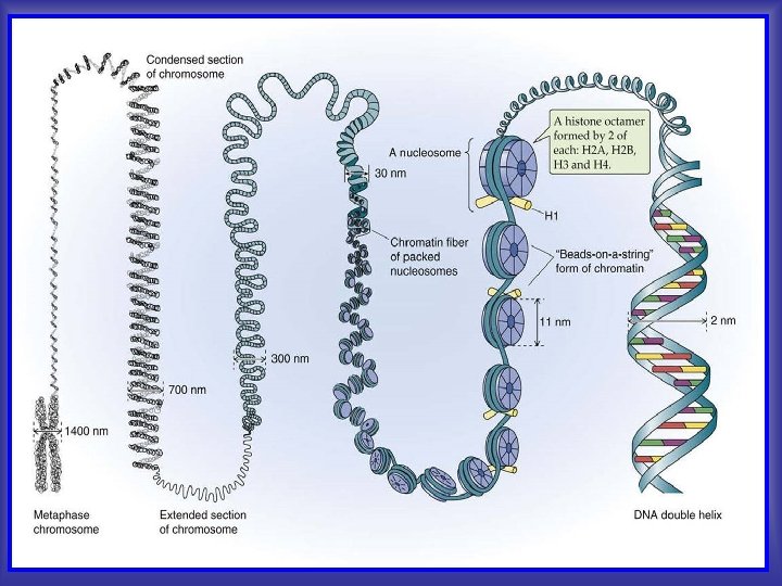
CHROMATIN Is the combination of DNA and proteins that make up the contents of the nucleus of a cell, that is usually dispersed in the interphase and condensed to form chromosomes in mitosis and meiosis. Functions ■Package DNA into a smaller volume to fit in the cell ■Strengthen and repair the DNA to allow mitosis and meiosis i. e. prevent DNA damage ■Regulate gene expression (transcription) and DNA replication Types (During interphase // no cell division) 1 - Euchromatin: is a lightly packed (less dense) form of chromatin that is rich in gene concentration, and is often under active transcription. It is found in both eukaryotes and prokaryotes. 2 - Hetrochromatin: is a tightly packed (dense) form of DNA that is inactive (no transcription) and remains compact during interphase. Heterochromatin plays a role in gene regulation and the protection of the integrity of chromosomes
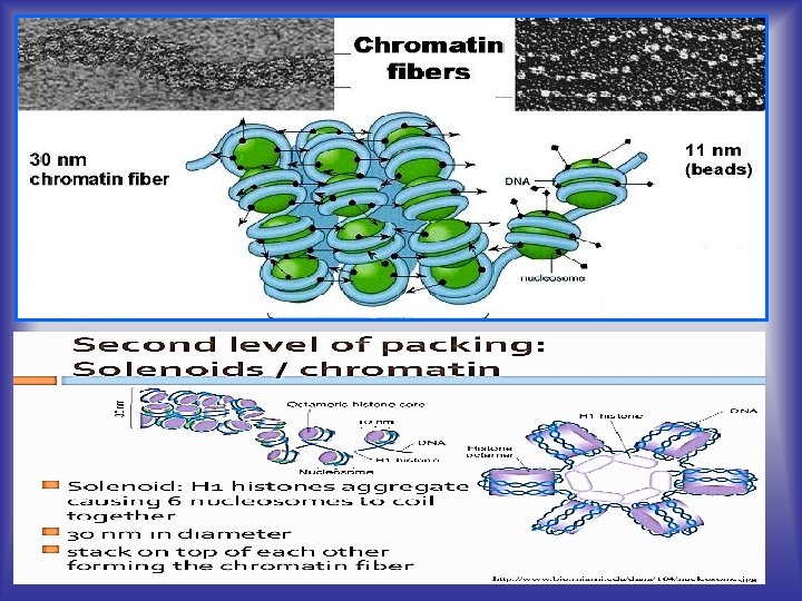
LEVELS OF CHROMATIN ORGANIZATION (Chromatin Packing) In general terms, there are three levels of chromatin organization: 1. the "beads on a string" structure, DNA wraps around histone proteins forming nucleosomes 2. 30 nm fiber, chromatin appears in interphase cells as tiny dots and fibers of 30 nm thickness 3. Higher level DNA packaging of the 30 nm fibre into the metaphase chromosome (during mitosis and meiosis).
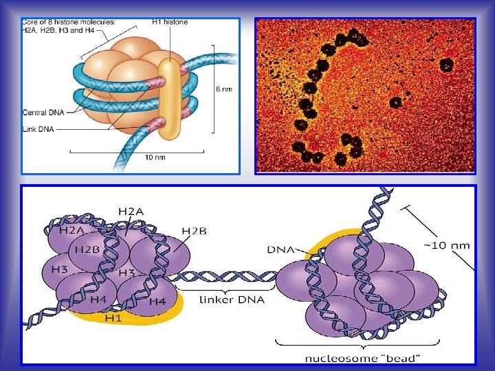
1. The "beads on a string” • DNA and histones are organized into repeating subunits called nucleosomes • nucleosomes composed of two loops of DNA wrapped around a protein core (eight histone molecules, two copies of H 2 A, H 2 B, H 3 and H 4) • A nucleosome core particle consists of 146 base pairs of supercoiled DNA wrapped almost twice around disk shaped complex of 8 histone molecules H 1 histone, the linker histone, resides outside the nucleosome and binds to the linker DNA that connects one neucleosome to the next. •
2. Fibers of 30 nm thickness The 30 nm fibers gather into larger supercoiled loops of thick fibers which normally spread through the interphase nucleus Strand of nucleosomes is coiled to produce 30 nm chromatin fibril Chromatin fiber is composed of the loops of chromatin fibril anchored into protein core of chromosome (composed of non histone proteins). During mitotic division chromatin fibers are highly condensed and form chromosomes
3. metaphase chromosome When cell prepares to divide, chromatin fibers coil up as separate structures, chromosomes In the early stages of mitosis or meiosis (cell division/ metaphase), the chromatin strands become more and more condensed. They cease to function as accessible genetic material (transcription stops) Chromosomes may exist as either duplicated or unduplicated. Unduplicated chromosomes are single linear strands, whereas duplicated chromosomes contain two identical copies (called chromatids) joined by a centromere. chromatin chromosome
Chromosome chromosome • A chromosome is an organized structure of DNA and protein found in cells. • Chromosomes are the physical carriers of genetic information. short arm centromere • The structure of chromosomes and chromatin varies through the cell cycle • Each human somatic cell contains 23 pairs i. e 46 chromosomes of different chromosomes (Diploid cells). GAMETES (sperm and egg cells) have one set of 23 chromosomes ( Haploid cells). One of the chromosome pairs consists of the sex chromosomes (X and Y), the other 22 pairs of chromosomes are termed autosomes long arm chromatid Replicated chromosome short arm • The members of each pair of autosomes are said to be homologs, or homologous, because their DNA is very similar. The X and Y long arm chromosomes are not homologs of one another. kinetochore centromere sister chrom
Chromatin Chromosome Definition The DNA molecules in the genome are packaged with histones, forming chromatin. The highest packaged structure of DNA appears in the metaphase of the cell division. Structure Chromatin is composed of nucleosomes. Condensed 50 timesthan the normal DNA double helix Chromatin fibres are thin, long, uncoiled structures Chromosomes are condensed into chromatin fibres. Condensed 10, 000 times the normal DNA double helix. Chromosomes are thick, compact, ribbon like structures. Pairs Chromatin is a single, unpaired fibres Chromosome exists as a pair. Metabolic activity Chromatin allows DNA replication, gene expression and recombination. Chromosomes do not show any metabolic activity Presence/ Period Chromatin appears in the interphase of the cell cycle Chromosomes appear during the metaphase and exist in the anaphase of the nuclear division Conformation Chromatin consists of two confirmations: euchromatin and heterochromatin. Chromosome is usually heterochromatic. Visualization Chromatin can be observed under the electron microscope as a bead and string structure Chromosome can be observed under the light microscope in its classic four arm structure Appearance
Deoxyribonucleic acid (DNA) DNA is a nucleic acid that contains the genetic instructions used in the development and functioning of all known living organisms. A gene is a unit of heredity in living organisms. DNA consists of thousands of genes. It specifies everything that is needed for the maintenance, function, and replication of the cell DNA consists of two (double helix) long polymers of simple units called nucleotides, with backbones made of sugars and phosphate groups joined by ester bonds. Nucleotides are molecules that, when joined together, make up the structural units of RNA and DNA. = nucleobase + sugar+ phosphate group Replication: DNA making a copy of itself Transcription: making of RNA from code of DNA Translation: making of protein coded by t. RNA via m. RNA via DNA
DNA “bases” – T, A, C, G (thymine, adenine, cytosine, guanine) RNA “bases” – U, A, C, G (uracil instead of thymine)
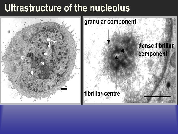
The Nucleolus The nucleolus (plural nucleoli) is a nonmembrane bound structure composed of proteins and nucleic acids found within the nucleus/ 1 -2 μm It is the most dense (prominent) structure of the cell, and frequently is located in central area of nucleus Function, site of r. RNA synthesis, initial ribosomal assembly Structure, -fibrillar centers, filaments of chromatin -pars fibrosa, newly transcribed r. RNA -pars granulosa, r. RNA bound to ribosomal proteins that are beginning to assemble into ribosomes