Nucleotides Nucleic Acids Watson Crick 1953 Franklin Wilkins
 Franklin &")
 Watson &")


 Axial")


- Slides: 21
Nucleotides & Nucleic Acids Watson & Crick 1953 (Franklin & Wilkins too) Franklin & Wilkins Heavy bands - indicates recurring bases X - indicates helical structure
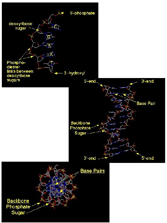
Nucleotides & Nucleic Acids Watson & Crick 1953 (Franklin & Wilkins too) Watson & Crick Used x-ray crystallography data Used Chargaff’s rules 3 D structure of DNA, strands antiparallel
Nucleotides & Nucleic Acids DNA structure Complementarity Strands not identical in base sequence or composition DNA replication
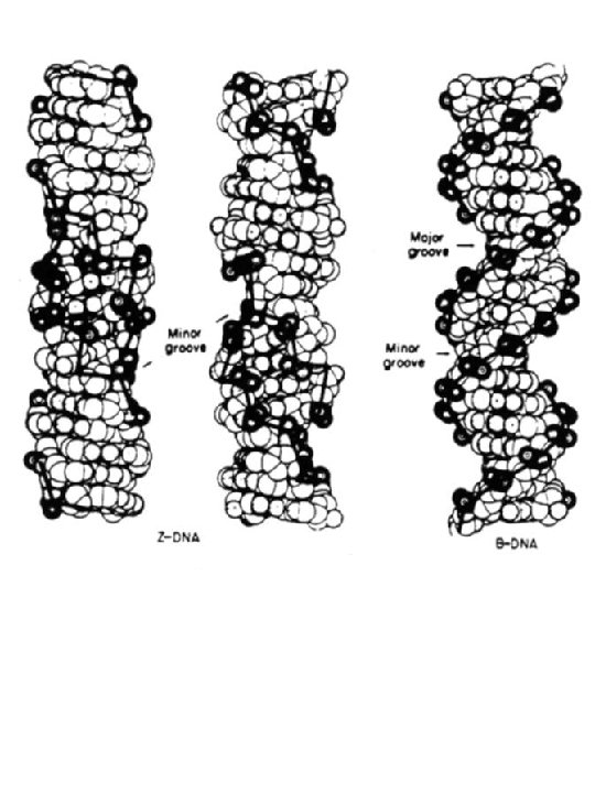
Nucleotides & Nucleic Acids DNA structure - different forms
A-DNA Z-DNA B-DNA
Nucleotides & Nucleic Acids DNA structure - different forms Structure type Pitch (Å) Axial rise (h) per nt (Å) Groove width (Å) minor major 11 2. 7 Groove depth (Å) minor major 2. 8 13. 5 A 28. 2 2. 56 B 33. 8 3. 38 5. 7 11. 7 7. 5 8. 5 Z 45 3. 7 8. 8 2 3. 7 13. 8 Z-DNA virtually no major groove due to syn-G, pulls C into major grv Pitch = axial rise x repeat (bp/turn) distance for complete turn of helix So for A-DNA 2. 56 Å x 11 bp/turn = 28. 2 Å axial rise (h) distance between bp
purine rotated into major groove
B-DNA A-DNA + bulge 32˚ tilted upward A-DNA
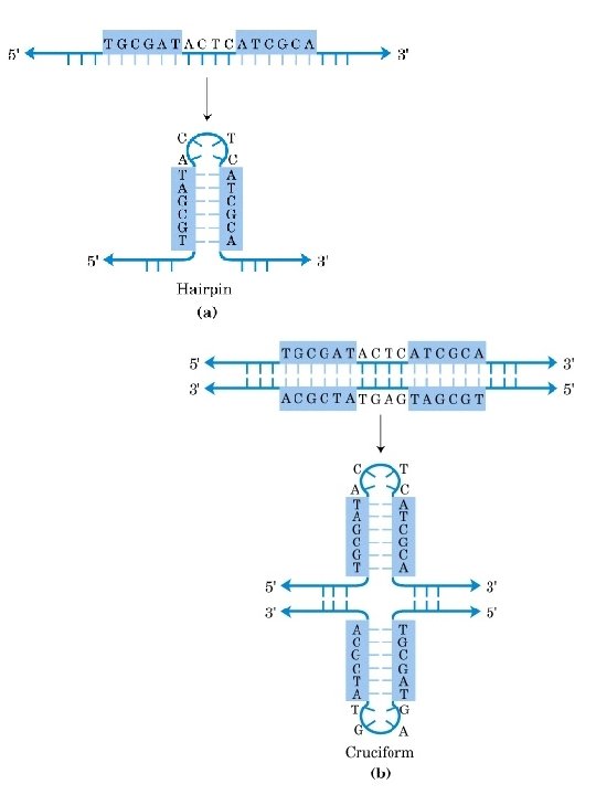
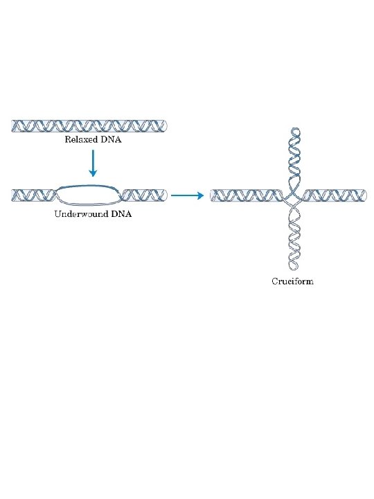
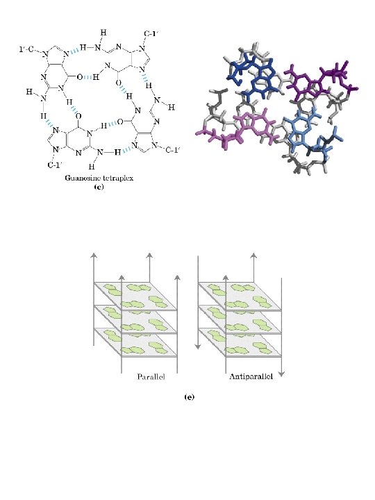
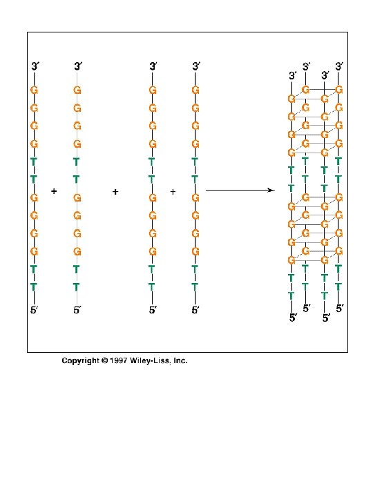
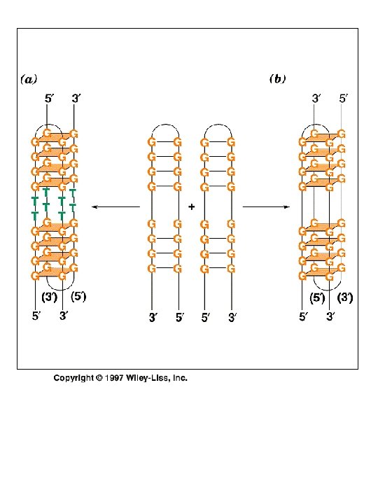
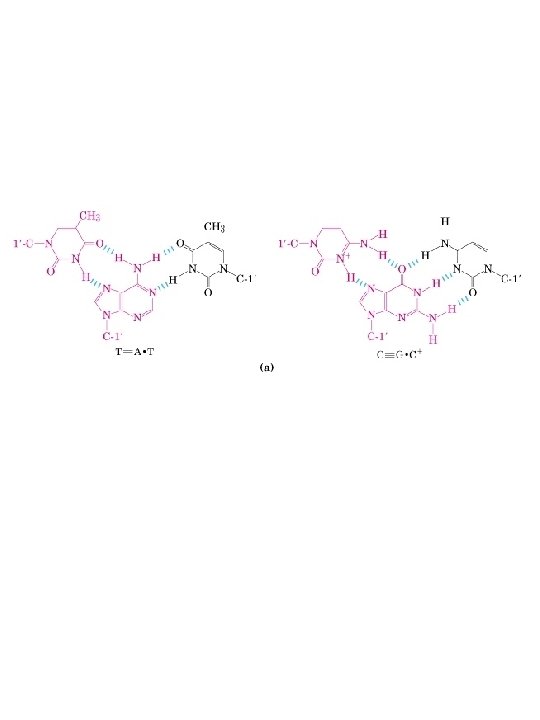
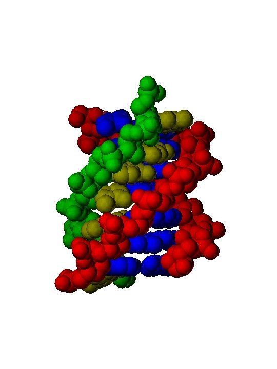
Triple Helix Intrastrand base pairing
Triple Helix using a third strand of DNA Interstrand base pairing Restriction endonuclease