NPDR IRMA Diabetes and Ocular Complications NVE CSME

NPDR IRMA Diabetes and Ocular Complications: NVE CSME NVD What Do Those Numbers Mean for the Eyes? FBS A 1 c Mindy J Dickinson, OD Midwest Eye Care, PC NIDDM CWS FA OCT

Objectives � � What is Diabetes ? What Types of Diabetes are there, and in general, how are they managed? What is Hemoglobin A 1 c ? What are the possible ocular complications associated with Diabetes ? ◦ Front to back of the eye approach Emphasis on non-proliferative and non-proliferative retinopathy and macular edema ◦ Images of complications Photos, fluorescein angiography, OCT, Visual Fields � What does Diabetic Retinopathy look like ? � How can these complications be treated/managed?

Diabetes Mellitus A group of metabolic diseases in which a person has high blood sugar, either because: 1) the body does not produce enough insulin … or because… 2) the cells of the body do not respond to the insulin that is produced
 Type II")
Types of Diabetes Type I ◦ Juvenile Onset ◦ Insulin Dependent (IDDM) Type II ◦ Adult Onset ◦ Non-Insulin Dependent (NIDDM) Gestational ◦ Develops during pregnancy ◦ ? Precursor to development of Type II

Type I Diabetes Body fails to produce enough insulin Due to death of cells in the pancreas Develops suddenly – kids in hospital ◦ auto-immune, ? Viral, genetics Accounts for 5 -10% of cases of diabetes Requires insulin treatment

Type I Diabetes Treatment Options ◦ Insulin 70/30 ◦ NPH/Regular ◦ Lantus/Humalog ◦ Insulin Pump ◦ Pancreas Transplant ◦ Islet Cell Transplant

Type II Diabetes Results from insulin resistance when cells fail to use insulin properly Sometimes combined with a degree of insulin insufficiency for a body’s needs Develops gradually

Type II Diabetes 3 core defects 1. Genetic: Poor production by the pancreas cells causes not enough insulin to be produced 2. Obesity: Increased resistance to insulin in the tissues 3. Liver sugar storage issues: Problems with hormone “glucagon” and it tells liver to release more sugar out into blood stream than it should

Type II Diabetes Treatment Options ◦ Lifestyle intervention - weight loss, nutrition ◦ Medications to reduces insulin resistance ◦ Medications to increase insulin secretion ◦ Insulin

Diabetes Treatment Education, Understanding, Participation Well-managed blood sugars Addressing other risks: ◦ ◦ ◦ Smoking Elevated cholesterol – (LDL < 100) High blood pressure – (< 130/80) Obesity Lack of exercise

Diabetes Challenges Blood glucose variability ◦ In a healthy patient – pancreas adjusts minute to keep blood sugar between 70 and 140 ◦ In a diabetic patient - no way for us to monitor glucose this frequently and to get medication in to the body to adjust it that quickly

Glucose Monitoring Serum glucose = immediate “snapshot” of blood sugar ◦ Can be measured “post-prandial” (after a meal) or “fasting” (usually 8 or 12 hours of not eating) Hemoglobin A 1 c = 3 month average blood sugar ◦ Looks at how much sugar has grown onto a RBC

Blood Sugars – high average and high variance A 1 C 14. 0 13. 0 12. 0 11. 0 10. 0 9. 0 8. 0 7. 0 6. 0 5. 0 BS 400 350 300 250 200 150 100 50 0 1 2 3 4 5 6 7 8 9 10 11 12 13 14 15 16 17 18 19 20 hours

Blood Sugars – lower average but high variance A 1 C 14. 0 13. 0 12. 0 11. 0 10. 0 9. 0 8. 0 7. 0 6. 0 5. 0 BS 400 350 300 250 200 150 100 50 0 1 2 3 4 5 6 7 8 9 10 11 12 13 14 15 16 17 18 19 20 hours

Blood Sugars – moderate average with low variance A 1 C 14. 0 13. 0 BS 400 350 12. 0 11. 0 10. 0 300 250 9. 0 8. 0 7. 0 200 150 6. 0 5. 0 100 50 0 1 2 3 4 5 6 7 8 9 10 11 12 13 14 15 16 17 18 19 20 hours

Blood Sugars – optimal average with low variance A 1 C 14. 0 13. 0 12. 0 11. 0 10. 0 9. 0 8. 0 7. 0 6. 0 5. 0 BS 400 350 300 250 200 150 100 50 0 1 2 3 4 5 6 7 8 9 10 11 12 13 14 15 16 17 18 19 20 hours
 Type I patients Compared Intensive treatment vs Conventional")
Diabetes Control and Complications Trial (DCCT) Type I patients Compared Intensive treatment vs Conventional treatment and their rate of complications Intensive Treatment Hb. A 1 c 7. 2 Conventional Treatment Hb. A 1 c 9. 1 Overall risk reduction 70%

United Kingdom Prospective Diabetes Study Newly diagnosed Type II Compared Intensive treatment vs Conventional treatment and their rate of complications Intensive treatment Hb. A 1 c Conventional treatment Hb. A 1 c 7. 0 7. 9 Risk reduction of 35% per 1% decrease in hemoglobin A 1 c

Diabetes Complications Heart Disease Stroke Kidney Disease Neuropathy of hands and feet Skin sores, amputation Gastointestinal Disease Genitourinary Complications Eye Disease

Mechanism of eye damage Poor blood sugar control = dilation/constriction of blood vessels Damaged lining = leak blood out into tissue Oxygen carried by blood does not get where it is supposed to Lack of oxygen = “hypoxia” or “ischemia” Hypoxia causes swelling of retina and loss of function Hypoxia causes release of hormone (VEG-F ) to try to grow new blood vessels

Neovascularization New blood vessels grow without a lining and do not grow in the retina but out in vitreous and in front of eye as well Leak blood constantly, act as tension cords, and can clog drain of eye New blood vessel growth can occur on optic nerve, retina, iris or in the drain in the front of eye

Ocular Complications ◦ ◦ ◦ Extraocular Muscle Palsy Dry Ocular Surface Neovascularization of iris and drain of eye Shift in refractive error (glasses Rx change) Vitreous gel hemorrhage Non-Proliferative Retinopathy Macular Swelling Retinal Non-Perfusion Tractional Retinal Detachment Neovascular Glaucoma Optic neuropathy

Cranial Nerve/Muscle Palsy Symptoms: ◦ ◦ ◦ Sudden onset of double vision May be worse in one position of gaze May be able to turn head to compensate Resolves when shut one eye Image may be side by side, up and down, or at a diagonal Usually gets better within 3 months 3 rd, 4 th or 6 th nerve palsy If does not resolve, may put prisms in glasses to realign images

Cranial Nerve/Muscle Palsy 6 th Nerve Palsy R eye

Dry Ocular Surface Symptoms: ◦ Burning, itchy, gritty, watering, red, foreign body sensation, tired, light sensitive, have to blink frequently, intermittently blurred vision

My glasses don’t work ! Lens of eye helps to focus light rays onto retina Typically lens prevents sugar from entering it When sugar breaches 200+ in blood stream, it can seep into lens To neutralize the sugar, water will be drawn in and lens swells Swollen lens bends light differently
 Non-Proliferative (NPDR) 2) Proliferative (PDR) Macular edema (CSME) ◦")
Diabetic Retinopathy 2 forms: 1) Non-Proliferative (NPDR) 2) Proliferative (PDR) Macular edema (CSME) ◦ Fluid seeping out in the most sensitive part of the retina and therefore usually affects visual acuity ◦ May occur on its own or with NPDR or PDR ** all of these forms start out painless, without any initial symptoms
 ◦ Tiny bulges off of retinal arterioles")
Retinal Findings in Diabetic Retinopathy Microaneurysms (MAs) ◦ Tiny bulges off of retinal arterioles Intraretinal hemorrhages ◦ “dot and blot” Cotton Wool Spots (CWS) ◦ Fuzzy white spots in retina due to infarct of retinal arteriole and lack of oxygen causing retina to swell Exudates ◦ Proteins seeping out of leaky blood vessels ◦ Well defined white or yellow deposits ◦ Usually form in a ring around central leaky vessel Retinal thickening/swelling

Retinal Findings in Diabetic Retinopathy Venous Beading ◦ “box-carring” or “sausage-linking” of retinal veins Intra. Retinal Microvascular Anomalies (IRMA) ◦ Anomalous fine caliber blood vessels that appear in pre-neovascular stage Retinal Non. Perfusion ◦ Drop-out of blood vessels in macular region that leads to vision loss Neovascularization of Disc (NVD) ◦ New blood vessels growing off of optic disc Neovascularization Elsewhere (NVE) ◦ New blood vessels growing any where else in retina

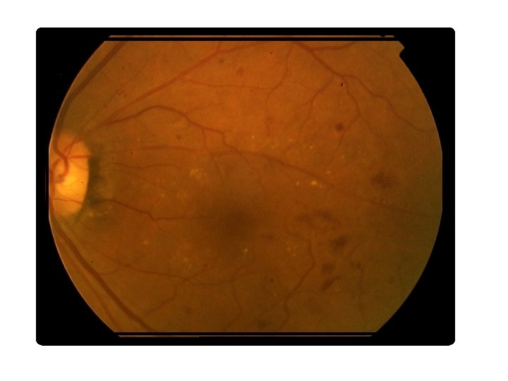
Non-Proliferative Retinopathy

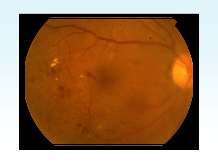
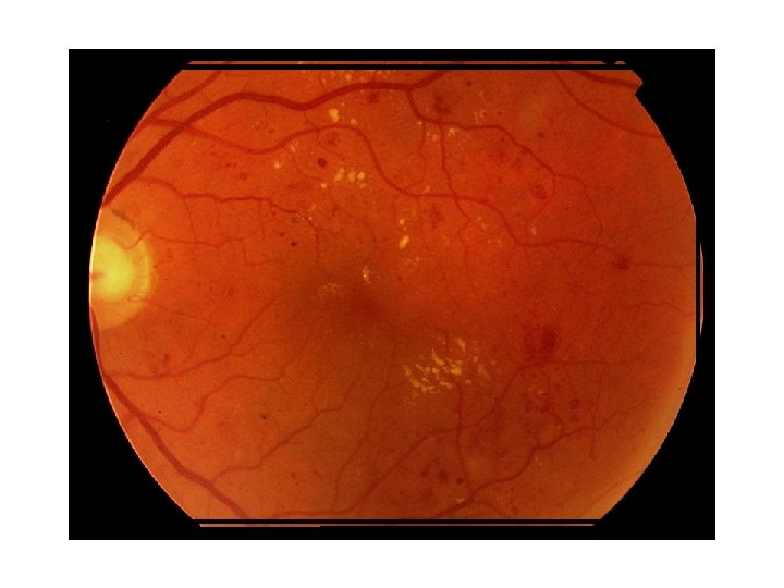
Exudates in NPDR

Exudates
- Slides: 35