NOTES CH 42 part 1 Circulation in Animals
")


 ● RIGHT: RIGHT")
 pumps")


 ●")


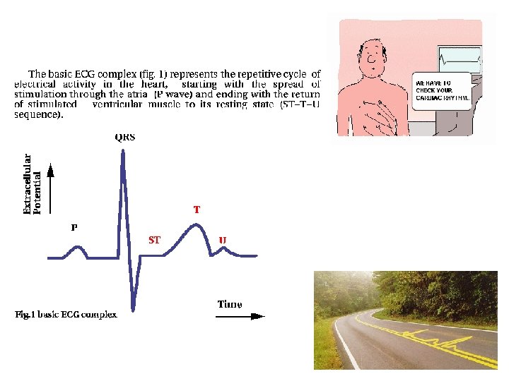
: ● records the electrical changes in the myocardium during a cardiac cycle")


 • contain HEMOGLOBIN, which combines with oxygen • hemoglobin synthesis")
 • function in defense against disease • also called: LEUKOCYTES")
 • normal WBC count is 5, 000 – 10, 000")


 Okazaki")


 / diastolic")

- Slides: 69
NOTES: CH 42, part 1 (Circulation in Animals)
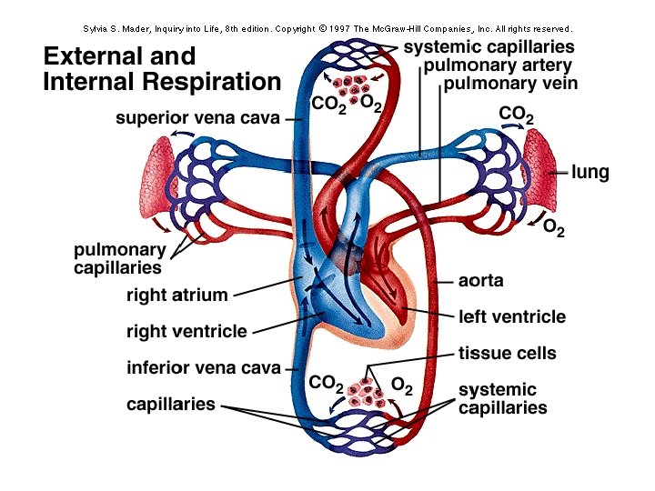
● Diffusion alone is NOT adequate for transporting chemicals over distances in animals ● The circulatory system ensures that no substance must diffuse very far to enter or leave a cell
The circulatory system… provides oxygen and nutrients to tissues and removes wastes.
STRUCTURE OF THE HEART Size: ● about 14 cm long and 9 cm wide Location: ● 2/3 left of midline ● below 2 nd rib and rests on diaphragm
STRUCTURE OF THE HEART Heart Covering: ● fibrous, outer pericardium: parietal pericardium ● serous, inner pericardium: visceral pericardium ● space between the layers = pericardial cavity (cushions and lubricates heart)
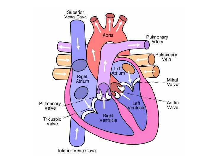
4 Heart Chambers: (heart is divided into right and left sides) ● RIGHT: RIGHT ATRIUM receives blood from the superior and inferior venae cavae and coronary sinus pumps blood into the RIGHT VENTRICLE right ventricle pumps blood out of the heart into the PULMONARY ARTERIES (take blood to the lungs)
● LEFT: LEFT ATRIUM receives blood from the pulmonary veins (from the lungs) pumps blood into the LEFT VENTRICLE left ventricle pumps blood out of the heart into the AORTA (takes blood to all of the body)
Heart Valves: designed to prevent backflow of blood! ATRIOVENTRICULAR VALVES: -TRICUSPID VALVE: separates R atrium from R ventricle -BICUSPID VALVE (a. k. a. MITRAL VALVE): separates L atrium from L ventricle
● Structure of AV valves: -CHORDAE TENDINAE: strong fibrous structures that attach to the flaps of the valves -PAPILLARY MUSCLES: muscles embedded in the endocardium; attach to the chordae tendinae
SEMILUNAR VALVES: -PULMONARY VALVE: separates R ventricle from pulmonary arteries -AORTIC VALVE: separates L ventricle from aorta
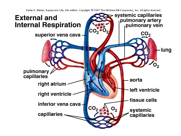
Path of Blood Through the Heart: ● blood low in oxygen and high in CO 2 enters the RIGHT SIDE through the venae cavae and is then pumped into pulmonary circulation (to the lungs)
Path of Blood Through the Heart: ● after blood is oxygenated in the lungs, it returns to the LEFT SIDE through the pulmonary veins and is then pumped into systemic circulation via the aorta (to the body)
Heart Blood Supply: ● the CORONARY ARTERIES supply blood to the myocardium ● blood returns to the R atrium through the cardiac veins and coronary sinus
**500, 000 Americans die per year of coronary artery disease **3. 5 million Americans have coronary problems
Cardiac Cycle ● ATRIAL SYSTOLE: atria contract while the ventricles relax (VENTRICULAR DIASTOLE) ● VENTRICULAR SYSTOLE: ventricles contract while the atria relax (ATRIAL DIASTOLE) ● all chambers relax for a brief period…; then the cycle repeats!
Diastole Systole
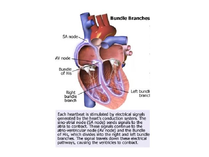
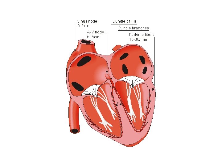
Initiation of Cardiac Cycle: ● electrical impulses originate in the SA node: stimulate the atria to contract ● impulses pass slowly to the AV node, then to the Purkinje fibers: stimulate the ventricles to contract
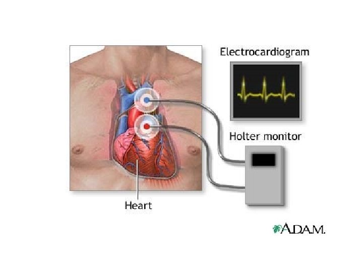
ELECTROCARDIOGRAM (ECG): ● records the electrical changes in the myocardium during a cardiac cycle ● the pattern has several characteristic waves: 1) P wave: atrial depolarization 2) QRS complex: ventricular depolarization 3) T wave: ventricular repolarization
Regulation of Cardiac Cycle: ● heartbeat is affected by: -physical exercise -body temperature -concentration of ions (calcium, potassium)
Blood Vessels and Blood Pressure
Blood vessels form a closed circuit of tubes that carry blood from the heart to body cells and back again.
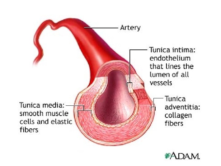
ARTERIES and ARTERIOLES ● Arteries are adapted to carry blood under HIGH PRESSURE away from the heart. ● Arteries eventually branch into smaller structures called ARTERIOLES
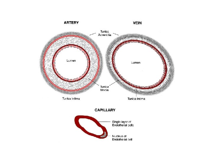
CAPILLARIES ● smallest diameter blood vessel ● connect arterioles to venules ● capillary wall = a single layer of cells; forms a semipermeable membrane
RBCs in a capillary!
CAPILLARIES ● tissues that use more energy/oxygen have the most capillaries (i. e. muscle tissue, nerve tissue) ● tissues that use less energy/oxygen have the fewest capillaries (i. e. epidermis, cartilage)
Exchanges in Capillaries: ● blood in capillaries drops off their nutrients and oxygen in exchange for metabolic wastes (CO 2, etc. ) ● large molecules (e. g. plasma proteins) remain in the blood ● most materials move across the capillary wall by DIFFUSION
VEINS and VENULES ● VENULES continue from capillaries and merge with VEINS ● veins carry blood TOWARD THE HEART ● venous walls are similar in structure to artery walls, but thinner and contain less muscle tissue
VEINS -carry blood to the heart -thinner walls -are less muscular than arteries ARTERIES -carry blood away from heart -largest artery = AORTA ARTERIOLES VENULES CAPILLARIES -walls are only 1 cell thick
BLOOD PRESSURE: Blood pressure is the force blood exerts against the sides of blood vessels.
BLOOD PRESSURE: ● SYSTOLIC PRESSURE: pressure produced in the arteries when the ventricles contract; highest pressure ● DIASTOLIC PRESSURE: pressure in the arteries when the ventricles are relaxed
Venous blood pressure is much lower than arterial blood pressure…how does blood flow through veins (in some cases, against gravity? ) skeletal muscle contraction squeezes blood from one segment to the next breathing movements change pressure in thoracic and abdominal cavities pulling/pushing blood upward toward the heart VALVES prevent blood from flowing backward into a previous segment
2 MAJOR PATHS OF CIRCULATION: ● PULMONARY CIRCUIT: vessels that carry blood from the heart to the lungs and back R ventricle pulmonary trunk R and L pulmonary arteries arterioles and capillaries in the R and L lungs venules veins pulmonary veins L atrium
2 MAJOR PATHS OF CIRCULATION: ● SYSTEMIC CIRCUIT: carries blood from the heart to all other body parts and back again; includes coronary circulation L atrium L ventricle aorta various arteries, arterioles, capillaries in body tissues venules veins superior and inferior venae cavae R atrium
BLOOD AND BLOOD CELLS Blood can be separated into: • Formed elements: -mostly red blood cells (RBCs) -include white blood cells (WBCs) and platelets • Liquid portions = PLASMA -transports water, gases, nutrients, hormones, electrolytes, and cellular wastes
Red Blood Cells (RBCs) • contain HEMOGLOBIN, which combines with oxygen • hemoglobin synthesis requires IRON
White Blood Cells (WBCs) • function in defense against disease • also called: LEUKOCYTES • WBCs include: neutrophils eosinophils basophils monocytes lymphocytes
White Blood Cells (WBCs) • normal WBC count is 5, 000 – 10, 000 cells per mm 3 of blood this number may vary in response to infection, emotional disturbances, loss of body fluids, leukemia
Platelets • fragments of larger cells • help close breaks in blood vessels / clot the blood
PLASMA Plasma transports gases and nutrients, helps regulate fluid and electrolyte balance, and helps maintain a stable p. H.
PLASMA Gases in plasma: -oxygen -carbon dioxide -nitrogen Plasma nutrients: -simple sugars -amino acids -lipids
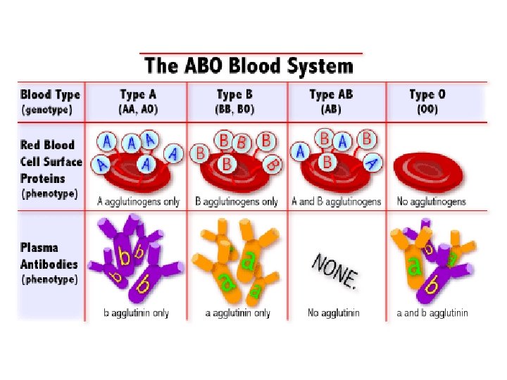
ABO Blood Group: • blood is grouped according to the presence or absence of antigens A and B. Type A = A antigens; plasma has anti-B antibodies Type B = B antigens; plasma has anti-A antibodies Type AB = A and B antigens; no antibodies Type O = neither antigen; both anti-A and B antibodies
ABO Blood Group: • mixing RBCs that contain an antigen with plasma that contains the corresponding antibody results in a negative reaction (AGGLUTINATION) *the anti-A and anti-B antibodies are too large to cross the placenta, so mother and child can safely have different ABO blood groups
Let’s play the game: “Are You My Blood Type? ”
The Blood Typing Game!
Rh Blood Group: • Rh-positive blood: RBCs possess the Rh antigens • Rh-negative blood: RBCs do not possess the Rh antigens, but DO possess anti-Rh antibodies RESULT: mixing Rh-positive RBCs with plasma that has the anti-Rh antibodies can result in agglutination
Rh Blood Group: *the anti-Rh antibodies are small enough to cross the placenta… So, the anti-Rh antibodies in a mother’s blood could react with the RBCs of an Rhpositive fetus
The “Rh Issue”… Mom = Rh-; Baby #1 = Rh+
A note about blood pressure…
How do we measure B. P. ? ● To measure arterial blood pressure, we use a device known as a sphygmomanometer … …GUESS WHAT…? ?
SPHYGMOMANOMETER! ***word #5 on my list!!!. . . the list is complete! 1) Okazaki fragments 2) plasmodesmata 3) cholecystokinin 4) acetylcholinesterase 5) sphygmomanometer 6) rubisco 7) oxaloacetate 8) Islets of Langerhans 9) Batesian mimicry 10) nodes of Ranvier
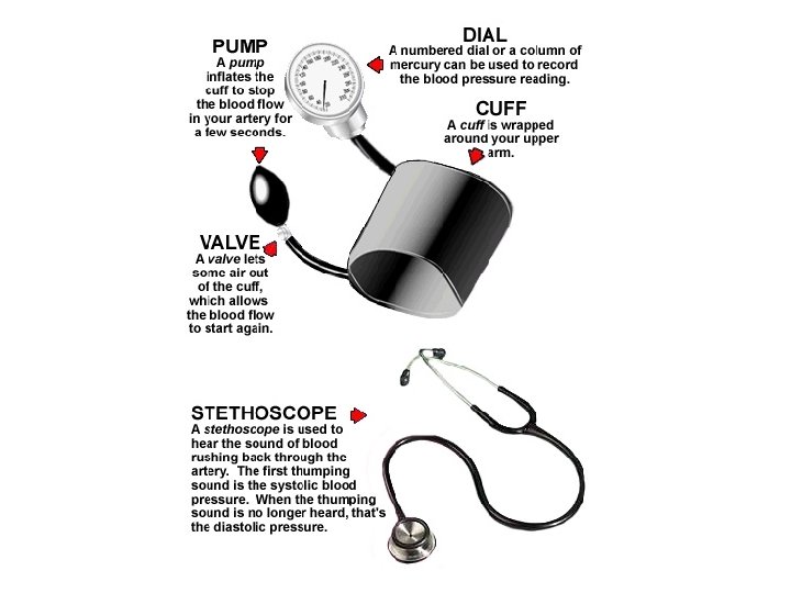
SPHYGMOMANOMETER: ● This device consists of an inflatable cuff connected by rubber hoses to a hand pump and to a pressure gauge. ● The cuff is wrapped around the upper arm and inflated to a pressure greater than the expected systolic pressure, thus closing down the brachial artery in the arm.
SPHYGMOMANOMETER: ● The examiner places the bell of a stethoscope in the inside of the elbow below the biceps muscle to pick up the sounds of the blood in the artery as the pressure in the cuff is allowed to fall. (This is done gradually by opening a screw valve next to the hand pump. )
SPHYGMOMANOMETER: ● At first there is no sound; then, a sharp, tapping sound of blood spurting through the partiallyopened artery at systolic pressure; ● and ultimately, no sound as even the lowpressure blood (@ diastolic pressure) can get through the completely open artery. **(The various sounds heard with the stethoscope are known as the sounds of Korotkoff. )
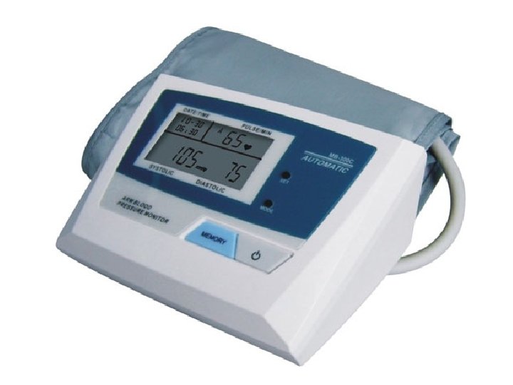
BLOOD PRESSURE: ● Blood pressure is recorded as: systolic pressure (mm Hg) / diastolic pressure (mm Hg). ● A normal blood pressure measurement for a given individual depends on the person’s age, sex, heredity, emotional state, body weight, etc. ● For young men and women at age 17 -18, the normal BP’s will range from 100 -120 mm Hg systolic pressure, and from 60 -80 mm Hg diastolic pressure.
“Is my blood pressure too high? ” ● One “high” BP reading may not indicate a health problem; ● BP needs to be measured over several days at different times of the day by a health care professional before a true measure is achieved. ● Blood pressure measurements that are chronically elevated may indicate poor cardiovascular health. ● This condition, called hypertension, is a major contributor to heart disease and stroke.