Normal and pathological development of the human skeleton


")


- Slides: 51
Normal and pathological development of the human skeleton before 21 weeks GA 2015 IPPA Update Course Australia, 22 nd-27 th November 2015 Inger Kjær Professor, Dr Odont et Dr Med Faculty of Health Sciences Institute of Odontology University of Copenhagen, Denmark
Spranger JW et al. 1982
Normal development • extremities • body axis, vertebral column • cranium Questions to ask: -Where does ossification begin? -Sequence: In what order do the bones of the upper extremities ossify? -Morphology: What is the initial appearance of the osseous primordia? -Developmental courses: How is the osseous morphology changed along the way?
GA 8 ½ W Normal development CRL 31 mm, GA 9 W
CRL 30 mm, GA 9 W Normal development CRL 40 mm, GA 10 W
Normal development CRL 55 mm, GA 11½ W CRL 87 mm, GA 14 W
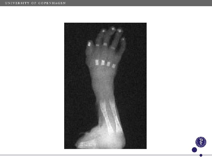
Normal development sequence, morphology and developmental courses The hand GA 16 weeks
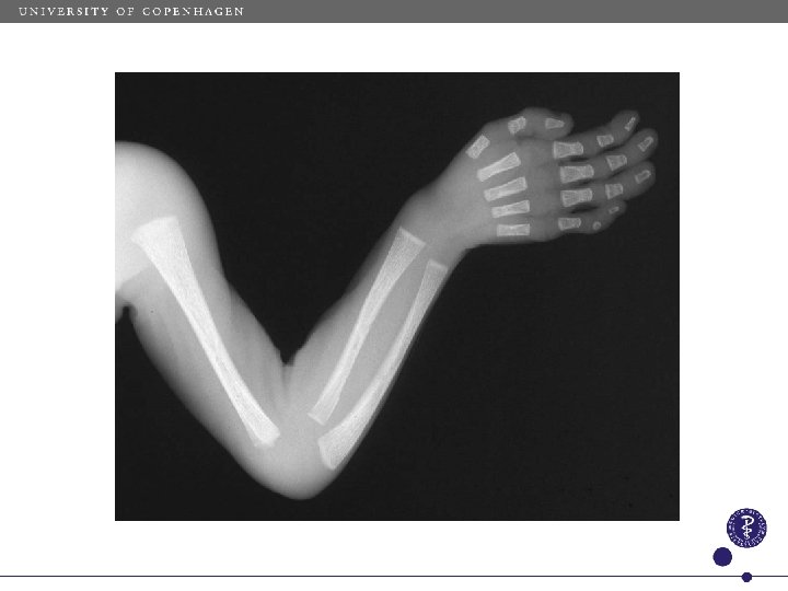
Normal development
CRL 31 mm, GA 9 W Normal development of • • • extremities body axis, vertebral column cranium Lower extremity
Normal development
Normal development
Normal development
Normal development Clavicula
GA 19 W Costae and columna vertebralis Normal development GA 11½ W 94 -02 GA 12½ W 1803 -88 HH 01 -94
Normal development Pattern of ossification
Normal development Os nasale
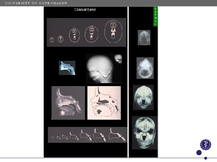
Normal development • • • extremities body axis, vertebral column cranium Cranium
Normal development Le Douarin, 1974 Larsen WJ. Human Embryology, 2001
Normal developmental fields
CRL=67 mm CRL=119 mm CRL=151 mm
Basilar part of occipital bone
Pars basilaris os occipitale
Spranger JW et al. 1982
Spontaneous abortions?
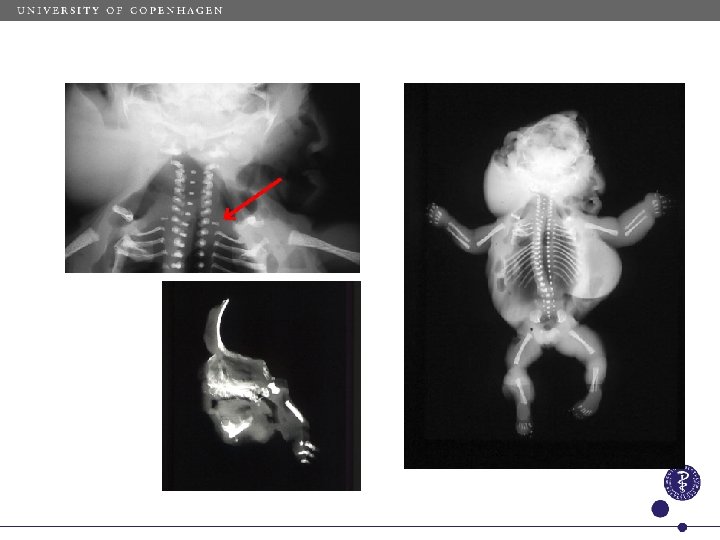
Hand malformations Meckel-Grubers syndrom, GA 23 W
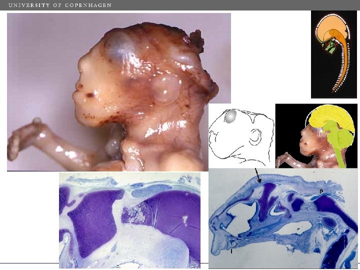
Holoprosencephaly Cleft lip
Holoprosencephaly face
Holoprosencephaly brain Jean Keeling
Holoprosencephali Holoprosencephaly skeleton
Holoprosencephaly cranium
Crouzon syndrome, 10 q 26
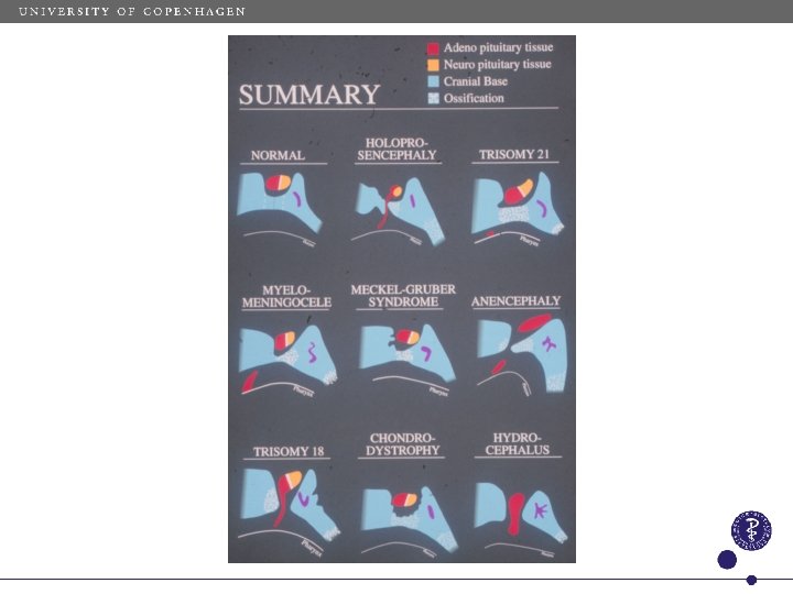
Body axes in different genotypes (pink: Always malformed)
Basial part of occipital bone in different malformations
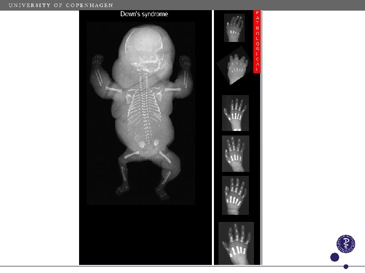
Trisomy 21
dfgjjklfgdyttrtyrybb uuuuiuyuuyuuuuuuuuuuuu
Turner syndrome Turner Syndrome
Turner syndrome
Turner syndrome hand
Anencephaly
Kjær et al, 1994
Fetal amniotic band sequence Keeling and Kjær, 1994
Keeling and Kjær, 1994
Thanks to fetal pathologists: Jean Keeling, UK Birgit Fischer Hansen, DK Powerpoint: Eva Marie Reinwald