
Nonvascular Interventions O Melih Topcuoglu M D Yeditepe



- Slides: 54
Nonvascular Interventions O. Melih Topcuoglu M. D. Yeditepe University Medical School
Nonvascular interventions • Image guided biopsies and drainage • Complex interventional radiological treatment of hepatic tumors • RF ablation in other organs (lung, renal and bone tumors) • Percutaneous (biliary) choledochal, cholecystic interventions (PTC, PTD, stent implant, choledochal stone removal, cholecystostomy) • Gastrointestinal interventions, endoluminal stent implantations • Percutaneous ethanol cyst treatments • Urinary tract interventions • Percutaneous interventional methods of the muscoluskeletal system
Image guided biopsies and drainage • Types of biopsies according to needle diameter – Fine Needle Aspiration Biopsy (FNAB) – Core biopsy
Image guided biopsies and drainage • Types of biopsies according to image guidance – US guiding – X-ray guiding – CT guiding – MR guiding – Hybrid imaging guiding
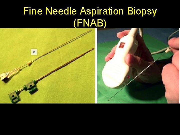
Fine Needle Aspiration Biopsy (FNAB) • With 20 G or thinner needles one can obtain cytological samples, thus smaller groups of cells can be aspired from a certain area
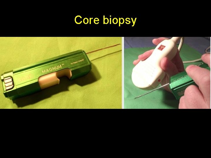
Core biopsy • Most often we use 14 -18 G needles for these tissue samplings • With the help of an automatic biopsy gun one (or more) tissue columns are acquired of the desired area
Types of biopsies according to image guidance
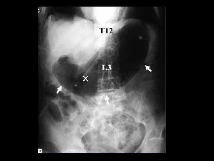
X-ray guiding
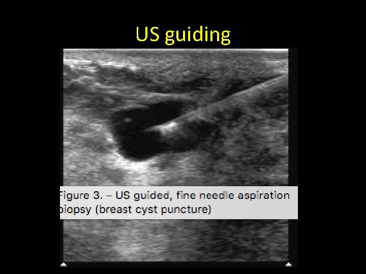
US guiding
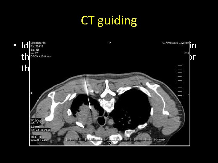
CT guiding • Ideal method if the lesion is located either in the chest, mediastinum, retroperitoneum or the pelvis.
MR guiding
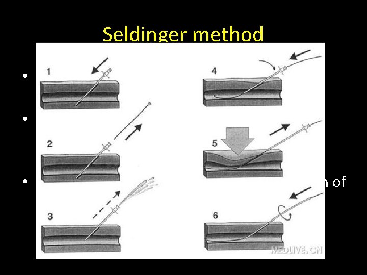
Seldinger method • US or CT guided puncture of the lesion is performed with a correct needle size • A guide wire of 0, 035” diameter is placed through the needle to the lesion, and the needle is removed • The guide wire is used to assist the insertion of a carefully selected, correct sized (6 -14 F) drainage catheter
Percutaneous drainage
Contraindications of biopsies and drainage • If no proper blood clotting parameters are met (it can be temporarily corrected for the time of the puncture with fresh frozen plasma (FFP) • If there is an unavoidable blood vessel (aorta, IVC) in the way to the lesion,
Complications of biopsy and drainage • Hemorrhage (subcapsular parenchymal, intraabdominal, intrathoracic, pseudoaneurysm) • Pneumothorax (thoracic, mediastinal, infraclavicular, in case of subdiaphragmatic intervention) • Perforation (it is FORBIDDEN to use core biopsy needle for lesions lying behind intestines) • Tumor spread in the cutaneous biopsy canal (the same risk factor applies in case of all guiding modalities)
Complex interventional radiological treatment of hepatic tumors • • • Percutaneous Ethanol Injection Radiofrequency ablation (RFA) Microwave tumor ablation Laser tumor ablation Cryoablation Chemoembolization
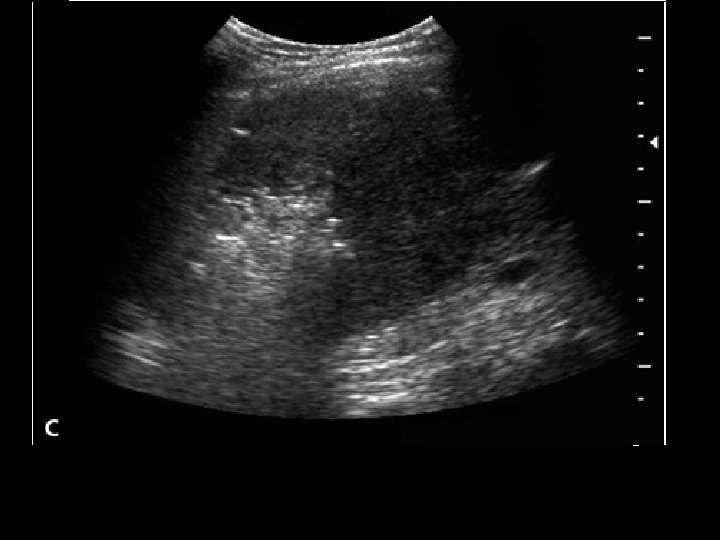
Percutaneous Ethanol Injection • It is the most commonly used and cheapest percutaneous method for the treatment of primary hepatic cancer (HCC) • Sterile, 95%, absolute alcohol is injected with US guidance to the tumor • Ethanol causes dehydration and coagulation necrosis of the tumor cells, followed by fibrotic degeneration. After alcohol injection, a typical “snow storm like”, hyperechogenic area can be seen in the treated area
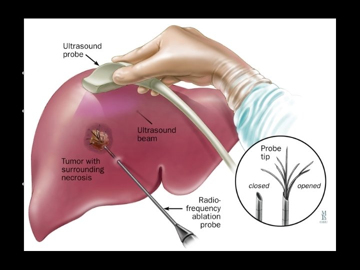
Radiofrequency ablation (RFA) • During RF ablation an electrode is positioned in the tumor. • The end of the electrode will produce extensive heat as ionizing current is generated at 460 k. Hz frequency, with alternating polarities • In a given (50 -200 Watt) energy range it is possible to produce 50 - 90 C degree heat under set circumstances
Radiofrequency ablation (RFA) • With the use of a special RF equipment and electrode (Berthold or Radionics equipment) the internal cooling of the needle is possible with physiologic saline solution, therefore carbonization is avoidable • The complete RF ablation in the vicinity of large venous branches (hepatic vein, portal vein, IVC) is difficult to achieve, since the flowing blood of the veins cools the nearby tumor tissues
Radiofrequency ablation (RFA) • Another difficulty is presented with lesions lying too close to the choledoch or the hepatic duct, since they poise the possibility of a serious biliary injury • The treatment of subcapsular tumors can lead to persisting pain, therefore in these cases combined methods are usually preferred (RF + chemoembolization, RF + PEI)
Radiofrequency ablation (RFA) • The most ideal scenario for RF ablation of liver tumors are the following: • 1. There are 4 or less lesions, • 2. lesions are equal to or less than 3 cm • 3. they are each located at least 1 cm below the hepatic capsule and • 4. any larger vein is located at minimum 2 cm of a distance from them
Radiofrequency ablation (RFA) • Therapeutic success can only be hoped to achieve with a tumor of maximum 5 cm diameter, however at this size multiple interventions are needed • With the help of hybrid guiding methods, larger lesions might be successfully treated in one session • Superficial lesions might be intraoperatively performed to avoid damage to the surrounding organs (diaphragm, gall bladder, large and small bowel)
Radiofrequency ablation (RFA) • Percutaneous RF ablation should only be performed with strong analgesia or in anesthesia. After treatment a 24 h clinical observation is necessary • The average time for the ablation of a lesion of 3 cm is 8 -10 minutes long. In case of 3 -4 lesions the procedure can last up to 40 -50 minutes • The effectiveness of the RF ablation is usually controlled by DWI
Microwave tumor ablation • Microwaves at a wavelength at 2450 MHzcreate a very fast rotation in the water molecules of the targeted lesion • This leads to the heating up of the tissues and the coagulation necrosis in a volume with elliptic crossection
Laser tumor ablation • After the development of laser ablation technique, it became possible to create reproducible tissue destruction with Nd YAG (Neodymium yttrium aluminium garnet) laser
Cryoablation • Cold under -20, -30 C degrees produces irreversible tissue destruction • The criteria for the treatment are basically the same as for RF ablation
Chemoembolization • Next lecture
RF ablation in other organs (lung, renal and bone tumors) • Lung – For inoperable pulmonary tumors it is well accessible, peripherally localized tumors that otherwise would carry significant surgical risk can be successfully treated with this alternative method – In the vicinity of larger blood vessels (branches of the pulmonary artery, SVC) the cooling effect of the flowing blood decreases the effectiveness of the RF procedure
RF ablation in other organs (lung, renal and bone tumors) • Kidney – 1. Elderly patient, with relatively large surgical risks – 2. Unilateral, solitaire RCC – 3. Palliative treatment in case of a centrally located tumor – 4. Patient denies surgery – 5. According to the latest recommendations tumors up to 5 cm in diameter can be treated successfully with RF
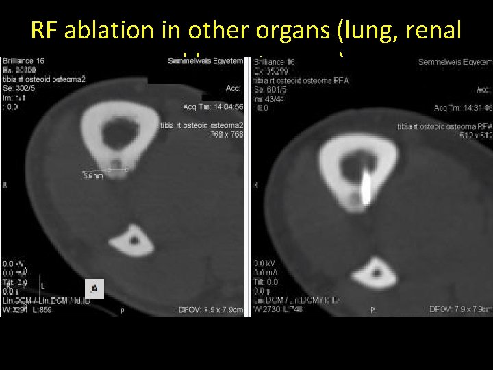
RF ablation in other organs (lung, renal and bone tumors) • Bone – The RF treatment of primary bone tumors and bone metastases have been the topic of several large scale studies from various centers – RF ablation is especially useful technique and has a good outcome in the direct treatment of osteiod osteomas – The invasiveness of the method is several folds smaller than orthopedic surgery
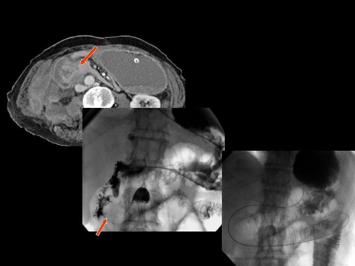
Percutaneous (biliary) choledochal, cholecystic interventions • Percutaneous interventions are usually necessary in case of malignant, inoperable pancreatic head tumors, if enlarged lymph nodes in the hepatic hilum compress the choledoch, or in case of the various types of cholangiocellular tumors (Klatskin tumor) • Among the benign lesions, primarily biliary strictures, inflammatory stenoses, and sclerotizing cholangitis cause indication for intervention
Percutaneous (biliary) choledochal, cholecystic interventions • In case of the failure of endoscopic biliary interventions or if there is a persisting Billroth II resection, percutaneous biliary intervention needs to be considered • It is important to note that due to the congestion of bile – which might serve as fertile ground for bacteria – one needs to provide prophylactic antibiotic therapy before the intervention.
Percutaneous transhepatic cholangiogrpahy (PTC) • During this procedure X-ray or US is used to guide the insertion of a 22 G (Chiba needle) from a right IX. or X. intercostals position into a dilated intrahepatic biliary branch • After the Chiba needle is well positioned the intra and extrahepatic biliary tree is filled with contrast material • Under special circumstances – when the left side of the biliary branches is affected primarily - PTC can be performed from an epigastric entry towards the left lobe of the liver
Percutaneous transhepatic drainage (PTD) • After a diagnostic PTC is successfully performed and a guide wire is positioned over the stenotic or occluded segment of the choledochal duct an external-internal drain (PTD) can be installed • If desired, a self expanding metallic stent could be used to override the stricture
Percutaneous transhepatic drainage (PTD) • If one cannot pass the biliary stenosis in the initial attempts, the placement of a temporary external drainage is advisory to control the biliary congestion and cholangitis • Later during a second session the insertion of an externalinternal drain will be possible • In special cases separate catheter insertion and stenting of both the left and right hepatic ducts might be necessary. This depends on the extent of the pathologic lesion (tumor or inflammation) • In case of a malignant stenosis the use of covered stents can prolong the passage in the tumor bound segment of the choledoch
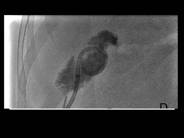
Percutaneous cholecystostomy • Usually in case of elderly patients and in case of certain types of acalculous cholecytitis US guided precutaneous drainage of the gall bladder proves to be a very effective technique
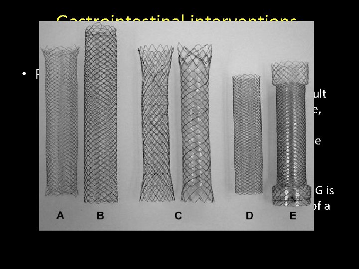
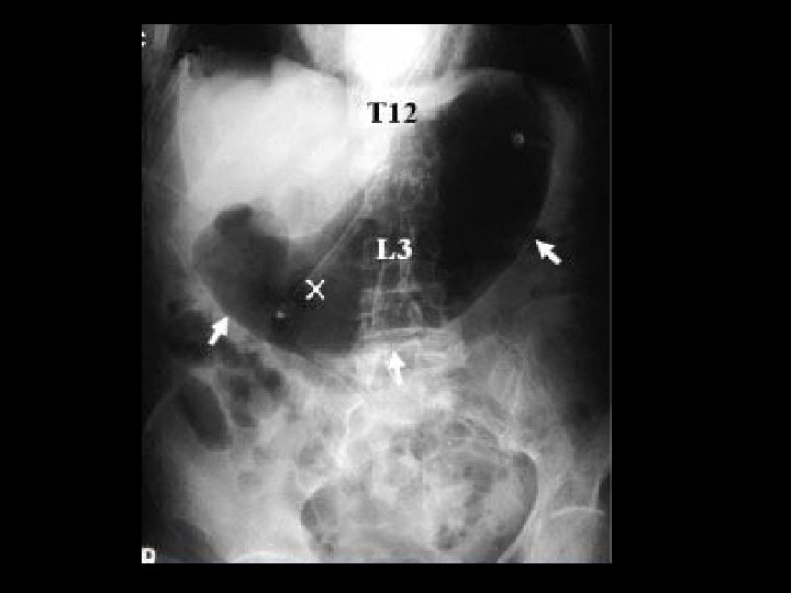
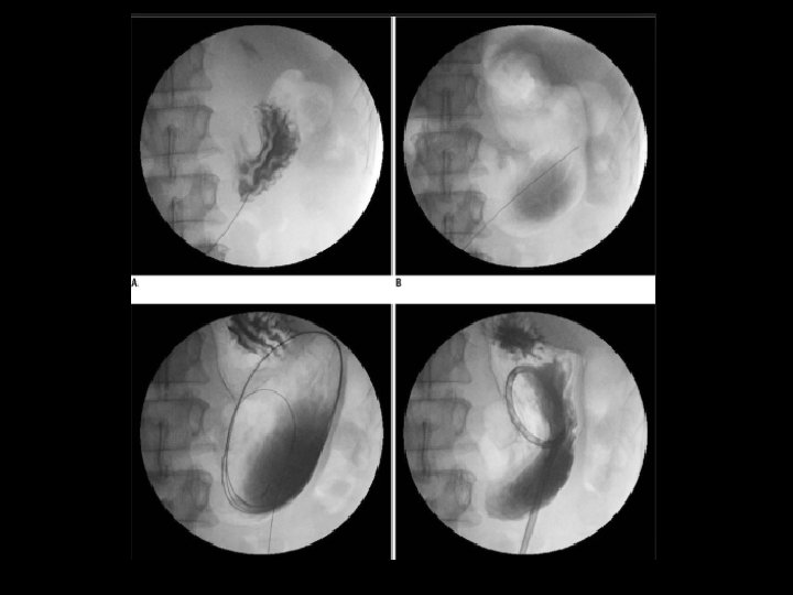
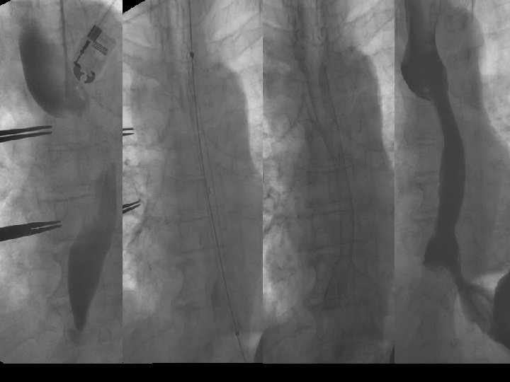
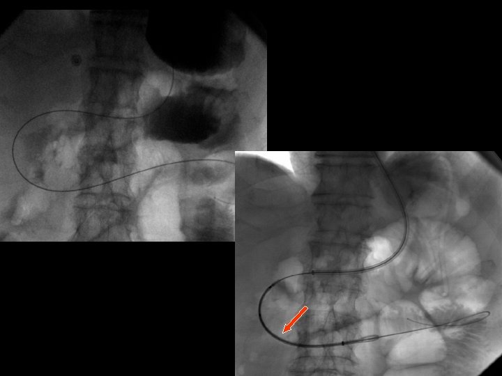
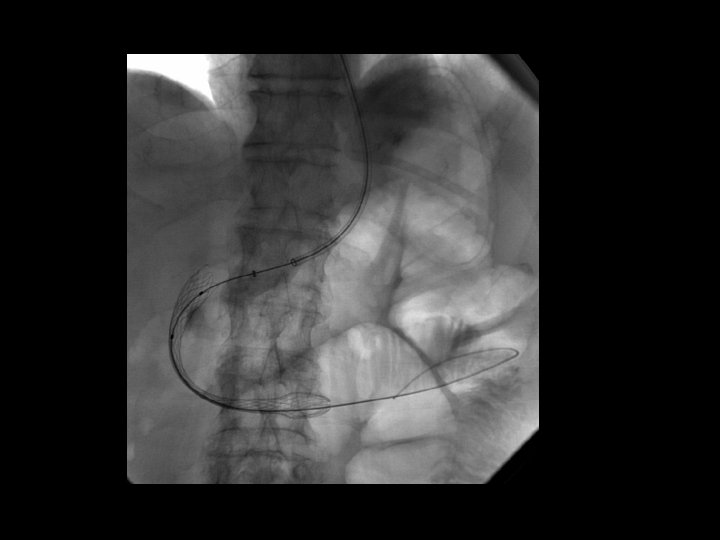
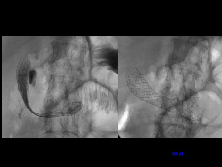
Gastrointestinal interventions, endoluminal stent implantations • GI stenting – In case of inoperable esophageal, gastric or duodenal cancer, large (15 -20 mm wide) endoluminal stents can be applied. – Moreover obstructive tumors on the descending colon or on the sigma can also be treated with these endoluminal stents. – The length of the stent has to be chosen so that its proximal and distal ends reach beyond the stenosis by about 2 -3 cm.
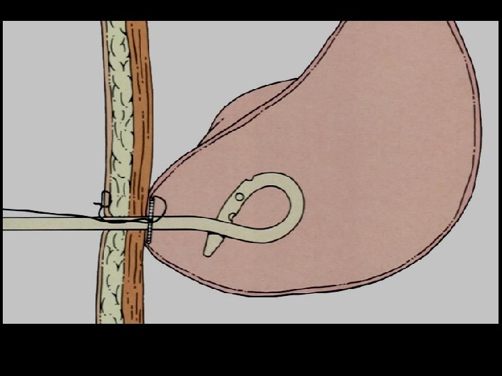
Gastrointestinal interventions, endoluminal stent implantations • Percutaneous gastrostomy – In case of swallowing impairment (usually as a result of neurological cause: severe stroke, brain damage, amyotrophic lateral sclerosis) percutaneous gastrostomy is used to temporality decompress the esophagus segment, to alleviate gastric emptying disorder or to cease intestinal obstruction. – Usually with mild sedation and local anesthesia PEG is inserted with endoscopic assistance with the use of a special catheter set.
24 st
Percutaneous ethanol cyst treatments • During US and CT examinations simplex cystic lesions are often accidentally encountered that do not cause any pain for the patient. • Cysts of the liver, spleen or the kidneys cause discomfort or pain for the patients need to be therapeutically addressed. • Compared to the surgical methods of past, nowadays these cystic lesions can be successfully treated percutaneously with alcoholic cyst sclerotization
Percutaneous ethanol cyst treatments • The injected ethanol is left in the cyst for 20 minutes. • Following this, the alcohol is drained as well. Cystic regression is then regularly controlled with US or CT examinations
Percutaneous ethanol cyst treatments • Percutaneous interventional treatment of Echinococcus cyst – The treatment of larger Echinococcus cysts in the liver is possible with percutaneous interventional methods. – The method is described best by its acronym PAIR (Puncture, Aspiration, Injection, Reaspiration). – Hyperosmogenic saline solution (15%) and/or absolute alcohol can be both successfully applied to treat the Echinnococus cysts. Therapeutic efficacy can also be controlled with US or CT exams.
Urinary tract interventions • The most commonly performed intervention is US guided percutaneous nephrostomy. • In special cases (obesity, visualization difficulties with US) CT guidance can also be chosen to create percutaneous nephrostomy • Ureteral stenting • Cystostomy
Percutaneous interventional methods of the muscoluskeletal system • Vertebroplasty – The goal is to stabilize the vertebral column and to prevent further destruction or collapse. Vertebroplasty is a CT guided procedure (CTfluoroscopy method) where a special, 10 G thick needle is applied. It is inserted transpedicularly in case of the lumbar vertebra, while in case of thoracic vertebrae insertion is through the intercostal space. – During the procedure the injection of bone cement is continuously controlled with CT-fluoroscopy and a final control CT examintation is performed to stage the status of the patient.