
NONINVASIVE VENTILATION Lijun Ding 23 Jan 2018 Learning

- Slides: 18
NON-INVASIVE VENTILATION Lijun Ding 23 Jan 2018
Learning objectives • What is NIV • The difference between CPAP and Bi. PAP • The indication of the use of NIV • Complication of NIV application • Patient monitoring and safety
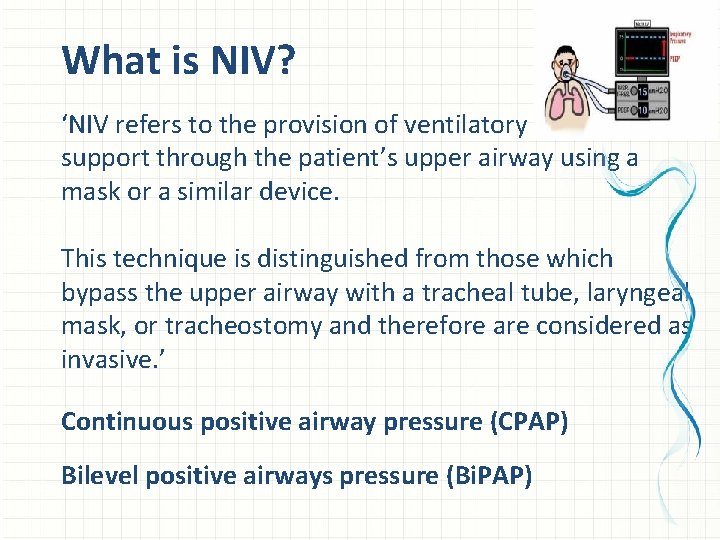
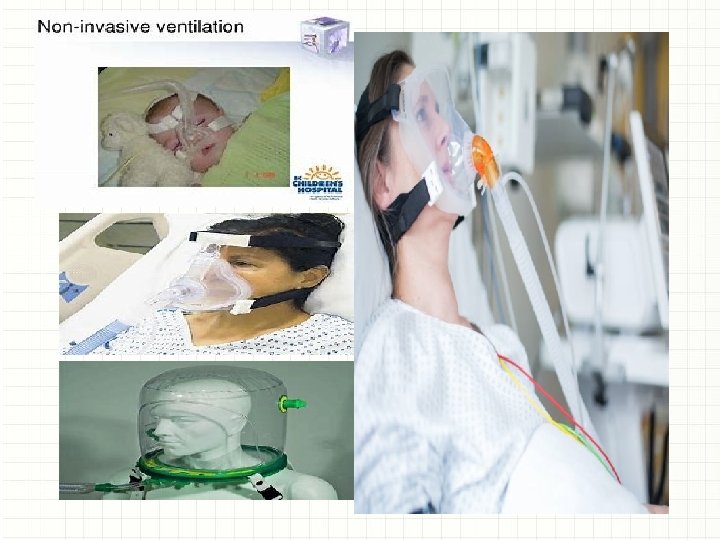
What is NIV? ‘NIV refers to the provision of ventilatory support through the patient’s upper airway using a mask or a similar device. This technique is distinguished from those which bypass the upper airway with a tracheal tube, laryngeal mask, or tracheostomy and therefore are considered as invasive. ’ Continuous positive airway pressure (CPAP) Bilevel positive airways pressure (Bi. PAP)
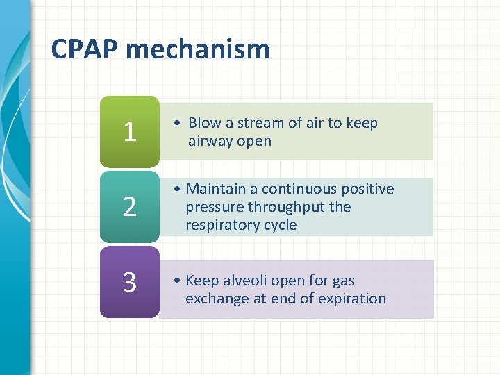
CPAP mechanism 1 • Blow a stream of air to keep airway open 2 • Maintain a continuous positive pressure throughput the respiratory cycle 3 • Keep alveoli open for gas exchange at end of expiration
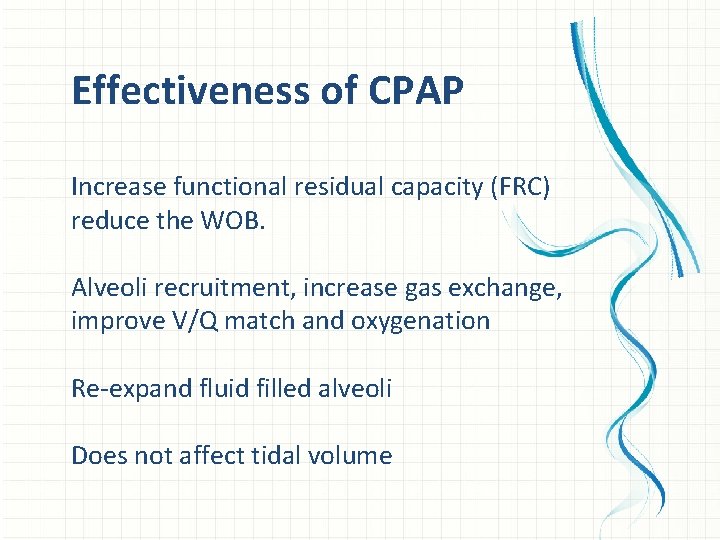
Effectiveness of CPAP Increase functional residual capacity (FRC) reduce the WOB. Alveoli recruitment, increase gas exchange, improve V/Q match and oxygenation Re-expand fluid filled alveoli Does not affect tidal volume
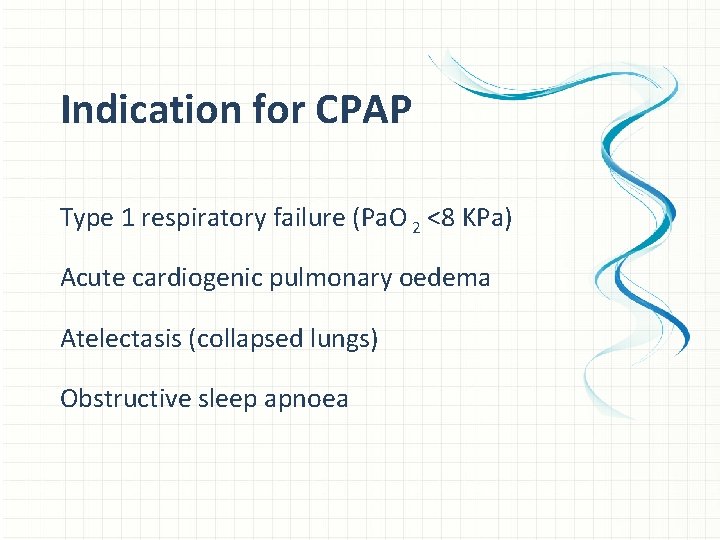
Indication for CPAP Type 1 respiratory failure (Pa. O 2 <8 KPa) Acute cardiogenic pulmonary oedema Atelectasis (collapsed lungs) Obstructive sleep apnoea
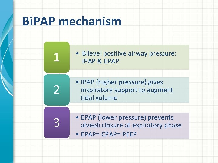
Bi. PAP mechanism 1 • Bilevel positive airway pressure: IPAP & EPAP 2 • IPAP (higher pressure) gives inspiratory support to augment tidal volume 3 • EPAP (lower pressure) prevents alveoli closure at expiratory phase • EPAP= CPAP= PEEP
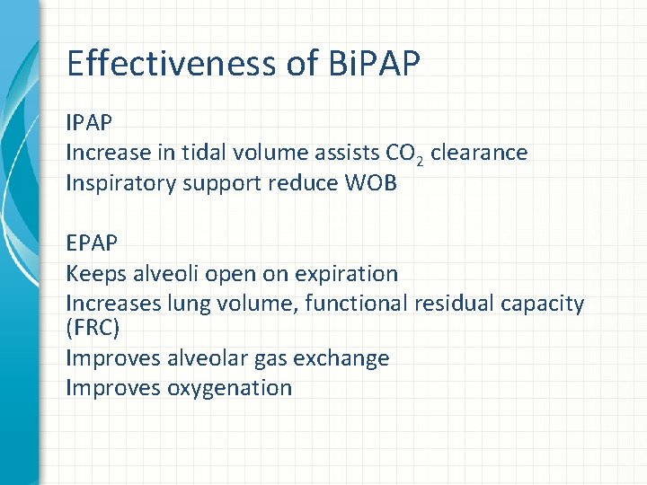
Effectiveness of Bi. PAP Increase in tidal volume assists CO 2 clearance Inspiratory support reduce WOB EPAP Keeps alveoli open on expiration Increases lung volume, functional residual capacity (FRC) Improves alveolar gas exchange Improves oxygenation
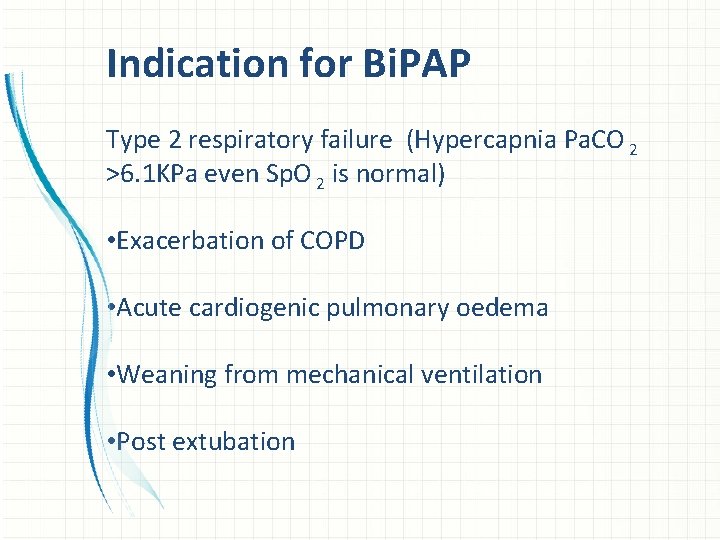
Indication for Bi. PAP Type 2 respiratory failure (Hypercapnia Pa. CO 2 >6. 1 KPa even Sp. O 2 is normal) • Exacerbation of COPD • Acute cardiogenic pulmonary oedema • Weaning from mechanical ventilation • Post extubation
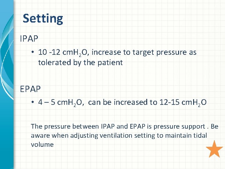
Setting IPAP • 10 -12 cm. H 2 O, increase to target pressure as tolerated by the patient EPAP • 4 – 5 cm. H 2 O, can be increased to 12 -15 cm. H 2 O The pressure between IPAP and EPAP is pressure support. Be aware when adjusting ventilation setting to maintain tidal volume
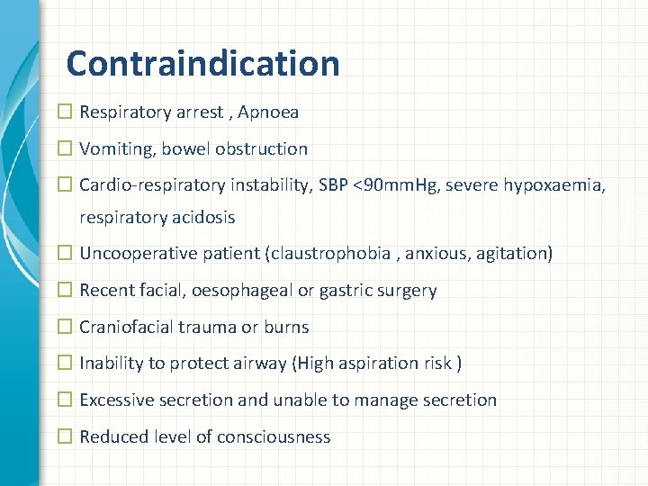
Contraindication � Respiratory arrest , Apnoea � Vomiting, bowel obstruction � Cardio-respiratory instability, SBP <90 mm. Hg, severe hypoxaemia, respiratory acidosis � Uncooperative patient (claustrophobia , anxious, agitation) � Recent facial, oesophageal or gastric surgery � Craniofacial trauma or burns � Inability to protect airway (High aspiration risk ) � Excessive secretion and unable to manage secretion � Reduced level of consciousness
Complications �Pressure sore �Air leak �Dry eyes �Gastro distension, vomiting and aspiration �Hypotension �Increased ICP �Secretion drying / retention �Difficulty in communication �Reduce in oral intake �Anxiety
Patient monitoring • Keep reassessing A and B, repeat ABG 30 -60 minutes after application • Alarms setting • Looking for improvement after 1 -2 hrs (no more than 4 hrs) • Always be prepared for intubation (NIV may fail, need management plan) • Need to recognise sings of failing (escalation when required)
Signs of failure • Unable to tolerate mask or failure of coordination with the ventilator • Development of new symptoms or complications such as pneumothorax, excessive sputum retention, nasal bridge erosion • Failure to improve in arterial blood gas • Respiratory acidosis worsening • Deterioration in patient's condition • CVS instability • reduced mental status
Patient care • Appropriate masks • Avoid high airway pressures • Gastric distension/aspiration • PUP • Sinus pain • Communication issues (psychological support) • Humidification/NEBS • Hydration and nutrition
References • Branson, R. (2013) ‘The Scientific Basis for Postoperative Respiratory Care’, Respiratory Care, 58 (11), pp. 1974 -1984. • British Thoracic Society (2002) ‘Non-invasive ventilation in acute respiratory failure’, Thorax , 57(3) , pp. 192 -211. • British Thoracic Society (2017) ‘BTS guideline for oxygen use in adults in healthcare and emergency settings, Thorax, 72, pp. i 1–i 90. doi: 10. 1136/thoraxjnl-2016 -209729 m. • Burns, K. , et al. (2014) ‘Noninvasive ventilation as a weaning strategy for mechanical ventilation in adults with respiratory failure: a Cochrane systematic review’, Canadian Medical Association Journal, 186(3). pp. E 112 -E 122. doi: 10. 1002/14651858. CD 004127. pub 3. • Chang, D. (2014) Clinical Application of Mechanical Ventilation. 4 th edn. New York: Delmar Health Care. • Esmond, G. and Mikelsons, C. (2009) Non-invasive respiratory support techniques: Oxygen therapy, Noninvasive ventilation and CPAP. Chichester: Wiley-Blackwell. Jaber, S. , Chanques, G. and Jung, B. (2010) ‘Postoperative non-invasive ventilation’, Anaesthesiology, 112 (2), pp. 453 -461. • • Mc. Neill, G. and Glossop, A. (2012) ‘Clinical applications of non-invasive ventilation in critical care’, Continuing Education in Anaesthesia, Critical Care & Pain, 12(1), pp. 33 -37. doi: 10. 1093/bjaceaccp/mkr 047. • Ornico, S. R. , et al. (2013) ‘ ventilation immediately after extubation improves weaning outcome after acute respiratory failure: a randomized controlled trial’, Critical Care, 17: R 39. doi: 10. 1186/cc 12549 • Vital, F. , Ladeira, M. and Atallah, Á. (2013) ‘Non-invasive positive pressure ventilation (CPAP or Bilevel NPPV) for cardiogenic pulmonary oedema (Review)’, Cochrane Database of Systematic Reviews, 5. doi: 10. 1002/14651858. CD 005351. pub 3.
ANY QUESTIONS?