NONEPITHELIALIZED PRIMARY BONE CYSTS The solitary bone cyst

NONEPITHELIALIZED PRIMARY BONE CYSTS

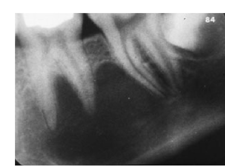
The solitary bone cyst occurs predominantly in children and adolescents with a peak incidence in the second decade. There is no definite sex predilection although some series have shown a slightly higher incidence in males.

presents as a radiolucency of variable size and irregular outline. Scalloping is a prominent feature particularly around and between the roots of standing teeth. The margins of the lesion are usually well defined.

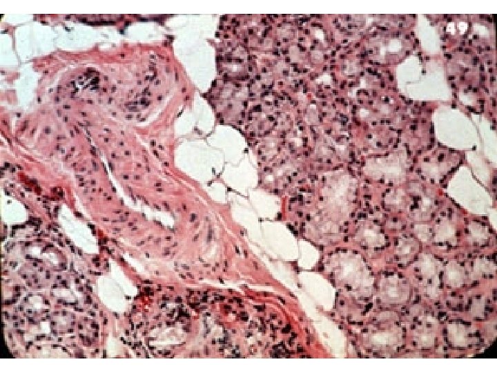
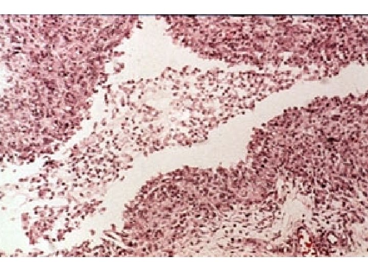
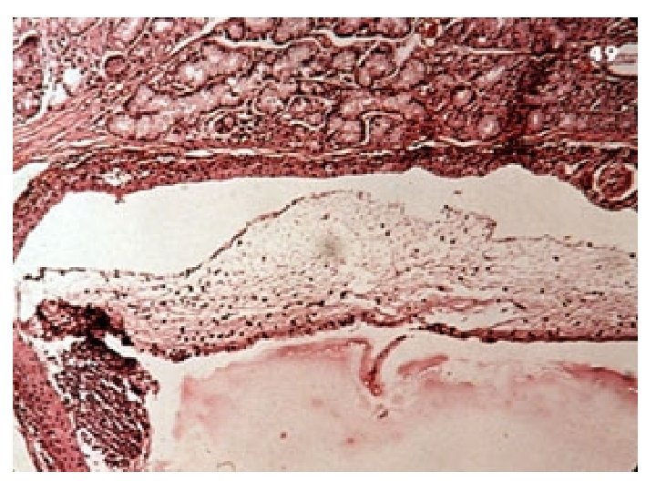
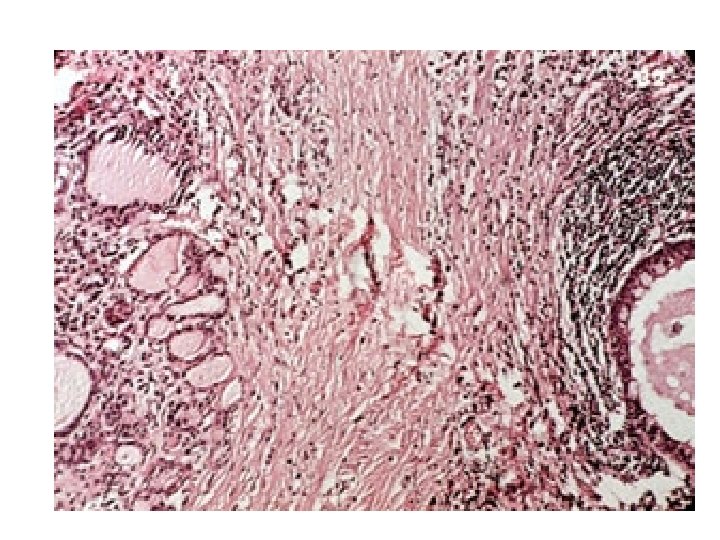
Microscopic examination - shows that the bony walls are covered by a delicate layer of loose, vascular fibrous tissue containing extravasated red blood cells and deposits of haemosiderin pigment.
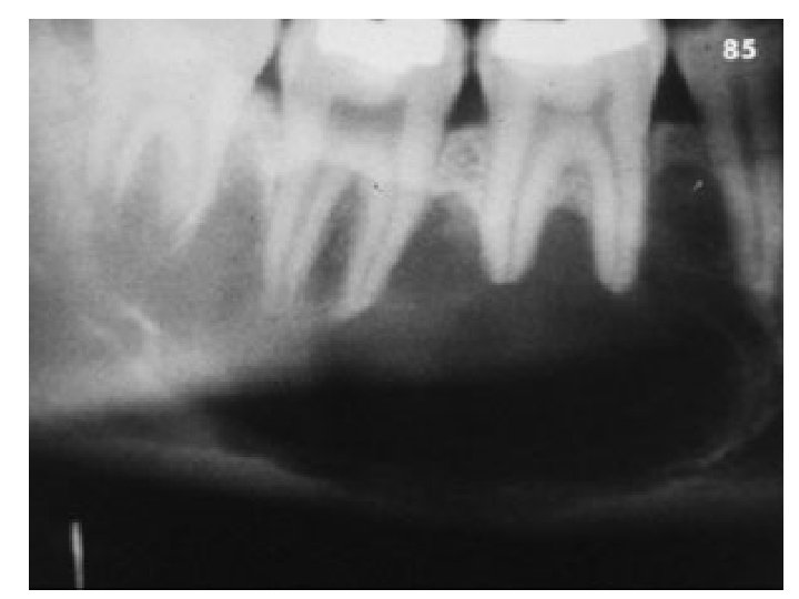

STAFNE’S IDIOPATHIC BONE CAVITY appears as a round or oval, welldemarcated radioluency between the premolar region and angle of the jaw, and is usually located beneath the inferior dental canal.

contains ectopic salivary tissue in continuity with the submandibular salivary gland. Sialography may be useful in identifying such salivary inclusions.

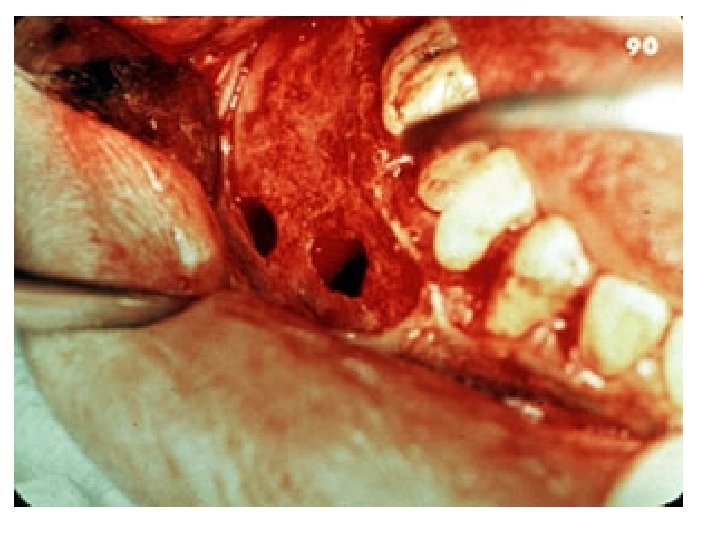
ANEURYSMAL BONE CYST Most of the reported cases have arisen in the mandible, usually the posterior part of the body or angle, and have occurred in children or young adults. It presents as a firm painless swelling.

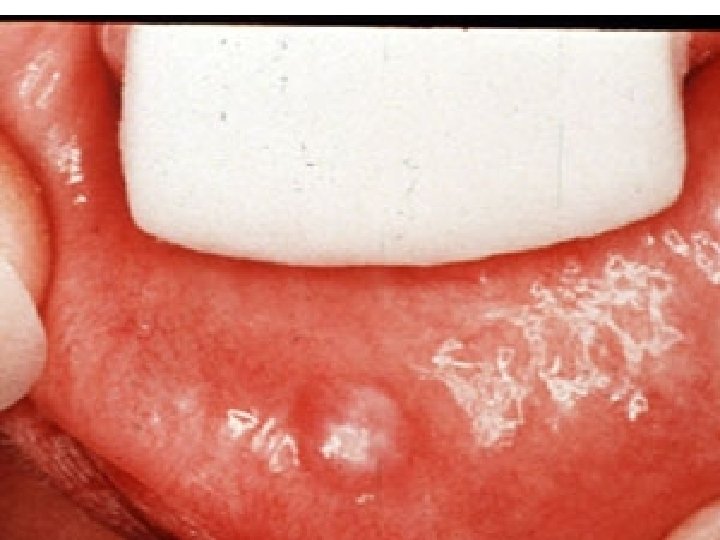
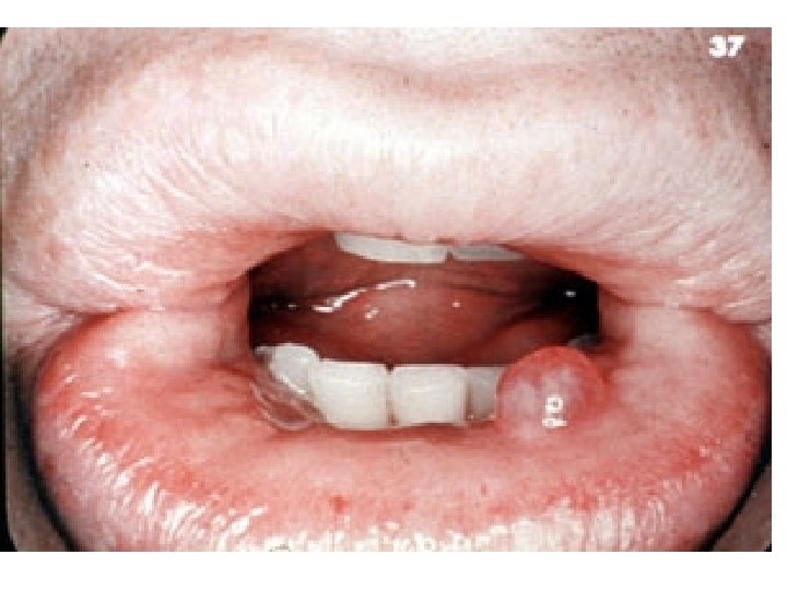
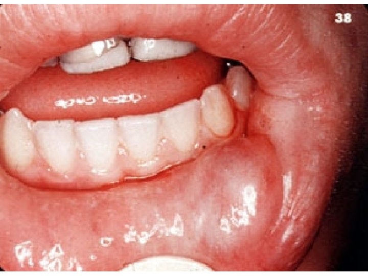
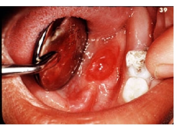
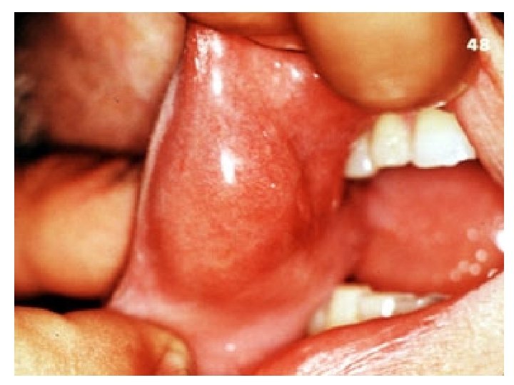
EXTRA VASATION MUCOCELES
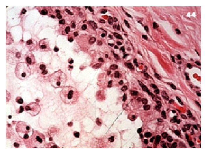

RETENTION MUCOCELES occur most frequently in patients over 50 years of age and are almost never found in the lower lip. They are derived from cystic dilatation of a duct and are lined by epithelium of ductal type.

DERMOID & EPIDERMOID CYST originate in the midline of the floor of the mouth above the mylohyoid muscle. They may present as intraoral or submental swellings
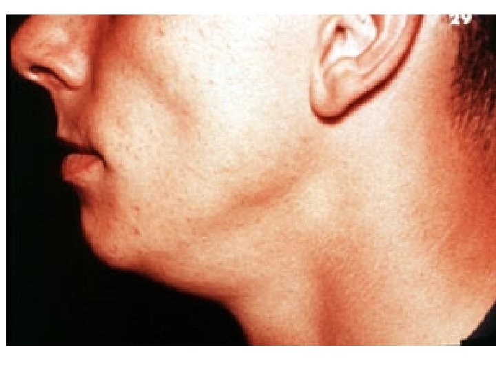

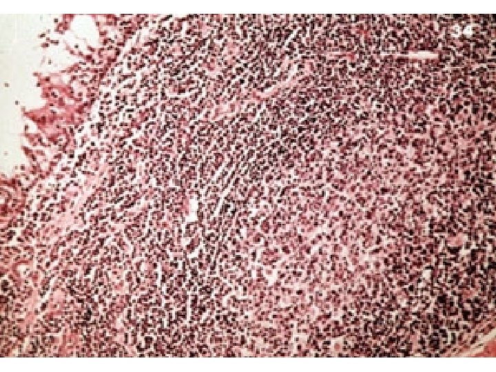
LYMPHOEPITHELIAL CYST The majority occur deep to sternomastoid or along its anterior border at the level of the angle of the mandible. It is an unusual lesion in the oral cavity, generally arising in the floor of the mouth.
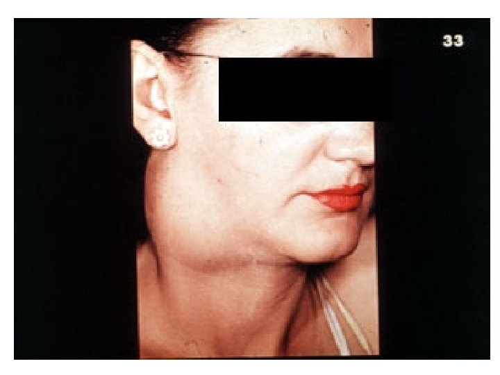

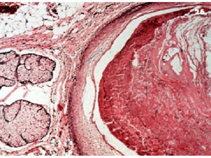
Histologically the cyst is lined by stratified squamous epithelium and its wall contains well-organized lymphoid tissue. The cysts are of developmental origin but their pathogenesis is uncertain.

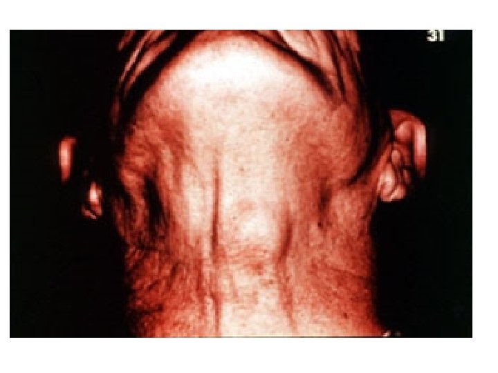
THYROGLOSSAL CYSt a developmental lesion derived from residues of the embryonic thyroglossal duct, the vestigeal remains of which are represented by the foramen caecum on the tongue.
- Slides: 33