Non enveloped DNA viruses ADENOVIRUS Mastadenovirus Mammalian Aviadenovirus


![Laboratory diagnosis�Isolation of virus in cell culture [different specimens] He. La cell line, Hep-2,](https://slidetodoc.com/presentation_image_h/1e0f2b8a2ced03bc50ca959881ada2d6/image-11.jpg "Laboratory diagnosis�Isolation of virus in cell culture [different specimens] He. La cell line, Hep-2,")


![Diseases – B 19 [Parvo B 19] �Erythema infectiosum or slapped cheek syndrome or](https://slidetodoc.com/presentation_image_h/1e0f2b8a2ced03bc50ca959881ada2d6/image-20.jpg "Diseases – B 19 [Parvo B 19] �Erythema infectiosum or slapped cheek syndrome or")

![Properties – �very small 22 nm �Non enveloped �ss DNA genome [-ve strand] �No](https://slidetodoc.com/presentation_image_h/1e0f2b8a2ced03bc50ca959881ada2d6/image-22.jpg "Properties – �very small 22 nm �Non enveloped �ss DNA genome [-ve strand] �No")


![B 19 Ig. G persists for years but not found in immunocompromised [eg. AIDS]](https://slidetodoc.com/presentation_image_h/1e0f2b8a2ced03bc50ca959881ada2d6/image-35.jpg "B 19 Ig. G persists for years but not found in immunocompromised [eg. AIDS]")


![Laboratory diagnosis�Clinically �Histopathology – Koilocytes in lesion �DNA hybridisation tests [commercially available] – detects](https://slidetodoc.com/presentation_image_h/1e0f2b8a2ced03bc50ca959881ada2d6/image-49.jpg "Laboratory diagnosis�Clinically �Histopathology – Koilocytes in lesion �DNA hybridisation tests [commercially available] – detects")


 �Human papova virus �Causes progressive multifocal leukoencephalopathy �This is")


- Slides: 59
Non enveloped DNA viruses
ADENOVIRUS – Mastadenovirus – Mammalian Aviadenovirus – Avians � 47 serotypes in 6 genera A, B, C, D, E & F – cause human disease. �Eg. - a. upper and lower respiratory diseases like pharyngitis, conjunctivitis, pneumonia, kerato-conjunctivitis can also occur. b. Gastroenteritis. c. Hemorrhagic cystitis d. In rodents it can cause sarcoma.
Properties - Non enveloped, ds-linear DNA, Icosahedral nucleocapsid. �Special feature – only virus with fiber - Protruding from each of 12 vertices of the capsid. Fiber is organ of attachment, it is a hemaggluttinin - also the main type of specific antigen. �Group specific antigen is also located on the hexon protein. �Serotypes 12, 18, 31 cause sarcomas at site of injection into lab rodents. Eg. Newborn hamsters. No evidence in humans.
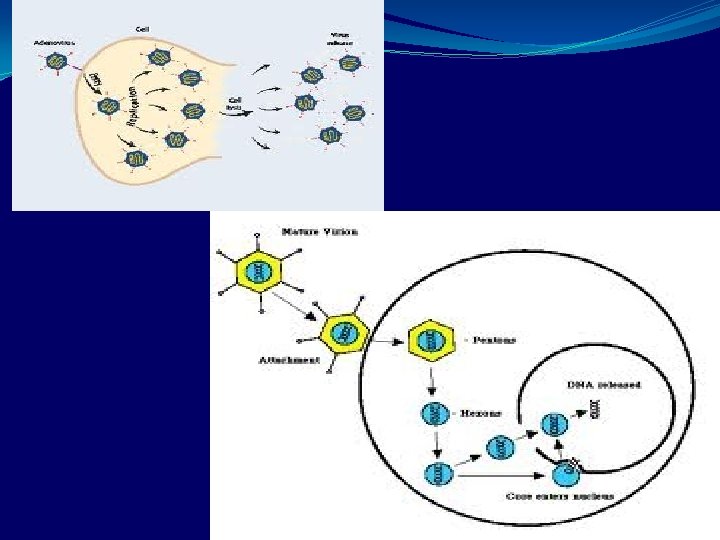
Replication – �Fiber attach to cell surface. �Penetration �Uncoating – viral DNA to Nucleus �Host cell DNA dependent RNA polymerase transcribes early genes. �Splicing enzymes remove the RNA representing the introns- Leads to formation of RNA[Introns and exons common in eukaryotic cell were first described for adenovirus DNA]
�Early RNA is translated into non-structural proteins in cytoplasm �Viral DNA then replicates in the nucleus. �Late MRNA is transcribed and translated into structural viral proteins. �Virus assembly in nucleus �Virus released from cell by cell lysis [not budding]
Transmission�Aerosol droplets �Fecal oral �Direct inoculation of conjunctiva by tonometers or fingers. �Animal strains not pathogenic to humans. �Endemic world wide. �Outbreaks can occur in military recruits, hostels etc. Meaning close living conditon favours transmission. �Eg. Types 3, 4, 7 & 21 can cause respiratory disease. � 8, 9 – epidemic keratoconjunctivitis � 11, 21 – Hemorrhagic cystitis. � 40, 41 – infantile gastroenteritis. � 37 – cervicitis, urethritis [sexual transmission]
Pathogenic and immunity�The name adenovirus is given because it was first isolated from adenoids in 1953 �Infect mucosal epithelium of several organs. Eg. Respiratory tract, G. I. tract and conjunctiva. �Neutralising antibody develops following infection but is type specific and may be life long. �It also causes death of cell in acute infection but has latency in adenoids and tonsils.
Clinical features. URT – Pharyngitis, pharyngoconjunctival fever, acute respiratory disease. Fever, sore throat, coryza and conjunctivitis.
�LRT – Bronchitis, atypical pneumonie. �Hemorrhagic cystitis – hematuria and dysuria �Children under 2 years – Gastro enteritis with non bloody diarrhoea. � Adenovirus infections usually resolve spontaneously. �Approx 50% infections are asymptomatic.
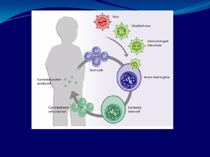
Laboratory diagnosis�Isolation of virus in cell culture [different specimens] He. La cell line, Hep-2, KB etc. to observe CPE. �It is also a exciting model for vector – so tried in gene therapy.
Direct – Inclusion body – basophilic
Electron Microscopy
�Immunofluorescence – using polyclonal or monoclonal antibody �For Feces – LA test, ELISA �Looking for viral DNA by electrophoresis. �Most imp. Serologic test – CF and haemagglutination inhibition. � 4 fold or greater Antibody titer increase [using the paired sera procedure].
Treatment- No antiviral therapy Prevention – �iatrogenic keratoconjunctivitis �maintain asepsis �hand washing.
�Vaccine – live non attenuated vaccine for types 4, 7, 21 given separately as enteric coated capsules for military only. �Since 1998 it is discontinued.
PARVO VIRUS-Parvo virinae – infects vertebrates -Densovirinae – infect insects
Parvovirinae – 3 genera 1. Parvo virus – diarrhoea in humans. Animal virus – Feline Pan leukopenia virus Canine parvo virus Cause serious veterinary disease 2. Erythrovirus – B 19 – divide rapidly in dividing cells autonomously. 3. Dependo virus – defective virus and they depend on a helper virus usually a adenovirus for replication. They are human adeno associated virus not known to cause disease
Diseases – B 19 [Parvo B 19] �Erythema infectiosum or slapped cheek syndrome or fifth disease. �Aplastic crises – especially in sickle cell anemia �Fetal infection – hydrops foetalis
�Pure red cell aplasia – suppression of bone marrow by virus. �Miscellaneous – doubtful – fulminant hepatitis, meningitis, encephalitis, vasculitis, myocarditis, cardiac allograft rejection and glomerulopathies in renal transplants.
Properties – �very small 22 nm �Non enveloped �ss DNA genome [-ve strand] �No virion polymerase �One serotype with icosahedral symmetric capsid.
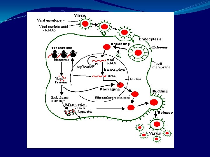
Replication�Adsorption to host cell receptor �Virion penetrate and moves to nucleus �Replication occurs �ss Genomic RNA is synthesized by the cellular RNA polymerase with DNA acting as intermediate. �Progeny virions assembled in the nucleus.
�B 19 replicates only when the cell is in the ‘S’ phase. �This is the reason why the virus replicates in red cell precursor and not in mature red cells.
Transmission�Respiratory route, blood transfusion. �World wide in distribution – half of population has antibodies to it. �Natural reservoir – humans. �Animals are not source for human infection
Pathogenesis and immunity 2 type of cells are infected. a. Red cell precursors in bone marrow – results in aplastic crisis. b. Cells of the endothelium – Partly contributes to the rash and immune complexes – virus + Ig. M or Ig. G contributes to rash and arthritis. �Infection and recovery gives life long immunity against re-infection.
Clinical features- 4 main presentations. a. Erythema infectiosum - slapped cheek syndrome, fifth disease. -Mild, mainly childhood disease. Bright red rash that is most prominent in the cheeks. -With low grade fever, running nose [coryza] and sore throat.
� A ‘Lacy’ less intense erythematous rash appears on the body. � Symptoms resolve in a week. � Main complication – B 19 arthritis. Mostly in adultswomen
b. Aplastic crisis – children with chronic anaemia, sickle cell anemia, thalassemia and spherocytosis have transient but severe aplastic anemia – aplastic crisis, when they are infected with B 19 virus. �People with normal RBC are not affected though their RBC precursors are infected.
c. Fetal infection – women infected during pregnancy virus may cross placenta and infect foetus. 1 st trimester – foetal death, <10% before 20 th week due to severe anemia. 2 nd trimester – hydrops foetalis. 3 rd trimester – No important clinical finding �B-19 is not a common cause of fetal abnormalities
d. Chronic B 19 infection - Immunodeficiencies especially HIV, chemotherapy or transplant patients – chronic anemia, Leukopenia, thrombocytopenia.
Laboratory diagnosis. Samples – serum, blood cells, tissue samples and respiratory secretions. �Most sensitive tests detect viral DNA Eg. -Dot blot hybridization of serum or tissue extracts. -In situ hybridization of fixed tissue. -PCR
Serological assays -based on recombinant parvovirus antigens derived from bacterial or baculovirus expression systems. Used to measure antibodies. �For fifth disease and aplastic crises. �Detect Ig. M antibody to B 19. �Indicates recent infection. �Present for 2 -3 months in the circulation after infection
B 19 Ig. G persists for years but not found in immunocompromised [eg. AIDS] So viral DNA in blood by PCR in these cases.
Fetal infection – �PCR of amniotic fluid detects virus. �Virus difficult to grow so not commonly done. �Diagnostic tests are available only in a few laboratories.
Treatment and prevention�No specific treatment �Pooled Ig have a beneficial effect on B 19 infection in immunodeficient patient. �Fifth disease and aplastic crises are treated symptomatically. �No vaccine or chemoprophylaxis.
PAPILLOMA VIRUS – �Papova viridae - Papilloma virus �Polyoma virus Causes – Papillomas – Benign tumors of squamous epithelial cells – eg. Wart on skin HPV – 16 – implicated to cause Cancer cervix
Properties – �Non enveloped �ds circular DNA �Icosahedral nucleocapsid �Belong to papova virus group. �Similar to polyoma virus and SV 40 virus. �But longer, larger genome and antigenically distinct.
�Two early genes E 6 & E 7 are implicated in carcinogenesis. � These genes encode proteins – that inactivate proteins encoded by tumor suppressor genes in human cells. Eg. p 53 gene proteins suppressed by E 6 RB [retinoblastoma] gene by E 7. �This inactivation of P 53 & RB gene is important step in the process by which normal cell becomes a cancer cell.
�DNA restriction fragmentation analysis – 100 types of papilloma viruses. �These viruses have a very pronounced predilection for certain tissue. eg. Skin warts caused by HPV – 1 to HPV 4. Genital warts HPV 6, HPV 11.
Replication- Little is known because virus grows very poorly in cell culture or not at all. �In human tissues infectious virus particles is situated in the terminally differentiated squamous cells rather than the basal cells. �In malignant cell viral DNA is integrated into host cell DNA near the cellular oncogenes.
�E 6 &E 7 are over expressed. �But in latently infected non-malignant cell –viral DNA is episomal, E 6 & E 7 not over expressed because of an early gene called E 2. This E 2 controls E 6 & E 7 expression. �This E 2 is functional only when the viral DNA is episomal but is inactivated when it is integrated.
Transmission and epidemiology�Mainly by skin to skin contact and by genital contact. �Genital warts is one of the most common STD. �Animal virus is not source of human infection.
Pathogenesis and immunity�Infected squamous cell show characteristic cytoplasmic vacuole – This process is called Koilocytoses. �So Koilocytes are hall mark of papilloma virus infection. �Most warts are benign- do not progress to malignancy.
�But HPV is implicated in carcinoma cervix. Protein encoded by viral genome E 6 & E 7 interfere with growth inhibiting protein produced by P 53 and RB gene. �So contributes to the oncogenicity of the virus. �Both CMI and AMI involved. �Spontaneous regression of warts can take place because of this. �More extensive warts in immunocompromised. Eg. AIDS.
Clinical features�Predominant finding is papillomas of various organs. �Specific HPV types cause. �Skin and plantar wart HPV 1, HPV 2, HPV 3 & HPV 4.
�Genital wart – condyloma accuminata – HPV 6, HPV 11. �Carcinoma of cervix, the penis, anus. �Premalignant lesion called intraepithelial neoplasia – HPV-16, HPV 18. �Occult premalignant lesion of cervix and penis can be revealed if you apply acetic acid to tissue.
Laboratory diagnosis�Clinically �Histopathology – Koilocytes in lesion �DNA hybridisation tests [commercially available] – detects presents of viral DNA. �Serology rarely done. �Culture unsuccessful.
Treatment and prevention�Genital warts – Podophyllin. �Alpha interferon – effective and better for preventing recurrences than antiviral treatment. �Liquid nitrogen commonly used for skin warts. �Plantar warts – surgical removal, topical salicylic acid. �Vaccine is marketed nowadays (recombinant vaccine containing antigen HPV 6, 11, 16 & 18 for adolescent & young women. �Common counseling for sexual behavior.
Polyoma and SV 40 Virus�Best characterised oncogenic papova viruses of animals �Poly=many, ma=tumour �Cause wide variety if histologically different tumor when inoculated into newborn rodents �Natural host is mouse
�SV 40 isolated from normal Rhesus monkey kidney cells �Causes sarcoma in new born hamsters
�Polyoma and SV 40 share many chemical and biological features - ds circular super coiled DNA of mol. wt. 3 x 10⁶ - 45 nm icosahedral nucleocapsid � sequence of DNA and antigens are different
�Both undergo a lytic or permissive cycle in cells of their natural host with production of progeny virus �If they infect cells of heterologous species, nonpermissive cycle occurs, no virus is produced , cell is malignantly transformed �In transformed cells, viral DNA integrates into the cell DNA and only early proteins are synthesized. �Some of the proteins like T antigen are required for induction and maintenance of transformed state
JC virus- (John Cunningham virus) �Human papova virus �Causes progressive multifocal leukoencephalopathy �This is a fatal demyelinating disease of white matter and multiple areas of brain involved �This occurs primarily in compromised CMI especially in AIDS
�Antigenically this virus is distinct from others like HPV � 75% of normal people have antibodies to JC virus meaning infection is widespread �Disease occurs when latent virus is activated in an immuno compromised
�Diagnosis- electron microscopy of diseased brain tissue -Cytology – exfoliated urine – enlarged cells with deeply stained basophilic nuclei with a single inclusion �Virus isolation- urine or brain biopsy material grown in fetal glial cell culture - Growth made out by the Haemagglutination inhibition test �Viral antigen – ELISA of urine sample, immunofluorescence of biopsy material �Viral nucleic acid – nucleic acid hybridisation and PCR �Autopsy- in situ hybridisation of brain biopsy material
BK virus�isolated from urine of renal transplant recipient �Subclinical infection seen in children before 10 yrs of age �Upper respiratory symptoms �Persists for life in kidneys �Reactivation can occur during the last trimester of pregnancy and also following immunosuppression for organ transplants �Leads to asymptomatic shedding in urine
�Diagnosis 1. Electron microscope – urine of renal transplant patient. Detection of viral antigen of ELISA 2. cytopathology- similar to JC virus 3. Virus isolation- human diploid fibroblasts 4. Detection of viral nucleic acid – PCR, DNA hybridization