Nociceptive and autonomic reflex arcs Spinal pathways injuries


 receptor is responsible for")


 pathways 1. PROTOPATIC SENSIBILITY • pain")

 pathways 1. EXTRAPYRAMIDAL TRACTS • Periodic")


- Slides: 24
Nociceptive and autonomic reflex arcs. Spinal pathways, injuries, symptoms Márk Kozsurek, M. D. , Ph. D. mark@kozsurek. hu EM II. , 28/09/2017
II. Spinal reflex arcs
Reflex: involuntary and automatic response to a stimulus A (1) receptor is responsible for detecting a stimulus and converting it into a neuronal signal. Evoked action potentials reach the central nervous system through the (2) afferent limb. Decission is made and response is generated by the (3) central processing neuronal networks. (4) Efferent limb exits the central nervous system and terminates on the (5) effector, which might be a muscle or a gland. (1) to (5) components altogether constitute the reflex arc! viscerosensory visceromotor central nervous system SENSORY GGL. skin, muscles, joints, tendons internal organs receptors somatomotor AUTONOMIC GGL. skeletal muscles smooth muscles , glands effectors v somatosensory
Spinal reflex: In the case of spinal reflex arcs the stimulus is received and processed by the spinal cord and the response is organized and performed by the spinal neuronal circuits. Under normal circumstances stimuli evoking spinal reflexes also reach the brain and descending fibres from higher centres may modulate spinal reflexes but experimentally spinal reflexes can be evoked even after the removal of the brain! • Proprioceptive reflex • Nociceptive reflex • Autonomic reflexes
1. Nociceptive reflex arc Synonyms: flexor reflex, withdrawal reflex, polysynaptic spinal reflex Ancient reflex that protects the body from potentially damaging, noxious stimuli.
Nociceptors of the skin and mucous membranes → Adelta and/or C fibres → spinal ganglion → spinal dorsal horn → numerous collaterals: excitacion of flexors and inhibition of extensor on the side of stimulus and stimulation of extensors together with inhibition of flexors on the contralateral side (flexor reflex with crossed extensor response).
2. Autonomic spinal reflex arcs Both proprioceptiv and nociceptive spinal reflexes were „somatic”: a somatosensory input evoked a somatomotor response. visceromotor viscerosensory central nervous system SENSORY GGL. skin, muscles, joints, tendons internal organs receptors somatomotor AUTONOMIC GGL. v somatosensory skeletal muscles smooth muscles , glands effectors In case of autonomic spinal reflexes either the afferent or the efferent limb of the reflex arc or both of them are „visceral”!
viscerosensory visceromotor central nervous system internal organs receptors AUTONOMIC GGL. v SENSORY GGL. smooth muscles , glands effectors Urinary or micturition reflex: after reaching the threshold of the stretch receptors within the wall of the urinary bladder, sphincters relax, the muscles of the bladder contract and the bladder discharges. (It is clearly seen in babies and after spinal cord trauma, as the supraspinal control is missing in both of these cases. Viscerocutaneous reflex: diseases of internal organs are frequently associated with local hyperemia (increased blood supply) of the skin due to the vasodilator innervation of the smooth muscles of vessels.
viscerosensory central nervous system somatomotor SENSORY GGL. AUTONOMIC GGL. skeletal muscles internal organs receptors effectors „Défense musculaire”: inflammation of abdominal organs will result in maximal contraction of abdominal muscles. This is protecting involved abdominal organs and immobilizes the patient.
visceromotor central nervous system somatosensory AUTONOMIC GGL. v SENSORY GGL. skin, muscles, joints, tendons receptors smooth muscles , glands effectors Cuti-visceral reflex: applying local heat may relax bowels and can reduce pain (but not in the case of inflammation!) or stimulation of the skin of the glans penis results in erection and ejaculation.
visceromotor viscerosensory central nervous system somatosensory skin, muscles, joints, tendons internal organs receptors AUTONOMIC GGL. v SENSORY GGL. smooth muscles, glands effectors Depending on the target, the neurotransmitter used and the physiological effect, different types of efferent limbs of the autonomic spinal reflex can be noted!
Two types of the visceromotor fibres can be distinguished: they can be parasympathetic or sympathetic. Sympathetic ones can be further classified as parietal (innervating the skin of limbs and the trunk with sudomotor, pilomotor and vasomotor fibres) and visceral (innervating internal organs).
1. All the internal organs have a double – sympathetic and parasympathetic – innervation. (Limbs and the skin of the trunk exclusively have sympathetic innervation!) 2. Sympathetic and parasympathetic fibres arise from completly different regions of the CNS. 3. Visceromotor efferents of spinal autonomic reflexes have to synapse once on the periphery in an autonomic ganglion. Parasympathetic (craniosacral): - Autonomic ganglia of cranial nerves - Plexuses within the walls of internal organs (thoracic, abdominal and pelvic viscera) Sympathetic (thoracolumbar): - parevertebral ganglia (sudo-, pilo- and vasomotor innervation of the trunk and limbs) - prevertebral ganglia (viscera)
S IML 3. white ramus communicans 5. gray ramus communicans parasympathetic – synapse in intramural plexus (or in the ganglia of the cranial nerves, but these are not related to the spinal reflexes)
Th-L IML 3. white ramus communicans 5. gray ramus communicans parietal sympathetic – synapse in the paravertebral ganglia of the sympathetic trunk
Th-L IML 3. white ramus communicans 5. gray ramus communicans visceral sympathetic – synapse in prevertebral ganglia (celiac, superior and inferior mesenteric ganglion)
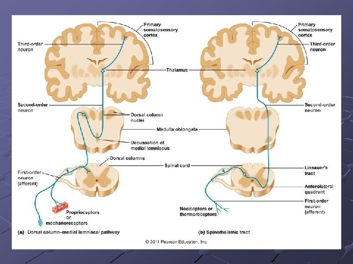
II. Tracts of the spinal cord Ascending (sensory) pathways 1. PROTOPATIC SENSIBILITY • pain and heat, potential harmful stimuli 2. EPICRITIC AND ÉS PROPRIOCEPTIVE SENSIBILITY • fine touch, vibration, two-point-discrimination • position and movement of joints and muscles
uncrossed spinobulba r tract Fl ec hs ig crossed Gowers ct ra ic t lam sp a oth in Spinothalamic tract situated in the anterior and lateral funiculi conveys protopathic (pain, heat) signals toward higher areas of the CNS. Spinobulbar tract carries epicritic and proprioceptiv information into the brainstem, while dorsal and ventral spinocerebellar tract (tract of Flechsig and Gowers, respectively) share epicritic and proprioceptiv signals with the cerebellum.
II. Tracts of the spinal cord Descending (motor) pathways 1. EXTRAPYRAMIDAL TRACTS • Periodic and automatic movements • Posture and body balance • Tone of muscles 2. PYRAMIDAL TRACT • Voluntary, individual movements
PYR e a b a: reticulospinal tract b: olivospinal tract c: vestibulospinal tract d: tectospinal tract e: rubrospinal tract c pyr a d extrapyramidal
Somatotopic arrangement of major sensory and motor pathways
Thank you for your attention!