
NIPAH and HENDRA VIRUS Henipavirus outbreak occurred in
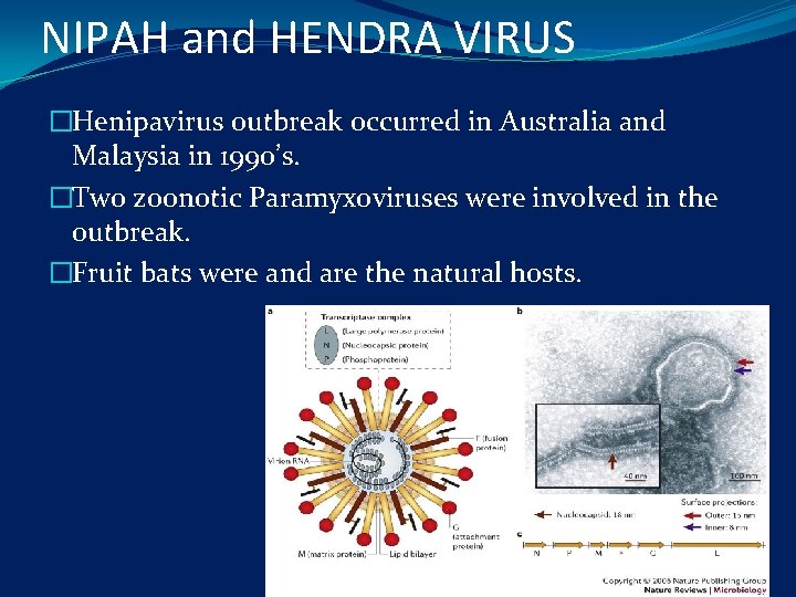
NIPAH and HENDRA VIRUS �Henipavirus outbreak occurred in Australia and Malaysia in 1990’s. �Two zoonotic Paramyxoviruses were involved in the outbreak. �Fruit bats were and are the natural hosts.

�Nipah virus-Severe encephalitis transmitted from pigs to humans directly occurred in Malaysia caused by this virus, with high mortality. �Hendra virus- caused deaths in many horses in Australia with some reported human cases. �Both the viruses caused high mortality and so these are classified as BIOSAFETY LEVEL 4 AGENTS.
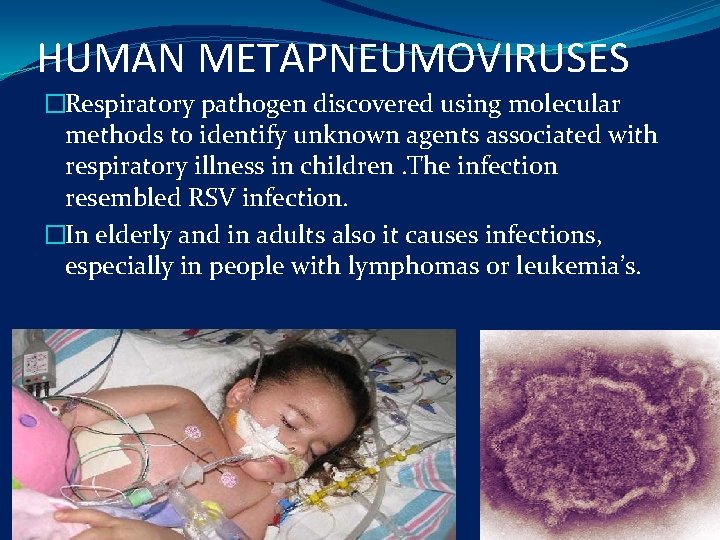
HUMAN METAPNEUMOVIRUSES �Respiratory pathogen discovered using molecular methods to identify unknown agents associated with respiratory illness in children. The infection resembled RSV infection. �In elderly and in adults also it causes infections, especially in people with lymphomas or leukemia’s.
RUBELLA VIRUS �GERMAN MEASLES or 3 DAY MEASLES � An acute febrile illness characterized by rash and with post-auricular, subocciputal lymphadenopathy �Effects adults and children. �Infection during early pregnancy may result in serious abnormalities of the fetus, including congenital malformation and mental retardation. �Consequences of rubella in-utero are referred to as congenital rubella syndrome.
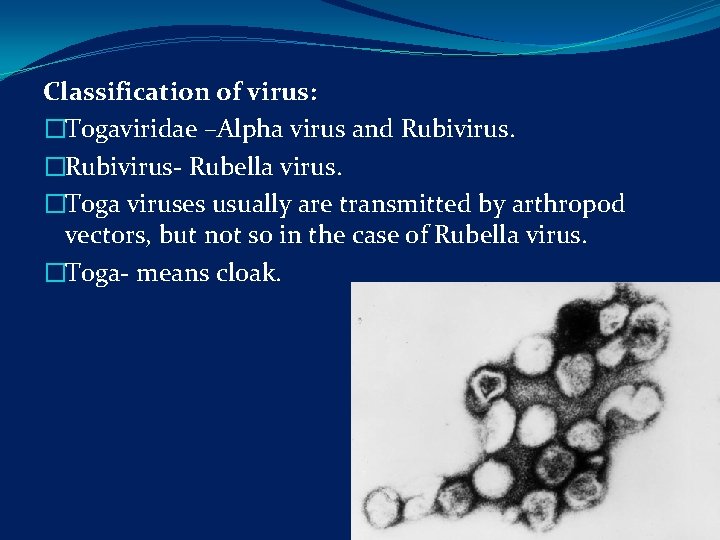
Classification of virus: �Togaviridae –Alpha virus and Rubivirus. �Rubivirus- Rubella virus. �Toga viruses usually are transmitted by arthropod vectors, but not so in the case of Rubella virus. �Toga- means cloak.
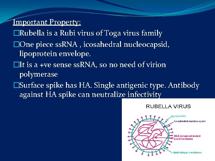
Important Property: �Rubella is a Rubi virus of Toga virus family �One piece ss. RNA , icosahedral nucleocapsid, lipoprotein envelope. �It is a +ve sense ss. RNA, so no need of virion polymerase �Surface spike has HA. Single antigenic type. Antibody against HA spike can neutralize infectivity
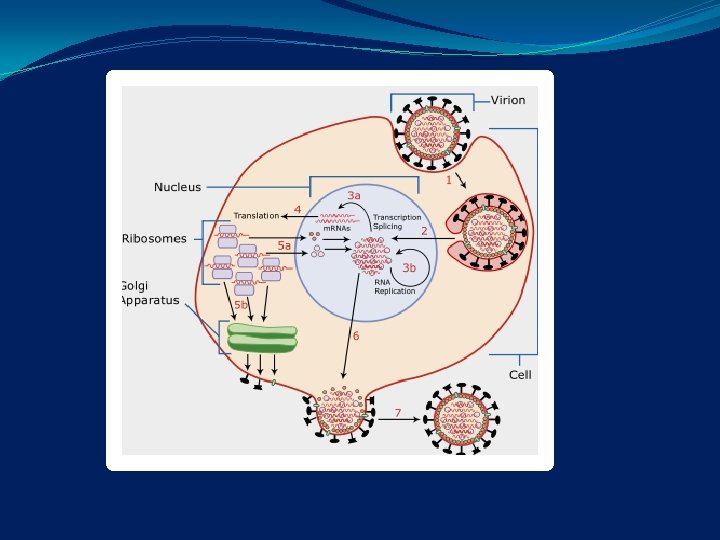
Summary of replication- not completely understood. May follow Toga virus cycle �Penetration of cell �Uncoating �+ss. RNA translated into several structural and nonstructural proteins �One of non-structural protein is RNA dependant RNA polymerase which replicates the genome first by making a -ve strand template and from that +ve strand progeny �Replication and assembly in cytoplasm and envelope acquired from outer membrane �Virus exits the cell
�Pathogenesis and pathology- 2 types �Post-natal Rubella �Congenital Rubella Syndrome
�Neonatal, childhood, adult infections occur through mucosa of upper respiratory tract. �Initial replication probably occurs in the mucosa of URT �Multiplication in cervical lymph node �Viremia after 5 -7 days and lasts till appearance of antibody on 13 -15 day �Appearance of antibody coincides with appearance of rash which has a immunological basis.
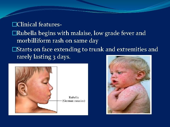
�Clinical features- �Rubella begins with malaise, low grade fever and morbilliform rash on same day �Starts on face extending to trunk and extremities and rarely lasting 3 days.
�This feature is not pathognomonic finding because other virus can also cause same type of rash. But posterior auricular and sub occipital lymphadenopathy are present. �Transient arthralgia and arthritis occur in women, maybe due to immune complexes. Rarely thrombocytopenic purpura and encephalitis
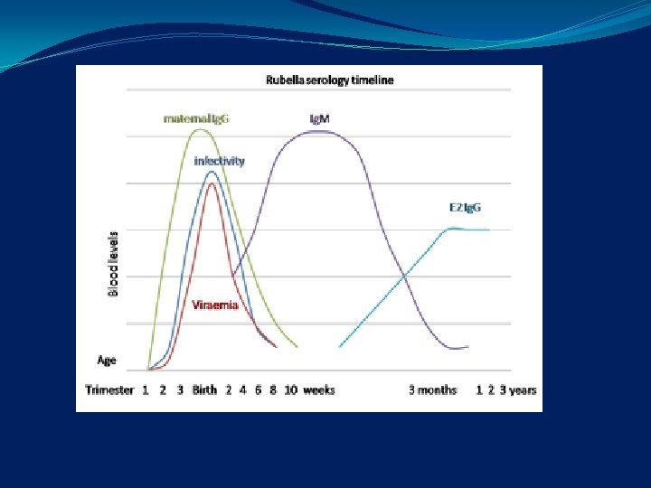
Immunity – �Ig. M and Ig. G appear as rash fades rapidly, rises over 1 -3 weeks �Ig. M upto 6 months �Rubella Ig. M is seen in serum and is detected after 2 weeks in single sample �Is Diagnostic of recent infection �Ig. G presents later and persists for life �One attack confers life long immunity �Immune mothers transfer immunity to offspring and these are protective for upto 4 -6 months

Lab diagnosis�By isolation of virus �Evidence of seroconversion
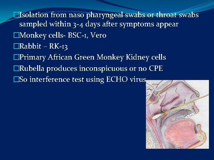
�Isolation from naso pharyngeal swabs or throat swabs sampled within 3 -4 days after symptoms appear �Monkey cells- BSC-1, Vero �Rabbit – RK-13 �Primary African Green Monkey Kidney cells �Rubella produces inconspicuous or no CPE �So interference test using ECHO virus
�Other methods – Immunofluorscence �Serology- �ELISA, Hemagglutination Inhibition test �Ig. M ELISA for recent infections �Ig. G ELISA for immunity �Paired sera for critical assessment of pregnant women, at least 10 days apart
Epidemiology- �post natal rubella- worldwide, infection all year round, peaks in spring �In India, during rainy season �Rubella is not as contagious as measles �Use of rubella vaccine eliminates epidemics which occurs 6 -10 yrs and pandemics 22 -25 yrs
Treatment- �Rubella is mild and self limiting and no specific treatment �Only in lab proved Rubella in first trimester of pregnancy, fetal infection is there �So Immune globulin serum can be given with unproven benefit. �They confuse serological reaction. So decision to have the baby or not rests with the family �Rubella status of the personnel involved in hospital work should be known when treating pregnant women.
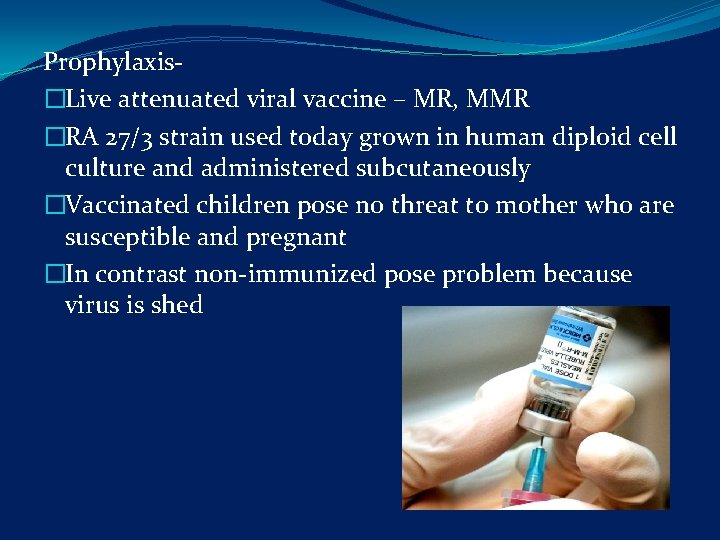
Prophylaxis- �Live attenuated viral vaccine – MR, MMR �RA 27/3 strain used today grown in human diploid cell culture and administered subcutaneously �Vaccinated children pose no threat to mother who are susceptible and pregnant �In contrast non-immunized pose problem because virus is shed
�Vaccine is good as it confers protection in 95% of vaccinees �Side effects- mild fever lymphadenopathy and fleeting rash but no residual effects �In adults only significant side effect is arthralgia �In post-pubertal girls, self limited arthralgia and arthritis is seen in one- third of vaccines �Rubella vaccine virus can cross placenta and infect fetus but is not teratogenic.
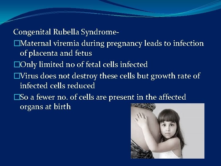
Congenital Rubella Syndrome- �Maternal viremia during pregnancy leads to infection of placenta and fetus �Only limited no of fetal cells infected �Virus does not destroy these cells but growth rate of infected cells reduced �So a fewer no. of cells are present in the affected organs at birth
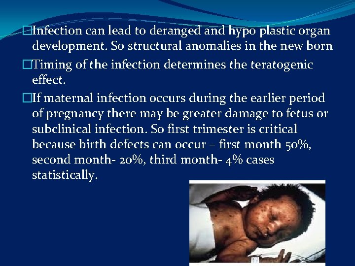
�Infection can lead to deranged and hypo plastic organ development. So structural anomalies in the new born �Timing of the infection determines the teratogenic effect. �If maternal infection occurs during the earlier period of pregnancy there may be greater damage to fetus or subclinical infection. So first trimester is critical because birth defects can occur – first month 50%, second month- 20%, third month- 4% cases statistically.
�Birth defects are uncommon after 18 weeks of pregnancy �Rubella rarely can cause fetal death and spontaneous abortion �Intra uterine infection also leads to chronic persistence of virus in new born �So virus is easily detected in pharyngeal secretions, multiple organs, CSF, urine and rectal swabs � 12 -18 months after birth viral shedding occurs and gradually decreases with age
Clinical features- �Virus isolated from many organs and damage is widespread Three types- �Transient effects �Permanent manifestations- apparent at birth or recognized during first yr �Developmental abnormalities that appear and progress during childhood and adolescence
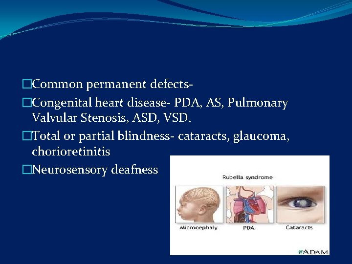
�Common permanent defects- �Congenital heart disease- PDA, AS, Pulmonary Valvular Stenosis, ASD, VSD. �Total or partial blindness- cataracts, glaucoma, chorioretinitis �Neurosensory deafness
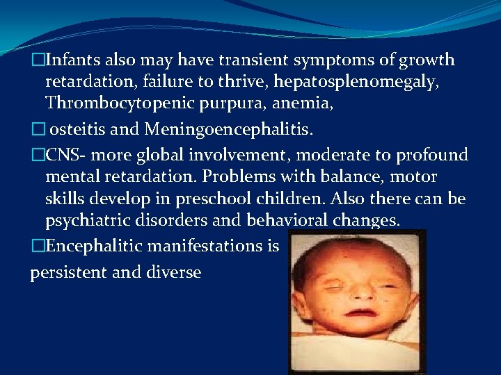
�Infants also may have transient symptoms of growth retardation, failure to thrive, hepatosplenomegaly, Thrombocytopenic purpura, anemia, � osteitis and Meningoencephalitis. �CNS- more global involvement, moderate to profound mental retardation. Problems with balance, motor skills develop in preschool children. Also there can be psychiatric disorders and behavioral changes. �Encephalitic manifestations is persistent and diverse
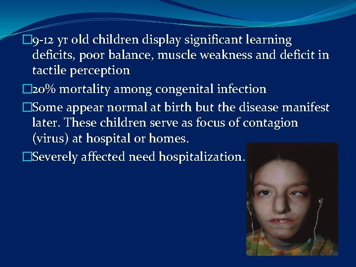
� 9 -12 yr old children display significant learning deficits, poor balance, muscle weakness and deficit in tactile perception � 20% mortality among congenital infection �Some appear normal at birth but the disease manifest later. These children serve as focus of contagion (virus) at hospital or homes. �Severely affected need hospitalization.
�Progressive rubella encephalitis is a rare complication occurring in second decade of life in children with congenital Rubella. �Severe neurological deterioration inevitably leading to death (high titer of antibody and virus isolated by cocultivation from brain in these cases).
�TORCH syndrome- one entity is rubella virus �Immunity - infants can be having rising titer of rubella specific Ig. M after in utero infection �Mothers Ab Ig. G can cross but gradually lost �Later Ig. G increases
Lab diagnosis- �Amniocentesis and detection of Fetal Rubella specific Ig M antibody �Virus isolation is definitive �Pregnant woman’s blood showing Ig. M Abs – denotes recent infection but 1: 8 or greater Ig. G means protected �Children with congenital rubella also have impaired CMI.
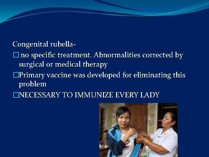
Congenital rubella� no specific treatment. Abnormalities corrected by surgical or medical therapy �Primary vaccine was developed for eliminating this problem �NECESSARY TO IMMUNIZE EVERY LADY
- Slides: 33