Newborn Examination Identifying Reportable Congenital Anomalies Disclaimer The

Newborn Examination Identifying Reportable Congenital Anomalies Disclaimer: The findings and conclusions in this presentation have not been formally disseminated by the Centers for Disease Control and Prevention and should not be construed to represent any agency determination or policy.

Supplies Needed for the Exam • • • Camera to take photographs Birth defects data collection form Clean gloves to use when examining the neonate Tape measure or a measuring board A scale to weigh the neonate, and paper/mats on which to place the neonate • Light source (e. g. , flashlight) to visualize the inside of neonate’s mouth and any other areas that need more light 2

Photographs • If parental consent is required for taking photographs, ensure that you have a consent form with you and ask the parents to sign it before taking photographs • If parents do not consent to photographs, the neonate should still be included in the surveillance program • When taking photographs, place tape measure beside the neonate or next to the defect, to serve as a reference scale, or place the neonate on the measuring board 3

The Exam • Perform the exam as soon after birth as possible • Note: If the neonate has a birth defect(s) that requires immediate attention, wait until the appropriate moment to do the examination • An exam usually takes 10 -15 minutes • Wear gloves to complete the exam • Wash your hands before and after putting on the gloves • The exam should be performed with the neonate undressed • Place the neonate on a clean, soft surface in a well-heated area • Never turn your back on the neonate or leave the neonate alone • Dress the neonate when the exam is complete • Consider having an assistant or family member present during the exam to help soothe or pacify the neonate 4

Describing Findings • If any defects are found during the exam, please make sure you include the following information in the newborn’s file: • A detailed description of the birth defect in the data collection form • Photographs, drawings, and/or illustrations • Any doctor's reports, radiographs, or tests 5

Performing the Exam: Overview • Measure infant weight • Measure infant length • Measure infant head circumference • Examine the head • Examine the body • Examine the limbs 6

The Exam: Measuring Weight • When weighing the neonate, make sure that all of the equipment is clean • Be extra careful with neonates who have open defects such as spina bifida, omphalocele, or gastroschisis • If possible, first place a barrier (e. g. , paper towel, blanket) between the scale and where the infant will lay • Zero the scale • Place the infant on the scale and record the weight in grams Source: http: //www. zuga. cl/que-es-el-percentil/ 7

The Exam: Measuring Length • Measure the neonate’s length with a tape measure or measuring board that is marked in centimeters • If neonate is flexing legs, gently extend legs as flat as possible lengthwise • Clean the measuring board or use a clean tape measure • If possible, place a barrier (paper towel, blanket, etc. ) between the measuring surface and the neonate • When measuring the neonate, record the length to the nearest tenth of a centimeter Source: http: //www. embarazoyparto. net/bebes/como-medir-la-talla-del-bebe/ 8

The Exam: Measuring Head Circumference • Look at the neonate’s head • Place a tape measure around the widest part of the head, above the eyebrows and ears • Take special care if the neonate has a defect affecting the head • Use gloves if defect is open • Record the head circumference to the nearest tenth of a centimeter Source: http: //www. crecerfeliz. es/El-bebe/Primer-mes/Primera-visita-al-pediatra 9

How to Examine the Head • Gently feel the top, back, and sides of the head • Note whethere is any absence of brain and/or skull bones • Note whether brain tissue is bulging • If yes, note the location of the bulge on the skull • Note whether the anterior fontanelle (front soft spot) is bulging • This can be an indication of hydrocephalus • Check the eyes • Note whether they are present • Note whether they are of equal size and appear normally spaced • Note whether cataracts, colobomas (fissure of the iris), cloudy corneas are present • Check the ears • Note whether they are present • Note any unusual shape and location • Check the mouth • Use a light source to check inside the mouth for any abnormalities • Use a gloved finger to feel the upper palate for any abnormalities • Note if there are fissures (opening/cleft) in the upper lip and/or at the top of the palate 10

How to Examine the Body • Check the baby’s chest • Note any unusual findings (e. g. , protruding or sunken breast bone) • Check the umbilical cord • Note if there is protrusion of organs into the cord or next to the cord • Check the baby’s back • Note if there any protrusions of the spinal cord • Check the baby’s genitalia • Note any unusual findings (e. g. , hypospadias) • Check the skin • Note any colored markings, or raised bumps – if present, note size, color, location, whether it's a flat discoloration or raised, whether hair is present 11

How to Examine the Limbs • Check for overall tone and movement • Note whether the neonate is moving all limbs • Check the upper limbs • Note whethere any missing segments • Note whether limb(s) are of an unusual shape • Note the number of fingers, position of hands, any abnormalities of hands/fingers • Check the lower limbs • Note whethere any missing segments • Note whether limb(s) are of an unusual shape • Note any abnormalities with feet or toes • Note position of feet • If abnormal position of foot, check to see if it is rigid or moveable 12

Acknowledgements q q International Clearinghouse on Birth Defects Surveillance and Research § Pierpaolo Mastroiacovo § Lorenzo Botto § Boris Groisman CDC National Center on Birth Defects and Developmental Disabilities § Surveillance Working Group Pictured top, left to right: Jaime Frías, RJ Berry, Joseph Mulinare, Jorge Rosenthal, Ibrahim Zaganjor Pictured bottom, left to right: Diana Valencia, Ahlia Sekkarie, Jennifer Williams, Hilda Razzaghi, Alina Flores, J. Fernando Arenas Not pictured: Michael Cannon, Yan Ping Qi, Christina Hillard 13

Newborn Examination Descriptions of Select Reportable Congenital Malformations that Might be Seen during Newborn Exam Advanced Slide-set Disclaimer: The findings and conclusions in this presentation have not been formally disseminated by the Centers for Disease Control and Prevention and should not be construed to represent any agency determination or policy. 14
 q")
Neural Tube Defects Checklist q Describe in detail (see below for specific recommendations) q Include photographs: useful for diagnosis and review q Describe additional malformations, if present q Specify consultations: particularly genetics and neurosurgery Specific recommendations q Spina bifida: q Level of lesion: e. g. , T 6 -T 9 or midthoracic, etc. q Presence/absence of hydrocephalus and talipes q Presence/absence of skin or membrane covering the defect q Associated anomalies q Report from neurosurgery, if available q Anencephaly: q Distinguish from acrania (absent skull bones with all brain present), and from acephaly in monozygotic twins (recipient twin has acardia-acephaly) q Associated anomalies (uncommon) q Encephalocele q Location: occipital, nasal, frontal, orbital, parietal q Associated anomalies: more common in encephalocele than in spina bifida or anencephaly q Report from neurosurgery, if available Coding: ICD-10 or ICD-10 RCPCH (Royal College of Paediatrics and Child Health) • anencephaly: Q 00. XX • encephalocele: Q 01. XX • spina bifida: Q 05. XX • General guidelines: include details for 4 th and 5 th digits, and avoid only using general codes (e. g. , “Q 00”, “Q 01”, “Q 05”) Notes • Talipes: often present with spina bifida, code it separately (Q 66. X) • Craniorachisis: anencephaly with a contiguous spinal defect without meninges covering the neural tissue is cranioraschisis: code as single defect (Q 00. 1). However, if the neural tube defects are not contiguous, code each defect separately. • Do not include: spina bifida occulta (Q 76. 0), atypical defects (usually due to amniotic bands), acrania, or acephaly. Examples Anencephaly (Q 00. 00) Lumbosacral spina bifida (Q 05. 7) Occipital encephalocele (Q 01. 2) 15

Microtia – Anotia Coding 2. 7 cm Normal: 3 -4 cm Microtia I Q 17. 21 Microtia II Q 17. 22 Microtia III Q 17. 23 Microtia IV /Anotia Q 16. 0 q Choose image (left) closest to case q If photo is unavailable, draw malformation (use drawing below as template) Photographs: http: //www. earmolding. org/photo-gallery/lidding/ & http: //en. atlaseclamc. org. q q Checklist Identify severity (type I-IV) and laterality (L-R-Bilateral) q Microtia type I – a small, but normally formed ear; usually considered a minor anomaly; check protocol Take photographs, side and front Check for (and if present, describe) q Preauricular tag or pits (describe, code) q Down-slanting palpebral fissures, small jaw, eyelid coloboma q Cervical vertebral anomalies (radiograph recommended) q Kidney ultrasound (only if multiple congenital anomalies or possible syndromes) q Atresia choanae, coloboma iris or retinae, genital anomalies, heart defects q Any other major anomaly Report any additional specialty consultation(s) and results http: //tiny. cc/ik 5 pby 16
, Cleft Lip (CL), Cleft Lip and Palate (CLP) Checklist q Describe")
Cleft Palate (CP), Cleft Lip (CL), Cleft Lip and Palate (CLP) Checklist q Describe in detail: q Laterality: right, left, or bilateral q Lower lip: pits present or absent q Extension of the cleft lip: minimum, partial or total; any involvement of the gum and nasal sill q Extension of cleft palate: hard and/or soft q Shape of cleft palate; “U” shape or “V” shape q Significant micrognathia or glossoptosis: present or absent q Additional anomalies: describe evaluations and findings q Photographs: very useful, can be crucial for review q Specialty consultations: report which were done (including genetics) and results Coding: ICD-10 or ICD-10 RCPCH • • Q 35. XX : cleft palate Q 36. XX : cleft lip Q 37. XX : cleft lip and palate Include 4 th and 5 th digits: avoid only using general codes (e. g. , “Q 35”, “Q 36” or “Q 37”) • Do not include in the group the following conditions: • median cleft lip (Q 36. 1) • cleft uvula (Q 35. 7) • facial fissures (oro-orbital, oro-auricular) • atypical facial clefts Unilateral CL (Q 36. 90) Cleft hard and soft palate with unilateral CL (Q 37. 50) Cleft hard palate (Q 35. 1) Cleft hard and soft palate with bilateral CL (Q 37. 4) Cleft soft palate (Q 35. 3) 17
: check the")
Hypospadias Checklist q Identify position of external meatus q Use illustration (right): check the box and use the associated term q Use most specific code: add 5 th digit if needed Coding: ICD-10 or ICD-10 RCPCH Q 54 Hypospadias Avoid using just the general code: be specific Q 54. 0 q Chordae: present (Q 54. 4) or absent q Cryptorchidism: present (unilateral Q 53. 1 or bilateral Q 53. 2) or absent Q 54. 1 q Take photograph: show clearly the urethral meatus (may be challenging) q Associated malformations: describe evaluations done and findings Q 54. 2 q Specialty consultation(s): report if done (including genetics, surgery, urology) and results Q 54. 3 18

Clubfoot: Talipes Equinovarus Checklist q Describe the presentation precisely, including: q Laterality: right, left, or bilateral q Mobility of foot: rigid vs. flexible (flexible is the same as positional, and excluded in most surveillance systems) q Photograph: useful for review, not sufficient as confirmation q Additional anomalies: describe evaluations and findings q Specialty consultations: report which were done (including genetics and orthopedics) and results Notes Coding: ICD-10 or ICD-10 RCPCH • Q 66 Congenital deformities of feet (avoid using this general code if more specific information is available) • Q 66. 0 Talipes equinovarus • Q 66. 1 Talipes calcaneovarus • Q 66. 4 Talipes calcaneovalgus • Q 66. 8 Other congenital deformities of feet; clubfoot unspecified q Flexible/positional talipes equivarus: exclude q Except if specifically included in protocol: inclusion discouraged because of variability, frequency, and minor health impact q Talipes calcaneovarus and calcaneovalgus: usually not considered an eligible birth defect (provide clear rule in protocol) Talipes equinovarus Talipes calcaneovalgus 19

Limb Deficiencies Checklist q Use figures provided: select type most similar to case q Describe in detail (not only “diagnostic term”): q Note each segment involved (see figures) for each limb affected; describe what is deficient or absent q Laterality: right, left, or bilateral q Soft tissue nubbins: present vs. absent (in transverse defects) q Amniotic band and/or soft tissue ring constriction: present vs. absent Coding ICD-10 or ICD-10 RCPCH Use figures on the next four slides to identify the most similar to the observed case q Photographs: crucial for review and classification q Radiographs: crucial for review and classification (at times even more than photographs) q Additional anomalies: describe evaluations and findings q Also note structures evaluated and found normal: (i. e. , heart, kidneys, gastrointestinal system, genitalia) q Specialty consultations: report which were done (including genetics and orthopedics) and results 20
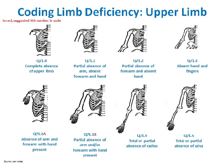
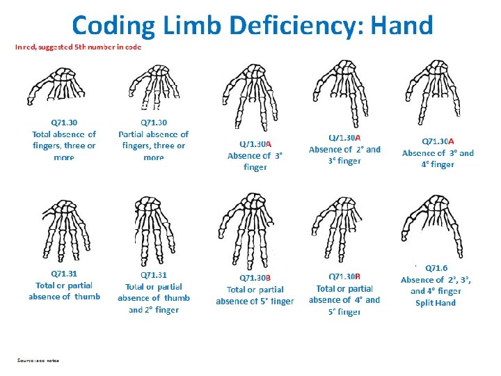
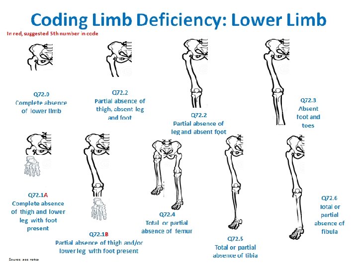
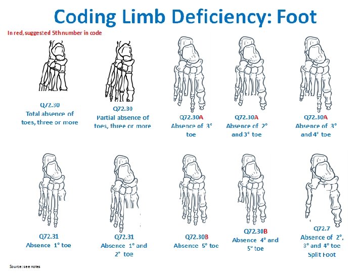

Limb Deficiency Coding and Classification Tool Provides a mapping from codes to the main classification groups for analysis Code Upper limb Code Lower limb Q 71. 0 Transverse terminal Q. 72 Transverse terminal Q 71. 1 A Transverse intercalary, typical phocomelia Q 72. 1 A Transverse intercalary, typical phocomelia Q 71. 1 B Transverse intercalary, atypical phocomelia Q 72. 1 B Transverse intercalary, atypical phocomelia Q 71. 2 Transverse terminal Q 72. 2 Transverse terminal Q 71. 3 Transverse terminal Q 72. 3 Transverse terminal Q 71. 30 Transverse terminal Q 72. 30 Transverse terminal Q 71. 30 A Transverse terminal Q 72. 30 A Transverse terminal Q 71. 30 B Transverse terminal Q 72. 30 B Transverse terminal Q 71. 4 Longitudinal preaxial Q 72. 4 Longitudinal preaxial Q 71. 31 Longitudinal preaxial Q 72. 31 Longitudinal preaxial Q 71. 5 Longitudinal postaxial Q 72. 5 Longitudinal preaxial Q 71. 6 Longitudinal axial, split hand Q 72. 6 Longitudinal postaxial Q 72. 7 Longitudinal axial, split foot 25

Omphalocele Checklist q Describe in detail: do not limit to using “omphalocele” but add detail on the following elements q q Code ICD-10 RCPCH Q 79. 2: Omphalocele Cord insertion: describe if midline, over umbilicus Covering membranes: yes/no, intact/ruptured Size: measure/estimate (in centimeters) Extruded organs: small intestine, liver, spleen, etc. q Photographs: show clearly umbilical cord/membrane q Associated defects: describe in detail q Syndromes: if known, describe (e. g. , Beckwith. Wiedemann syndrome) or, if not known, describe relevant minor anomalies q Procedures: describe procedures to assess additional malformations q Specialty consultations: report which were done (including genetics, surgery) and results Describe three most important features: midline defect, insertion through cord, membrane 26

Gastroschisis Checklist q Describe in detail: do not limit to using “gastroschisis” but add detail on the following elements q q Code ICD-10 RCPCH Q 79. 3: Gastroschisis Side relative to the umbilical cord: right/left Covering membranes: yes/no Size: extension of the abdominal defect (in cm) Extruded organs: specify also bowel segment involved q Associated defects: describe q Photographs: show clearly the umbilical cord q Procedures: describe procedures to assess additional malformations q Specialty consultations: report which were done (including genetics and surgery) and results Describe the two most important features: defect and umbilical cord 27

Down Syndrome Checklist q Karyotype available: report results q Karyotype not available: q Check clinical signs (see right) on which diagnosis was based q Take photographs Check for the presence of the following signs General q Hypotonia Head/Neck q Brachycephaly q Large anterior fontanel q Short neck In all cases, report : q Associated malformations q Specialty consultations: including genetics, cardiology and results q q Nystagmus Brushfield spots on iris Small ears (<3 cm) Overfolded helix (ear) Chest q Absent breast buds q Excess nuchal skin q Protruding tongue q Narrow palate q Flat nasal bridge q Upslanting palpebral fissures q Epicanthal folds Extremities q Short broad hands q 5 th finger clinodactyly q 5 th finger single flexion crease q Single palmar crease q Wide gap between 1 st and 2 nd toes Coding ICD-10 RCPCH Karyotype available Clinical diagnosis, only Q 90. 0 Q 90. 1 Q 90. 2 Q 90. 9 Down syndrome Trisomy 21, meiotic nondisjunction Trisomy 21, mosaicism (mitotic nondisjunction) Trisomy 21, translocation Down syndrome, unspecified 28

Acknowledgements q q International Clearinghouse on Birth Defects Surveillance and Research § Pierpaolo Mastroiacovo § Lorenzo Botto § Boris Groisman CDC National Center on Birth Defects and Developmental Disabilities § Surveillance Working Group Pictured top, left to right: Jaime Frías, RJ Berry, Joseph Mulinare, Jorge Rosenthal, Ibrahim Zaganjor Pictured bottom, left to right: Diana Valencia, Ahlia Sekkarie, Jennifer Williams, Hilda Razzaghi, Alina Flores, J. Fernando Arenas Not pictured: Michael Cannon, Yan Ping Qi, Christina Hillard 29
- Slides: 29