
NEW IMAGING TECHNIQUES IN THE EVALUATION OF CROHNS



- Slides: 63
NEW IMAGING TECHNIQUES IN THE EVALUATION OF CROHNS DISEASE Barry Daly, M. D. Department of Radiology University of Maryland School of Medicine
University Logo…
Unofficial Abdominal Imaging Logo
Imaging for Crohn Disease Traditional Techniques Newer Techniques
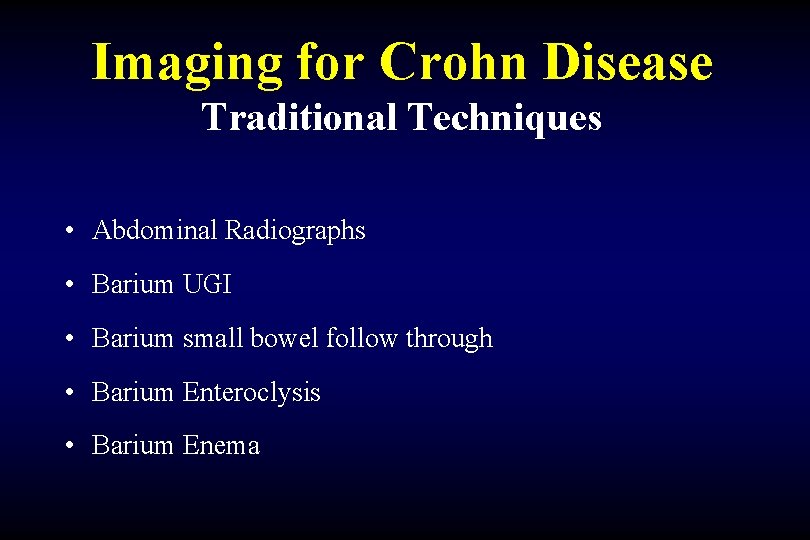
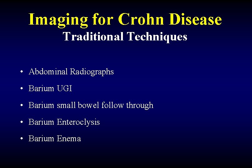
Imaging for Crohn Disease Traditional Techniques • Abdominal Radiographs • Barium UGI • Barium small bowel follow through • Barium Enteroclysis • Barium Enema
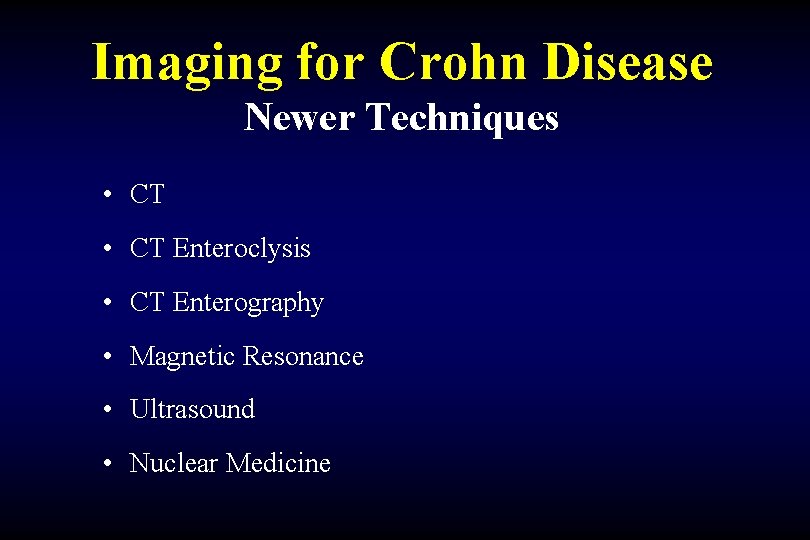
Imaging for Crohn Disease Newer Techniques • CT Enteroclysis • CT Enterography • Magnetic Resonance • Ultrasound • Nuclear Medicine
Imaging for Crohn Disease Traditional Techniques • Abdominal Radiographs – Use for initial evaluation of acute pain – Bowel obstruction – Perforation – Limited value
Imaging for Crohn Disease Traditional Techniques • Barium UGI – limited in the evaluation of milder cases of mucosal and transluminal inflammation in EGD region
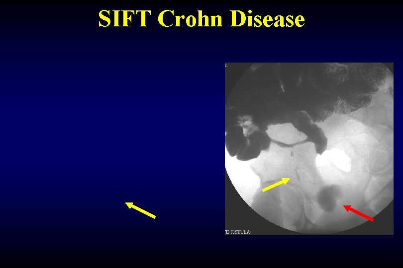
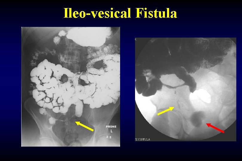
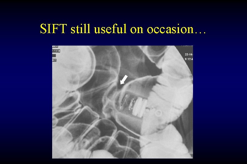
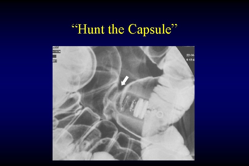
Imaging for Crohn Disease Traditional Techniques • Barium small bowel follow through – Distention of small bowel with contrast material is essential for proper evaluation - poor distension of the lumen causes subtle lesions to be overlooked – Must use intermittent compression to find lesions – Role in 2005: pre capsule endoscopy evaluation for strictures ?
SIFT Crohn Disease
Ileo-vesical Fistula
SIFT still useful on occasion…
“Hunt the Capsule”
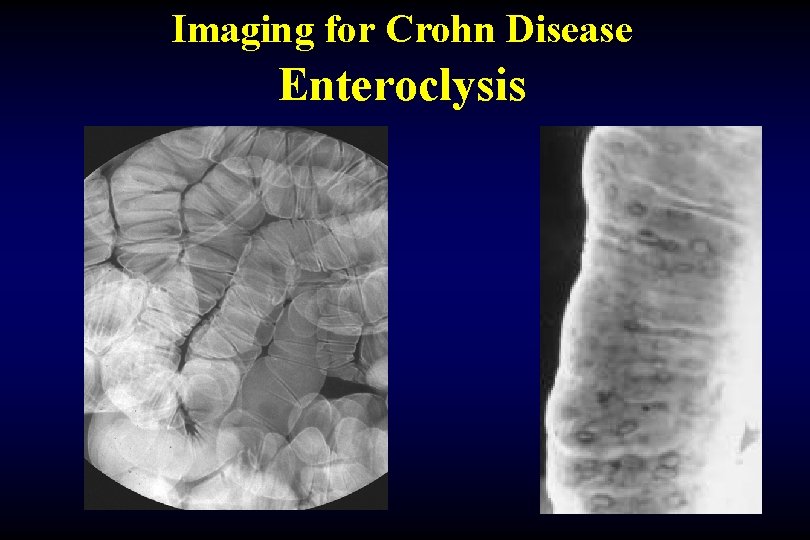
Imaging for Crohn Disease Traditional Techniques • Enteroclysis – Enteroclysis can improve small bowel distension by infusing barium contrast rapidly via a duodenal tube – Unfortunately, the passing of the enteroclysis catheter into the distal duodenum is often difficult and unpleasant for the patient – Time consuming procedure, difficult technique
Imaging for Crohn Disease Enteroclysis
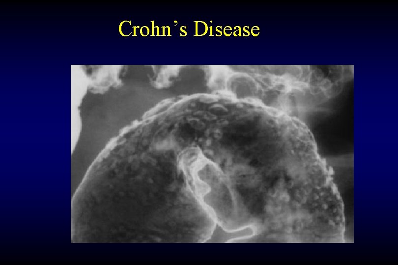
Imaging for Crohn Disease Traditional Techniques • Barium Enema – Used less frequently in recent years – helpful in patients who have strictures that preclude endoscopy • Asymmetric colonic wall involvement • Punched-out ulcers (aphthous, rose thorn, collar stud) • Discontinuous bowel inflammation • Terminal ileum often involved
Crohn’s Disease
Imaging for Crohn Disease Newer Techniques • CT Enteroclysis • CT Enterography • Magnetic Resonance • Ultrasound • Nuclear Medicine
Imaging for Crohn Disease Newer Techniques • CT Enteroclysis • CT Enterography • Magnetic Resonance • Ultrasound • Nuclear Medicine
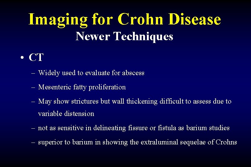
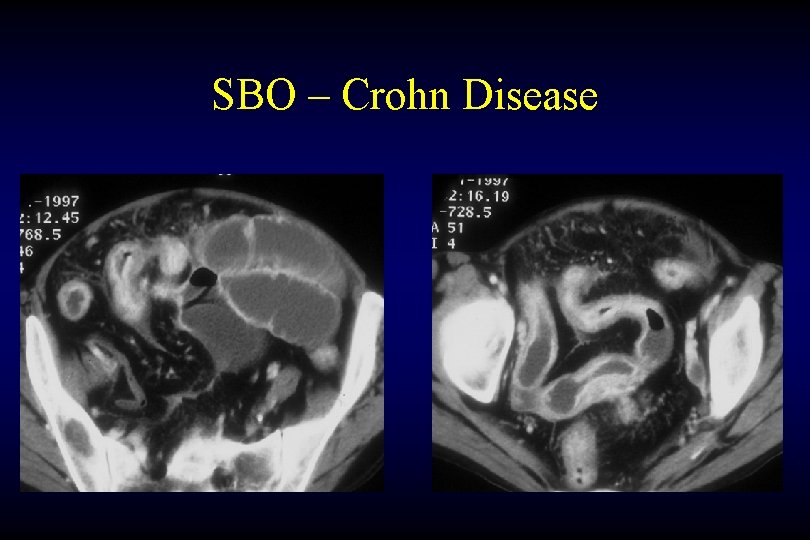
Imaging for Crohn Disease Newer Techniques • CT – Widely used to evaluate for abscess – Mesenteric fatty proliferation – May show strictures but wall thickening difficult to assess due to variable distension – not as sensitive in delineating fissure or fistula as barium studies – superior to barium in showing the extraluminal sequelae of Crohns
SBO – Crohn Disease
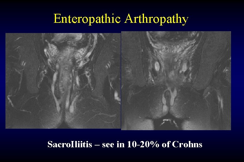
Enteropathic Arthropathy Sacro. Iliitis – see in 10 -20% of Crohns
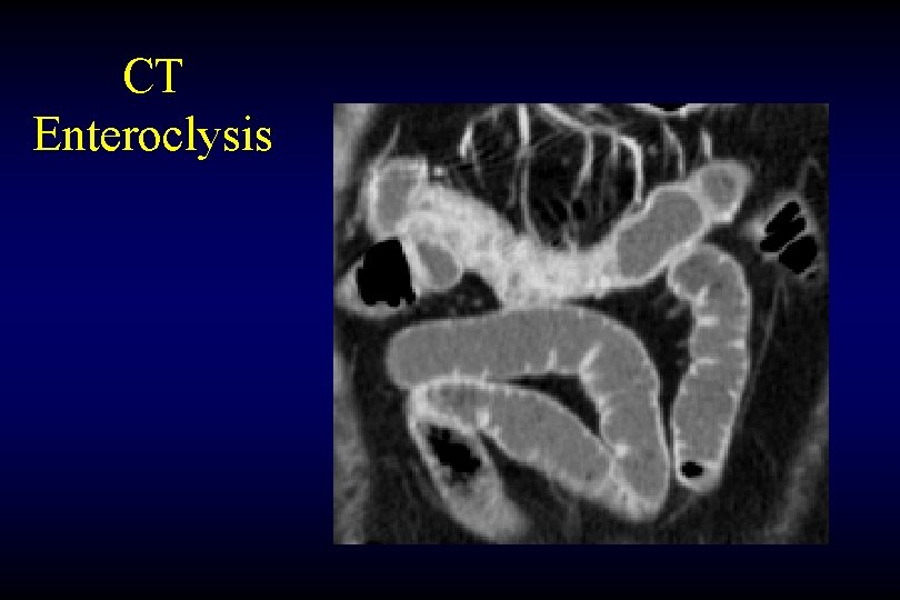
Imaging for Crohn Disease Newer Techniques • CT Enteroclysis – High volume positive contrast infused rapidly via tube – improves small bowel distension – sensitive for small lesions – Time consuming procedure to pass Enteroclysis tube – Need to use Fluoro room & CT scanner – Unpopular with patients (and radiologists !)
CT Enteroclysis
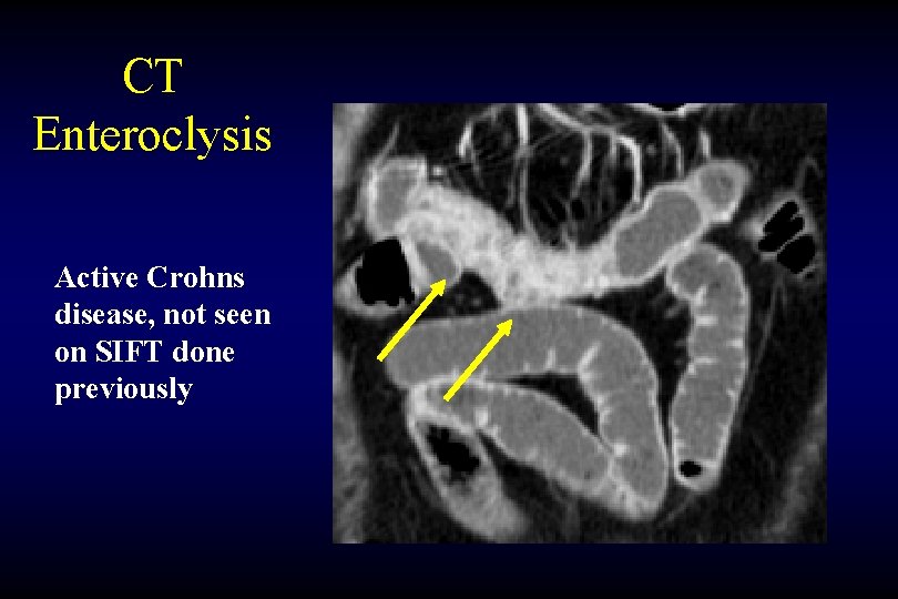
CT Enteroclysis Active Crohns disease, not seen on SIFT done previously
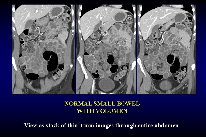
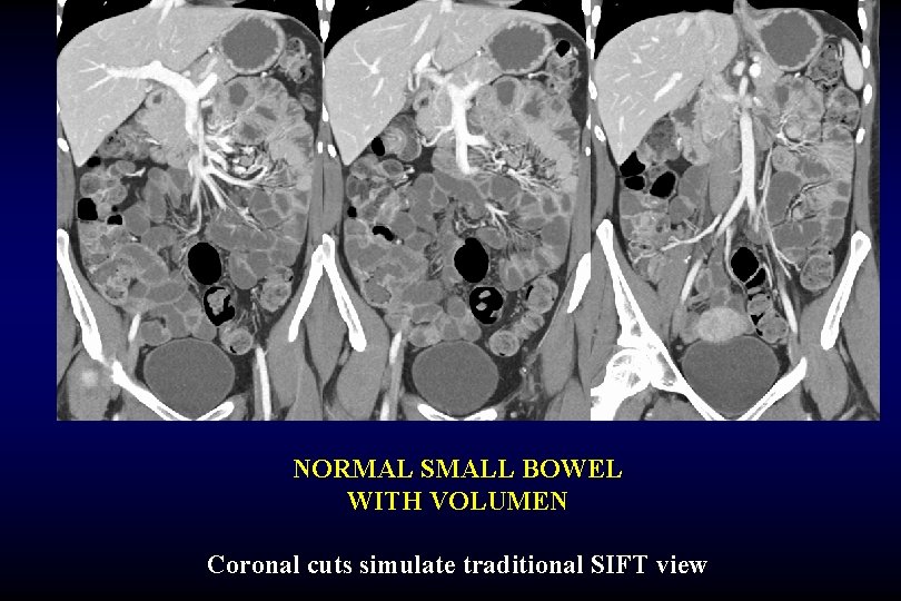
Imaging for Crohn Disease Newer Techniques • CT Enterography – High volume (1200 ml) negative oral contrast (Vo. Lumen) over 1 hour – improves small bowel distension c/w regular CT or SIFT – Give IV contrast to evaluate bowel wall – Use thin section multislice CT cuts to generate 3 D coronal and sagital views also – Well tolerated by patients, no need for jejunal tube
NORMAL SMALL BOWEL WITH VOLUMEN View as stack of thin 4 mm images through entire abdomen
NORMAL SMALL BOWEL WITH VOLUMEN Coronal cuts simulate traditional SIFT view
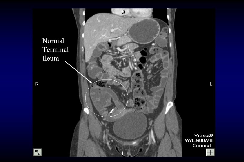
Normal Terminal Ileum
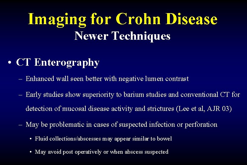
Imaging for Crohn Disease Newer Techniques • CT Enterography – Enhanced wall seen better with negative lumen contrast – Early studies show superiority to barium studies and conventional CT for detection of mucosal disease activity and strictures (Lee et al, AJR 03) – May be problematic in cases of suspected infection or perforation • Fluid collections/abscesses may appear similar to bowel • May avoid post operatively or when abscess suspected
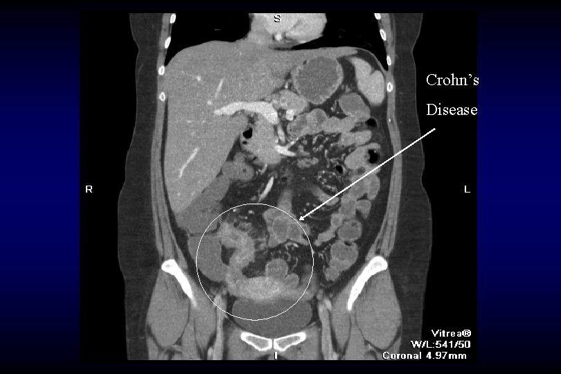
Crohn’s Disease
Crohn’s Disease Inflammatory Hyperemia and Reactive adenopathy
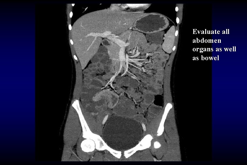
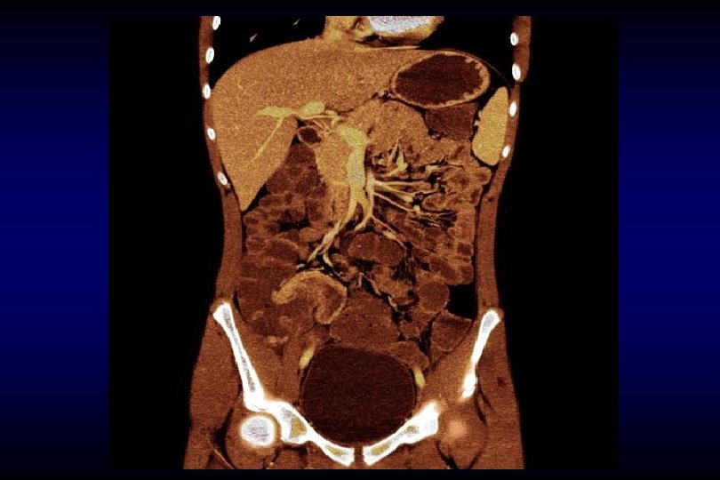
Evaluate all abdomen organs as well as bowel
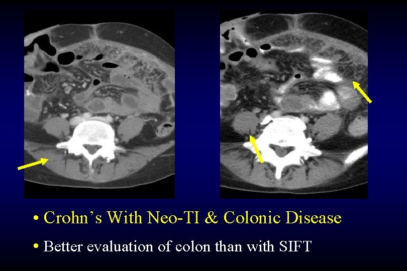
• Crohn’s With Neo-TI & Colonic Disease • Better evaluation of colon than with SIFT
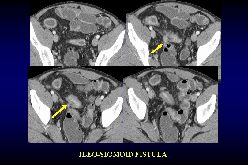
ILEO-SIGMOID FISTULA
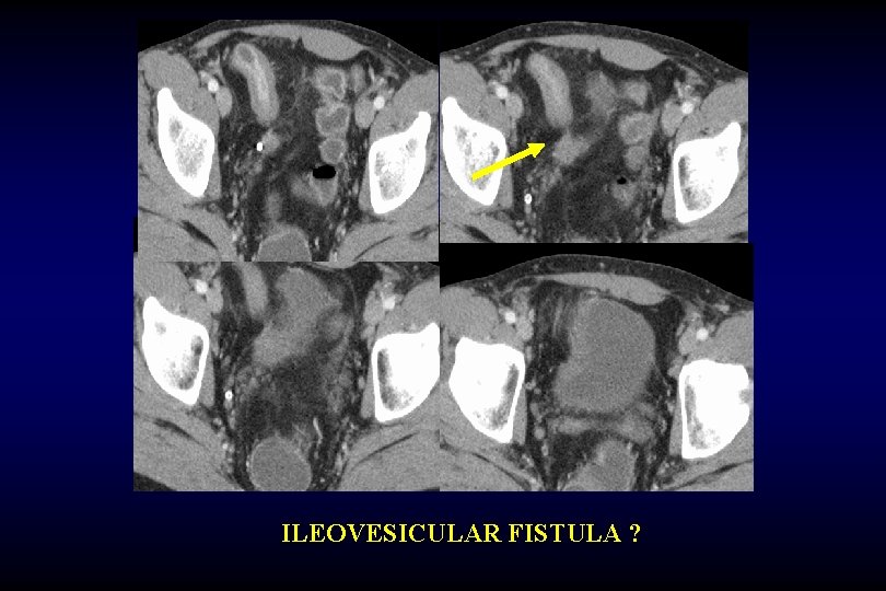
ILEOVESICULAR FISTULA ?
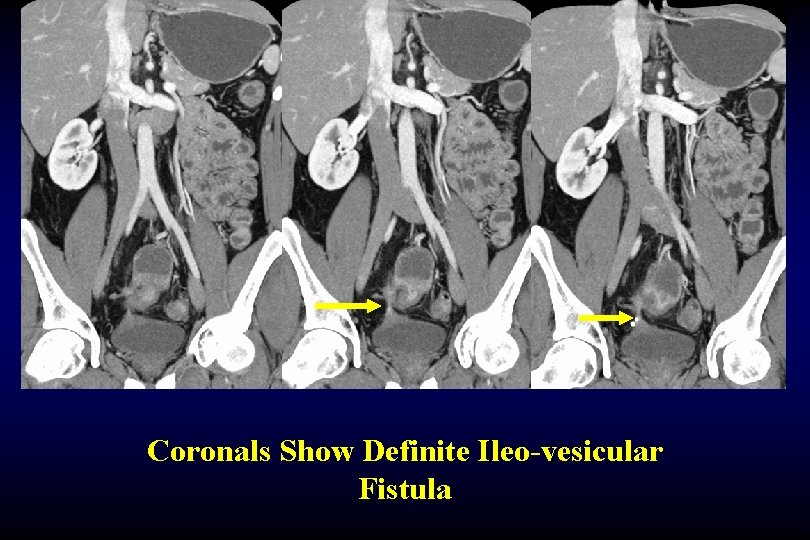
Coronals Show Definite Ileo-vesicular Fistula
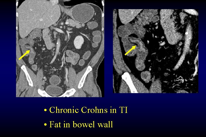
• Chronic Crohns in TI • Fat in bowel wall
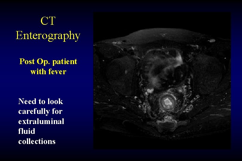
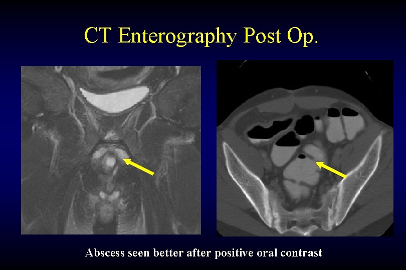
CT Enterography Post Op. patient with fever Need to look carefully for extraluminal fluid collections
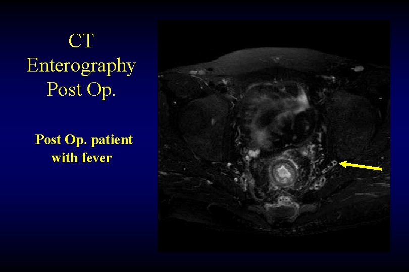
CT Enterography Post Op. patient with fever
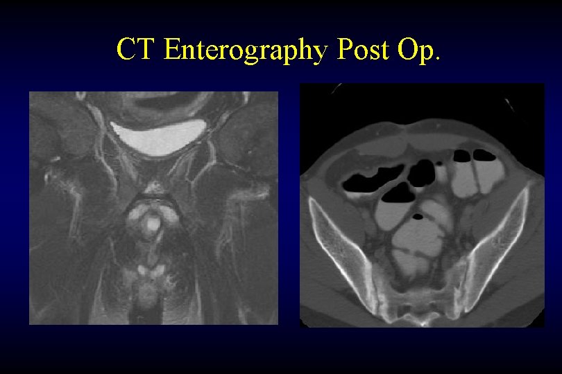
CT Enterography Post Op.
CT Enterography Post Op. Abscess seen better after positive oral contrast
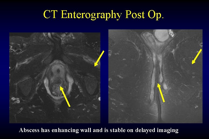
CT Enterography Post Op. Abscess has enhancing wall and is stable on delayed imaging
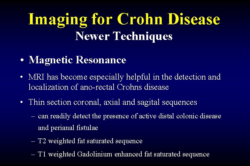
Imaging for Crohn Disease Newer Techniques • Magnetic Resonance • MRI has become especially helpful in the detection and localization of ano-rectal Crohns disease • Thin section coronal, axial and sagital sequences – can readily detect the presence of active distal colonic disease and perianal fistulae – T 2 weighted fat saturated sequence – T 1 weighted Gadolinium enhanced fat saturated sequence
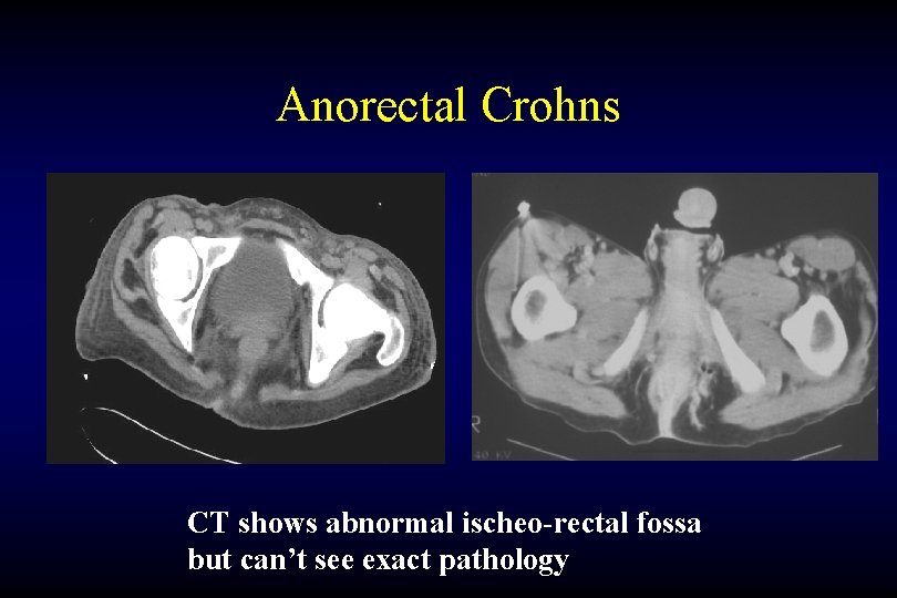
Anorectal Crohns CT shows abnormal ischeo-rectal fossa but can’t see exact pathology
MR of Ano-rectal disease Coronal Axial
MR of Ano-rectal disease Axial
MR of Ano-rectal disease Coronal
MR of Ano-rectal disease Bilateral severe complex trans-sphincteric fistulae
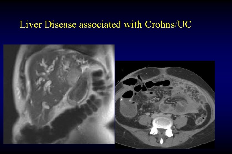
Liver Disease associated with Crohns/UC
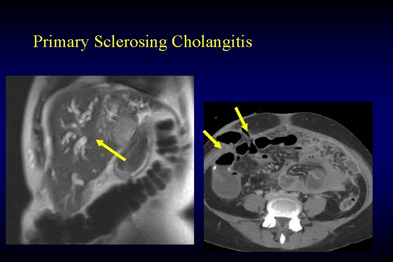
Primary Sclerosing Cholangitis
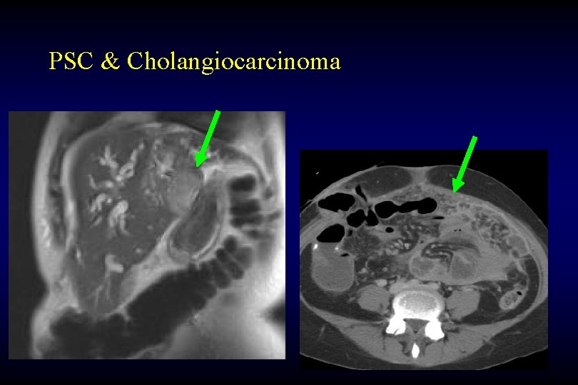
PSC & Cholangiocarcinoma
Imaging for Crohn Disease Newer Techniques • Ultrasound – difficult to do, inconsistent results – May be used to monitor therapy in kids • Nuclear Medicine – Indium scan – Not often used – May be incidental finding of increased activity in bowel
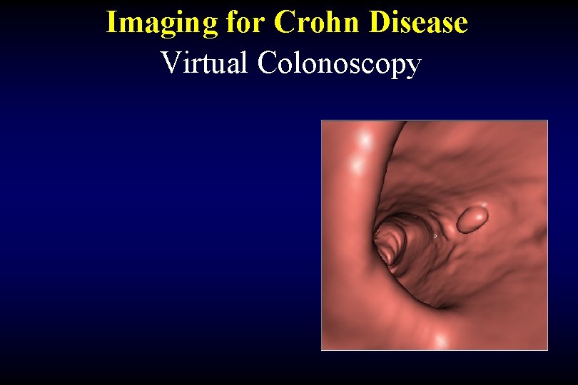
Imaging for Crohn Disease Newer Techniques CT Colography (Virtual Colonoscopy)
Imaging for Crohn Disease Virtual Colonoscopy
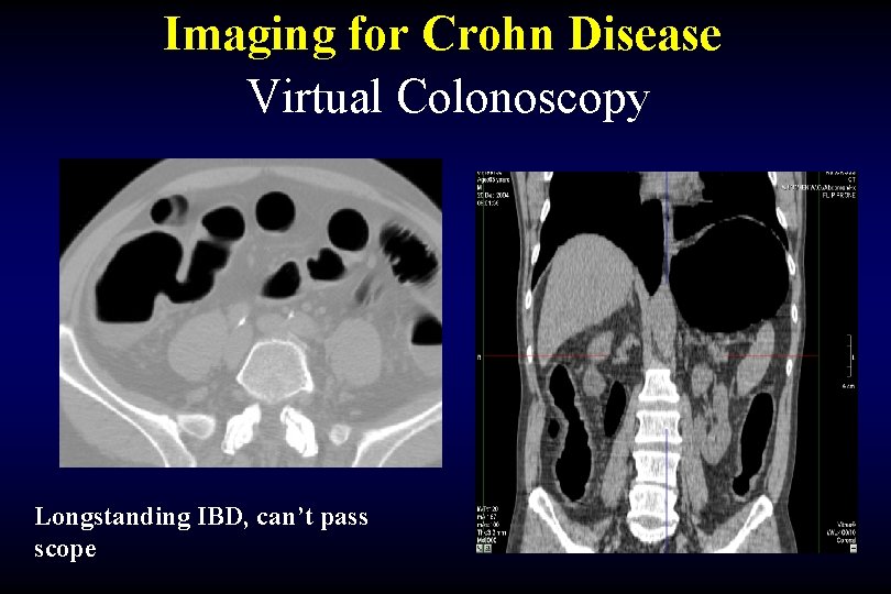
Imaging for Crohn Disease Virtual Colonoscopy Longstanding IBD, can’t pass scope
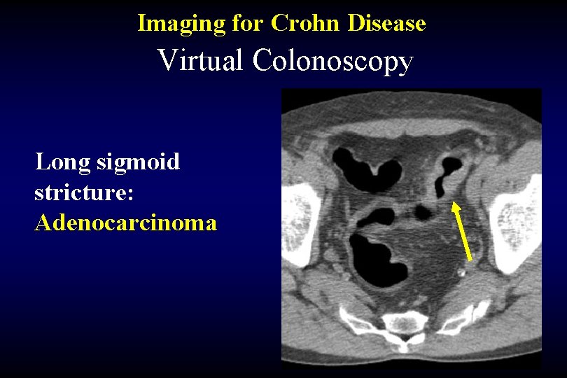
Imaging for Crohn Disease Virtual Colonoscopy Long sigmoid stricture: Adenocarcinoma
Imaging for Crohn Disease Conclusions Traditional Techniques Newer Techniques
Imaging for Crohn Disease Traditional Techniques • Abdominal Radiographs • Barium UGI • Barium small bowel follow through • Barium Enteroclysis • Barium Enema
Imaging for Crohn Disease Newer Techniques • CT Enteroclysis • CT Enterography • Magnetic Resonance • Ultrasound • Nuclear Medicine
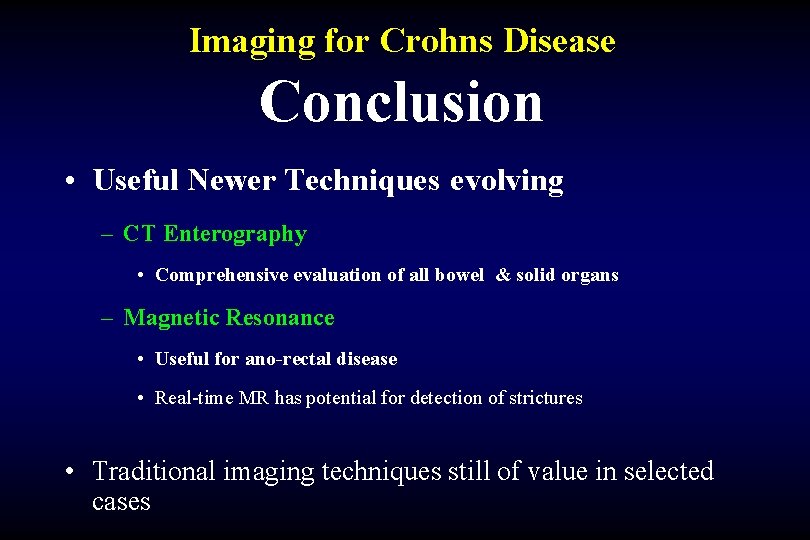
Imaging for Crohns Disease Conclusion • Useful Newer Techniques evolving – CT Enterography • Comprehensive evaluation of all bowel & solid organs – Magnetic Resonance • Useful for ano-rectal disease • Real-time MR has potential for detection of strictures • Traditional imaging techniques still of value in selected cases
THANK YOU