Neurosurgical Management of Intraspinal Lesions Considerations for the


- Slides: 36
Neurosurgical Management of Intraspinal Lesions: Considerations for the Deformity Surgeon ICEOS November 18, 2011 Amer Samdani, MD Director Pediatric Spine Surgery Shriners Hospitals for Children Philadelphia, PA
Objectives Main intraspinal lesions n Chiari malformation n Tethered cord n Diastematomyelia n Patient with myelomeningocele n Timing and effect of neurosurgical intervention n
Chiari Malformation n n Chiari 1 n Herniation of cerebellar tonsils below foramen magnum n Usually asymptomatic n Associated syrinx Chiari 2 n Almost exclusively seen in children with MM n Herniation of tonsils, vermis, 4 th ventricle Chiari 3 Chiari 4 Hankinson et al Neurosurg Clin North Am 2007
Case Presentation n 8 yo girl presented with 30° right thoracic curve n MRI scan demonstrated Chiari malformation with holocord syrinx Underwent Chiari decompression What are chances of curve improvement or stabilization?
Result of Chiari Decompression on Spine Deformity Eula et al Spine 2002 Brock Meyer et al Spine 2003 Flynn et al Spine 2004 Bangor et al Childs Nervous System 2006
SRS 2009: The Natural History of Scoliosis Secondary to Chiari I Malformation and Syringomyelia after Suboccipital Decompression in Young Patients n n n Li Wei-guo, MD; Prof. Quiz Yong Largest series to date of 121 patients with scoliosis and Chiari decompression Progressors older than nonprogressors (14. 2 vs. 9. 2 years) Progressors mean Cobb angle of 43° versus 31° for nonprogressors
Chiari with No Syrinx n 12 yo girl with AIS n MRI probably not necessary n Do et al JBJS 2002 n 2% (7/327) n None required neurosurgical intervention n MRI reserved for high risk n Richards et al Spine 2011 n 529 patients n 6. 8% with intraspinal anomalies n Risk factors: increased
MRI n n n ‘Low lying tonsils’ No syrinx Underwent uneventful surgery
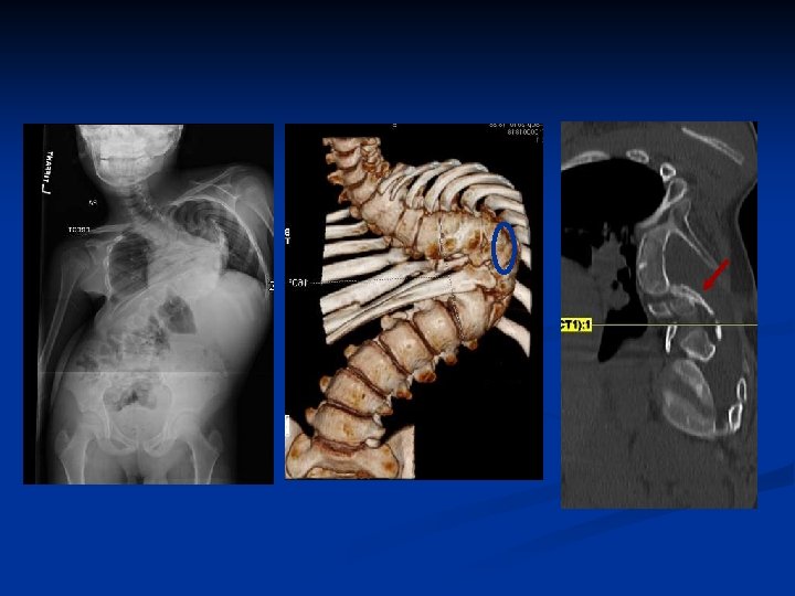
Chiari with Syrinx n n n 14 yo girl with a Lenke 2 curve n MRI revealed a Chiari with large syrinx Underwent Chiari decompression How long should one wait prior to deformity correction? n Repeat MRI 4 -6 months to document decrease in syrinx Witten et al J Neurosurg Pediatr 2008
Postoperative Films
What if the syrinx does not resolve? n n n Wait longer if deformity permits n Atenello et al Neurosurgery 2008 n Median time to resolution of syrinx = 10 months Drainage of syrinx n Morbidity n Aghakhani et al Neurosurgery 2010 Increased risk of not obtaining reliable neuromonitoring
Tethered Cord n n Conus ends at L 1 -L 2 Variable etiologies n Fatty filum n Lipoma n Myelomeningocele n All radiographically tethered Mc. Clone Pediatr Neurosurg 1992
Tethered Cord n n 3 year old with presumed idiopathic scoliosis Very high incidence of intraspinal anomalies n Dobbs et al JBJS 2002 n 21. 7% (10/46) neural axis abnormalities n Philadelphia Shriners n Pahys, Samdani, Betz Spine 2009 n 13% (7/54)
Tethered Cord MRI reveals a low-lying conus with a fatty filum n Recommend untethering n Low morbidity n. Bowman et al J Neurosurg Pediatr 2009 n
Intraspinal Anomaly and Scoliosis Correction n Timing of surgery n Traditionally 2 stages operated on three months apart n. CSF leak n. Two hospitalizations n. Risk of retethering
Intraspinal Anomaly and Scoliosis Correction n Improved neuromonitoring allows for one stage procedures n Samdani et al Spine 2007 n Hamzaoglu et al Spine 2007 n 21 patients with congenital scoliosis/kyphosis n No infections,
Myelomeningocele and Tethered Cord Hudgins & Gilreath, 2004 n All patients with MM radiographically tethered BUT 10 -30% symptomatic n Weakness n Gait n Pain n Scoliosis n Worsening of foot and hip deformities n Urologic
Outcomes Following Detethering n n n Bowman et al J Pediatr Neurosurg 2009 n 114 patients Pierz et al J Pediatr Orthop 2000 n 21 patients Herman et al Pediatr Neurosurg 1993 n 153 patients Fone et al J Urol n 39 patients Reigel et al Pediatr Neurosurg 1994 n 262 patients Palmer et al J Urol 1998
Untethering in Patients with MM n Risks n Neurologic worsening n CSF leak n Wound problems
Aim n Is untethering necessary in the asymptomatic patient with MM prior to deformity correction?
Methods n n n We retrospectively identified 19 patients with MM who had: n no evidence of a clinically symptomatic tethered cord n a spinal fusion for deformity correction n no untethering for at least one year prior to surgery Minimum follow-up after fusion was 2 years Charts and radiographs were reviewed for neurologic or shunt complications intra-op and within 3 months of surgery
Patient Demographics Total Patients Males Females Avg. age at surgery 19 8 11 12. 2 years (10 -17) Follow-up Shunt present (%) Motor level Thoracic L 1 or L 2 L 3 L 4 3. 9 years (2 -8) 14/19 (74%) 8 7 2 2
Results n n Untethering 1 year prior to spine correction New neuro deficit Pre-op major Cobb Post-op major Cobb 0% 1 patient * 81° 33° * Transient lower extremity weakness which returned to baseline within one month of surgery Samdani et al Neurosurg Focus 2010
Conclusion n n Our results suggest that spinal cord untethering may not be necessary in all patients with myelomeningocele undergoing spinal deformity surgery Future studies with larger numbers are needed
Myelomeningocele: Always Consider the Shunt n n Majority of patients with MM will have a VP shunt n Talamonti et al J Neurosurg 2007 Early postoperative death following deformity surgery from shunt malfunction n Geger et al Eur Spine J 2007
Patient KP n n 15 you boy with MM and VPS n Severe curvature and pelvic obliquity n VPS placed at birth and not revised Multiple stage procedure planned n Stage 1: Halo, instrumentation, osteotomies n Stage 2: VCR with completion
Stage 1 One hour after being positioned prone n Bradycardia with hypertension n Emergent shunt tap performed n Elevated intracranial pressure n Surgery aborted n Head CT no change n Neurologically intact n
Possible Reasons for Increased Intracranial Pressure Coincidental shunt malfunction n Unlikely n Increased abdominal pressure resulting in shunt malfunction n Miele et al 2004 Neurosurgery n
Options n n Cancel surgery Externalize the shunt n Would require multiple neurosurgical procedures Intracranial monitoring and CSF drainage Procedure completed n Required CSF drainage to maintain normal ICP
Recommendations Consider baseline HCT n Aggressive bowel prep pre-op n Abdomen free during surgery n Prep patient in a manner to allow easy access to the shunt n Samdani et al JBJS in press
Diastematomyelia n n Split cord syndrome n Associated with congenital scoliosis n Type 1: two separate dural sacs n Type 2: one dural sac Can cause tethering
Patient GF n 12 yo boy with congenital scoliosis, diastematomyelia n Laminectomy and partial resection of diastematomyelia at age 4 n Progressive, severe scoliosis and increased pain with ambulation
Options n Considered resection of diastematomyelia; however… n Shen et al SRS 2010 n 95 patients with diastematomyelia underwent deformity surgery n None prophylactically removed n No neurological
Post-op X-rays
Summary n n n Chiari malformation with syrinx should be decompressed. A repeat MRI 4 -6 months obtained to document a decrease in syrinx size. Not all MM patients need to be dethered prior to scoliosis correction. Not all patients with a diastat need removal prior deformity correction.