Neuroscience Nurse Intracranial Pressure Monitoring Alida Lorenz RN


 • Treatment – Ventricular drain –")


- Slides: 50
Neuroscience Nurse: Intracranial Pressure Monitoring Alida Lorenz, RN, MSN Neuroscience Program Manager
Learning Objectives • Review pathophysiology of increased ICP. • Discuss management of patients with increased ICP. 2
3
CPP: Cerebral Perfusion Pressure 4
5
Pressure Volume Curve 6
Pathophysiology • Increase in brain tissue by space occupying masses • Cerebral edema • Increase in CSF volume • Increase in blood volume • Failure of compensatory mechanisms 7
8
Presentation: Herniation • Earliest clinical signs of transtentorial herniation – Headache and altered level of consciousness • Followed by pupillary changes – Bradycardia is another early sign in children
Hydrocephalus • Types – Communicating – Non-communicating (obstructive) • Treatment – Ventricular drain – Lumbar drain – Ventriculoperitoneal shunt 10
Presentation: Symptoms • Global symptoms of elevated ICP – Headache • Probably mediated via the pain fibers of cranial nerve (CN) V in the dura and blood vessels – Depressed global consciousness • Due to either the local effect of mass lesions or pressure on the midbrain reticular formation – Vomiting • Focal symptoms – May be caused by local effects in patients with mass lesions or herniation syndromes
Presentation: Symptoms 12
Presentation: Symptoms 13
Presentation: Signs • Dilated pupil – Usually on the side of the lesion • Cranial nerve palsies of the third, fourth, and sixth cranial nerves can occur – 3 rd nerve palsy most common – May cause double vision or abnormal head posture
Presentation: Signs • Papilledema – If present can confirm the diagnosis – Papilledema may be absent in acute ICP elevations because it takes several days to become apparent – Is not invariably present in patients with intracranial hypertension
Presentation: Signs • Retinal hemorrhages – may be present in patients with increased intracranial pressure, and should raise the suspicion of nonaccidental head trauma
Late Signs: Posturing 17
Respiratory Patterns 18
Motor Responses 19
Monitoring Concepts • Indications – Head injury – GCS ≤ 8 – SAH – aneurysm – Tumor • Associated complications – Infection most common 20
ICP Monitoring 21
EVD vs. Subarachnoid Bolt In addition to ICP monitoring, what else can a ventricular catheter facilitate? 22
Waveform interpretation • A waves - plateau waves – • • Pressure waves - occur with increased ICP • • If stays 30 – 70 mm. Hg over 20 minutes means CPP severely • compromised • • B & C waves not clinically significant • Condition • • P 1 (percussion wave) –sharp peak and a fairly constant amplitude. • • P 2 (tidal wave) –ends on the dicrotic notch. (elevation is indicative of poor • compliance and therefore outcome) • • P 3 (dicrotic wave)–follows the dicrotic notch. 23
ICP Monitoring 24
Why is it important to monitor ICP? 25
26
Troubleshooting Equipment 27
Complications of Elevated ICP 28
Monitoring ICP 29
Monitoring ICP: Cause For Concern 30
Interventions for ICP Patient Transport 31
Nursing Goals 32
Evaluation: Neuroimaging • Head CT may demonstrate: – Underlying etiology of elevated ICP (eg, mass lesion, hemorrhage) – Findings consistent with elevated ICP (eg, midline shift, effacement of the basilar cisterns – And/or effacement of the sulci) • Patients without these findings on initial CT may have elevated ICP – This was demonstrated in a prospective study of 753 patients treated at four major head injury research centers in the United States, in which patients whose initial CT scan did not show a mass lesion, midline shift, or abnormal cisterns had a 10 to 15 percent chance of developing elevated ICP during their hospitalization Eisenberg, HM, Gary, HE Jr, Aldrich, EF, et al. Initial CT findings in 753 patients with severe head injury. A report from the NIH Traumatic Coma Data Bank. J Neurosurg 1990; 73: 688.
Evaluation: Lumbar Puncture • LP, if necessary, should be deferred until after head CT scan in any patient in whom intracranial hypertension is suspected – Due to the possibility of precipitating herniation across the tentorial notch or into the foramen magnum by increasing the pressure gradient between compartments • In patients in whom central nervous system infection is a strong consideration, deferral of lumbar puncture should not delay the initiation of empiric antibiotic therapy
Medical Management Nursing Care & Priorities Ventriculostomy Sedation, analgesia, neuromuscular blockade Prophylactic anticonvulsant Electrolyte balance Barbiturate coma Proper positioning Temperature control Adequate nutrition VAP bundle 35
36
Management: Intubate and Sedate • Maintaining the head in a midline position • Avoiding high positive pressures and expiratory pressures – May increase intrathoracic pressure and impede venous drainage • Maintaining adequate sedation to permit controlled ventilation – Neuromuscular blockade may be required if ICP remains elevated despite adequate sedation • Muscle relaxation also can prevent fighting against the ventilator and permit hyperventilation if it is required; short-acting agents are preferred, and can be withheld periodically to permit neurologic evaluation – Administration of lidocaine before endotracheal tube suctioning to blunt the gag and cough responses 37
Initial Stabilization: Circulation • Cerebral perfusion must be maintained to prevent secondary ischemic injuries • Hypovolemia should be treated with hypertonic fluids with a goal of attaining a state of normal volume • Excess intravascular volume may exacerbate the development of cerebral edema
39
Management: Mannitol • Establishes an osmotic gradient between plasma and parenchymal tissue, resulting in a net reduction in brain water content • Rapid onset of action and maintains its effect for a period of hours • Can be used to decrease ICP and improve CPP include acute herniation, acute elevation of ICP, and ICP elevation that does not respond to otherapies
Management: Mannitol • Recommended dose is 0. 25 to 1 g/kg IV bolus • Repeat doses can be administered every six to eight hours to increase serum osmolarity to 300 to 310 m. Osm/L • Carefully evaluate in patients who have renal insufficiency
Management: Mannitol Controversies • Mannitol administration has the potential side effects of hyperosmolarity, hypovolemia, electrolyte imbalance, and acute renal failure • More common with chronic or high-dose administration • Serum osmolarity, serum electrolytes, and renal function should be measured at least every six to eight hours • When administered chronically and in high doses, mannitol may cross the injured blood-brain barrier at the site of the cerebral lesion and cause an exacerbation of cerebral edema
Management: Hypertonic Saline • Has been shown to decrease ICP and increase CPP in patients with elevated ICP that is refractory to conventional therapy – Acts by establishing an osmotic gradient that reduces brain water content – Appears to maintain efficacy with repeat dosing even in patients who have stopped responding to mannitol • Unlike mannitol, hypertonic saline does not cause profound osmotic diuresis, and the risk of hypovolemia as a complication is decreased – Theoretical complications, such as hyperosmolarity, central pontine myelinolysis, and congestive heart failure, have not been reported
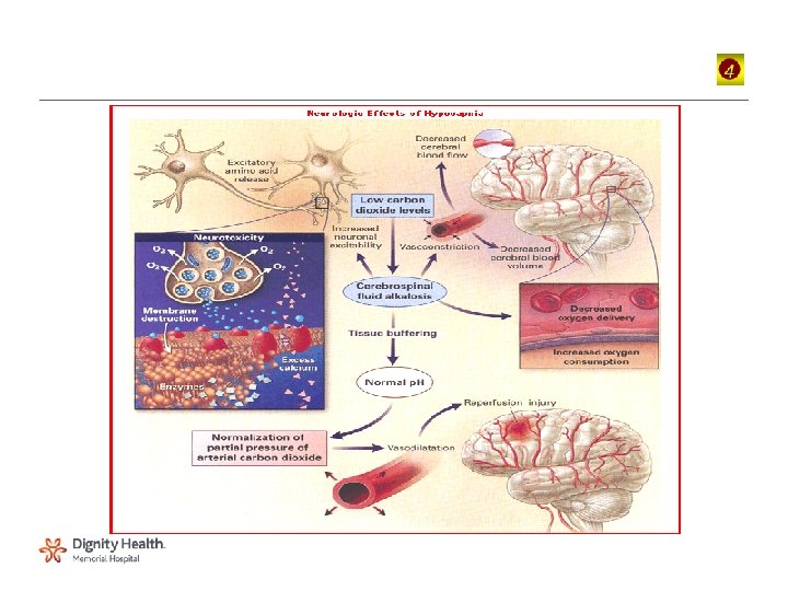
Management: Hyperventilation • Can effectively lower ICP via its effect on Pa. CO 2 – Low Pa. CO 2 causes cerebral vasoconstriction, decreased CBF, and consequently, decreased cerebral blood volume and ICP • Aggressive hyperventilation may decrease CBF enough to cause cerebral ischemia and actually increase the extent of brain injury – In one study of 21 patients with severe traumatic brain injury, forced hyperventilation to an end-tidal PCO 2 of 21 mm. Hg normalized ICP and CPP, but significantly reduced cerebral oxygenation Unterberg, AW, Kiening, KL, Hartl, R, et al. Multimodal monitoring in patients with head injury: evaluation of the effects of treatment on cerebral oxygenation. J Trauma 1997; 42: S 32. – Reserved for episodes of acute brain herniation or ICP elevation that fail to respond to otherapies
Management: Barbiturate Coma • Barbiturates are used to treat intracranial hypertension that is refractory to other modalities – Pentobarbital is the barbiturate that is best studied and most commonly used • Works by decreasing the cerebral metabolic rate, which causes a reduction in CBF and thus, in ICP • May also provide some protective effect for the brain tissue during periods of hypoxia or hypoperfusion • Ability to control ICP elevations with barbiturates is associated with a decreased mortality rate
Management: Barbiturate Coma • Barbiturates produce cardiac suppression, which may result in hypotension – Should be anticipated and treated promptly with fluids and inotropic support if necessary • Invasive cardiopulmonary monitoring may be needed • May also benefit from EEG monitoring to maintain a burst suppression pattern and to monitor for underlying seizures
Management: Hypothermia • Controlled hypothermia has been shown to help reduce ICP in some patients with refractory intracranial hypertension and may improve outcome • Aggressively treating fever with antipyretics and cooling blankets – Hyperpyrexia increases cerebral metabolism and increases CBF, further elevating ICP – Controlling shivering in intubated patients with muscle relaxants
Management: CSF Drainage • In cases of uncontrolled intracranial hypertension, an intracranial drain can be placed to remove CSF and monitor ICP – As the ICP increases, the compliance of the brain decreases, and small changes in volume (eg, the removal of as little as 1 m. L of CSF) can significantly reduce ICP
Craniotomy • Last resort • Allows brain to swell 50