Neuroscience and Behavior Chapter 2 The Brain Studying

Neuroscience and Behavior Chapter 2 – The Brain

Studying the Brain • Clinical Observations • Lesion – Tissue destruction – Can occur naturally or we can selectively destroy clusters of normal or defective brain cells • Electroencephalogram (EEG) – Electrodes measure electrical waves sweeping across surface of the brain – electrodes placed on the scalp
 • Uses X-rays taken")
Studying the Brain • Computerized Axial Tomography (CT or CAT-scan) • Uses X-rays taken from different angles • Often show the size & locations of brain abnormalities caused by tumors, blood vessel defects, blood clots, strokes and other problems. • Can’t view the mental processes
 scan – visual display of brain")
Studying the Brain • Positron emission tomography (PET) scan – visual display of brain activity • Injected with low & harmless dose of radioactive glucose. • Maps the brain at work - Shows the brain’s consumption of glucose; finds brain activity by locating the radioactivity or “hot spots”
 – Uses magnetic fields & radio")
Studying the Brain • Magnetic Resonance Imaging (MRI) – Uses magnetic fields & radio waves to produce computer-generated images that distinguish among different types of soft tissue • Allows us to see structures in the brain (doesn’t reveal functions) • Head put in strong magnetic field. Aligns atoms in brain, then disorients them, measures detectable signals as they move back into place
 – technique for revealing blood flow")
Studying the Brain • Functional MRI (f. MRI) – technique for revealing blood flow and, therefore, brain activity by comparing successive MRI scans • Can reveal functioning as well as structure • Active part of brain = more blood there • Those parts light up during certain mental functions • Gives insight into how brain divides jobs among different parts of brain

Studying the Brain Differences between CT Scan & MRI • CT scans use X-rays • MRI scans use powerful magnetic fields and radio frequency pulses to produce detailed pictures • Differences between normal and abnormal tissue is often clearer on an MRI image than a CT. • No radiation in an MRI scan, but it can be a noisy exam and takes longer than a CT

Inside the Brain • Divided into 3 parts: • Hindbrain – most primitive part – responsible for our basic life functioning (heartbeat, digestion, arousal, balance) • Midbrain – sends signals from the hindbrain to the forebrain – helps process info relating to the senses • Forebrain – last part of the brain to form – most complex – regulates emotions, hunger, formation of long-term memories, growth hormones, & sense of smell

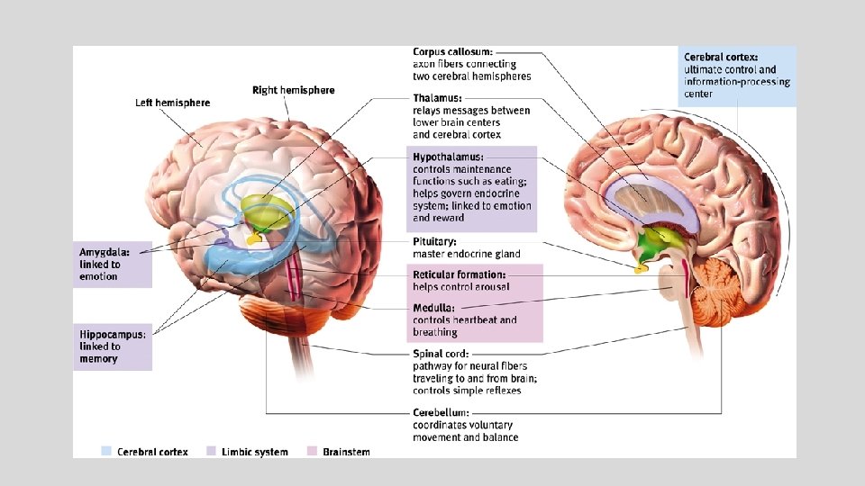
Older Brain Structures • Brainstem - the oldest & innermost region, beginning where the spinal cord swells and enters the skull. • Responsible for automatic survival functions • Medulla – base of brainstem • controls basic life support functions heartbeat, blood pressure, and breathing • Regulates reflexes such as swallowing, sneezing, etc.

Older Brain Structures Pons – above the medulla • mass of nerve fibers that help relay messages from the cortex & cerebellum • involved in facial movement, sleep & arousal Reticular Formation – finger-shaped nerve network and cross-wiring area, extends from spinal cord to the thalamus • filters and relays info from body to brain • Involved in arousal – wake & sleep

Older Brain Structures • Thalamus – top of brain stem, eggshaped structure • “Gateway to the cortex” • Sensory switchboard of the brain • Receives sensory signals (except smell) from the spinal cord and sends them to brain regions associated with seeing, hearing, tasting, touching

Figure 2. 16 The brainstem and thalamus Myers: Psychology, Eighth Edition Copyright © 2007 by Worth Publishers

Older Brain Structures • Cerebellum – “little brain” – two lobes attached to the rear of the brainstem • Helps coordinate voluntary movements and balance

Older Brain Structures - The Limbic System • The Limbic System includes the hypothalamus, amygdala, and the hippocampus • Doughnut-shaped system of neural structures at the border of the brainstem • Core of the forebrain • Regulates memory, fear, aggression, hunger, & thirst

Older Brain Structures - The Limbic System Amygdala – a ring of structures at the border of the brainstem and cerebrum (lima bean shaped) • Associated with emotional processing such as fear, aggression and drives for food and sex • Coordinates fight or flight response

Older Brain Structures - The Limbic System • Hypothalamus – sits just below the thalamus • Links the brain to the endocrine system through control of the pituitary gland = hormone release • Involved in motivation & homeostasis • Regulates body functions - hunger, thirst, body temp, biological rhythms

Older Brain Structures - The Limbic System • Hippocampus • Enables the formation of long term memory - “file cabinet” • Involved in the storage & retrieval of memories located elsewhere in the brain • Largest concentration of acetylcholine – lack of = Alzheimer’s Hippocampus

Structure of the Cortex • Cerebrum – topmost layer of the brain; the bulbous cap over the limbic system • Cerebral Cortex – layer of thin, gray matter covering the cerebrum (wrinkles are called fissures) • Most highly evolved part of the human brain – enables perceiving, thinking, & speaking • Cerebral hemispheres – the left and right side of the cerebral cortex • Corpus callosum – large bundle of axons that connects the two hemispheres; relays info between the two

Structure of the Cortex • Part of the cerebrum, the two large hemispheres comprising 85% of brain weight • The cerebral cortex is made up of four lobes
 • Most evolved, largest lobe •")
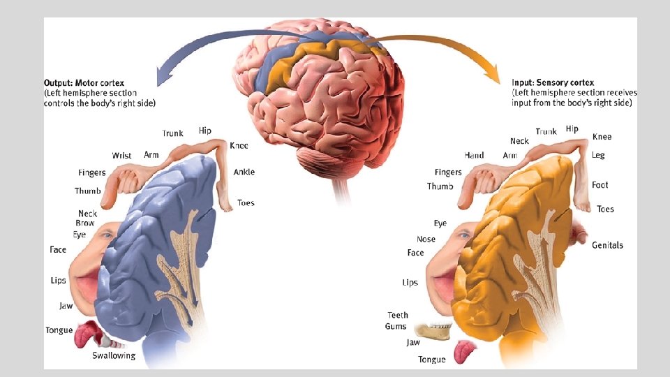
Structure of the Cortex Frontal Lobes - (forehead) • Most evolved, largest lobe • Controls the motor cortex – voluntary muscle movements • Involved in planning, making judgements, creative thinking, emotional control Parietal Lobes - (top to rear head) • Receives & processes sensory information • Integrates in the visual senses from the occipital lobe • Contains sensory cortex – registers & processes body touch and movement sensations
 • Visual cortex -")
Structure of the Cortex Occipital Lobes - (back of head) • Visual cortex - Processes visual information • Damage = Vision problems Temporal Lobes – (side of head, above ears) • Process sound sensed by our ears • Interpreted in Auditory Cortex

Structure of the Cortex • Association Areas – large areas of cerebral cortex that haven’t been specifically labeled for anything motor or sensory (present in all 4 lobes) • Interpret, integrate, & act on info. processed by the sensory areas • Responsible for higher level mental functions: Learning, remembering, thinking, speaking, planning, forming judgments

Phineas Gage • A 19 th century American railroad construction foreman • In 1848, survived an accident in which a large iron rod was driven completely through his head, destroying much of his brain's left frontal lobe • The first case suggesting that damage to specific regions of the brain might affect personality and behavior.

Language

Language Acquisition Areas • Aphasia – impairment of language usually caused by damage to the Broca’s area or the Wernicke’s area. • Broca’s area – left frontal lobe; responsible for transferring thoughts into spoken language • Damage = inability to speak coherently • Wernicke’s area – left temporal lobe; responsible for transferring spoken words into thoughts • Damage = inability to understand spoken language • Angular gyrus – an area of the left occipital lobe that transforms visual representation into an auditory code

Language Acquisition Areas

Plasticity • Severed neurons usually do not regenerate, but some neural tissues can reorganize in response to damage. • Plasticity refers to the brain’s ability to modify or reorganize its neural pathways after some types of injury or illness. • Neurogenesis is the formation of new neurons. • When we are young our brains are more plastic.

Splitting the Brain • A form of psychosurgery in which the two hemispheres of the brain are isolated by cutting the connecting fibers between them (mainly those of the corpus callosum). • Roger Sperry conducted research on this procedure to help people suffering from grand mal seizures

Splitting the Brain • The 2 hemispheres compliment each other • Info from left half of vision field goes to right hemisphere; info from right half goes to left • Each receives sensory info from both the right & left visual fields • Data transmitted to the other hemisphere via the corpus callosum…but not in a person with a severed corpus callosum

Splitting the Brain • Sperry & Gazzaniga - tested split-brain patients • HE-ART flashed on screen • HE goes to right hemisphere, ART to left • Patients said they saw ART (speaking = Broca’s in the left hemisphere) • But pointed w/ left hand to HE (right visual field communicates with left part of body)

Splitting the Brain • With a split brain, both hemispheres can comprehend & follow an instruction. • Gazzaniga – concluded that the conscious left hemisphere is an “interpreter” that constructs theories to explain our behavior • Left hemisphere more active when a person deliberates over a decision • Right hemisphere better than left at recognizing faces, perceiving emotion, and expressing emotion Researchers say people choose the on the right, because the right hemisphere, which is skilled in emotion processing, receives info from the left half of each face (the left half looks happier on the right)

Hemispheric Differences • Hemispheric specialization – lateralization • Our brain is divided into two hemispheres, but both work together equally. • No activity in which only 1 hemisphere is truly, responsible • Logic is not confined to only the left hemisphere • Creativity is not only in the right • No one is purely left or right-brained

The Divided Brain

The Divided Brain • Brain scans show normal individuals engage their right brain when completing a perceptual task and their left brain when carrying out a linguistic task • Left hemisphere is good at making quick, literal interpretations of language • Right hemisphere excels in making subtle inferences

Brain Organization & Handedness • 90% of humans are right-handed • The 10% of left-handers show less predictable patterns of hemispheric dominance • Causes? • Genetics? • Fetal testosterone levels? • Learned? • Handedness and sexual orientation?
- Slides: 37