Neurophysiology II PHS 415 by Adedayo Lawrence Department
 by Adedayo, Lawrence Department of Physiology Faculty of Basic Medical")
Neurophysiology II (PHS 415) by Adedayo, Lawrence Department of Physiology Faculty of Basic Medical and Health Sciences Bowen University Iwo First Semester 2018/19 session

CONTROL OF POSTURE AND MOVEMENT, MAJOR CORTEX, MAJOR PATHWAY LECTURE 4

INTRODUCTION • Somatic motor activity depends ultimately on the pattern and rate of discharge of the spinal motor neurons and homologous neurons in the motor nuclei of the cranial nerves. • These neurons, the final common paths to skeletal muscle, are bombarded by impulses from an immense array of pathways. • The inputs converging on the motor neurons subserve three semi-distinct functions: a. They bring about voluntary activity

CONT’D B. They adjust body posture to provide a stable background for movement C. They coordinate the action of the various muscles to make movements smooth and precise. The patterns of voluntary activity are planned within the brain, and the commands are sent to the muscles primarily via the corticospinal and corticobulbar systems. Posture is continually adjusted not only before but also during movement by posture-regulating systems.

CONT’D § Movement is smoothed and coordinated by the medial and intermediate portions of the cerebellum (spinocerebellum) and its connections. § The basal ganglia and the lateral portions of the cerebellum (neocerebellum) are part of a feedback circuit to the premotor and motor cortex that is concerned with planning and organizing voluntary movement.

TYPES OF MOVEMENT • Involuntary or reflexive • Voluntary Involuntary or reflexive: Some would add as a subdivision of reflex responses rhythmic responses such scratching, and as swallowing, walking, which chewing, are largely involuntary but subject to voluntary adjustment and control. Voluntary: these may be a) gross b) fine discrete (isolated) c) learned (skilled)

Nervous control of voluntary movements The proper performance of voluntary movements requires integrity of the following: Ø a) motor and premotor cortex (area 4 & 6) Ø b) cerebellum Ø c) basal ganglia Ø d) UMN & LMN. It is carried out in two stages as shown in the next slide

Cont’d

Processes involve in the control 1. Planning occurs as follows: • The idea and purpose of the movement originates in the cortical association areas, from which signals are discharged to both the basal ganglia via the caudate and putamen circuits as well as the lateral cerebellum i. e neocerebellum or cerebrocerebellum via the cortico-ponto-cerebellar pathway. • The basal ganglia convert thoughts into a motor plan, and put the program of the performance especially of slow movements.

Cont’d § Then discharge to the cortical motor areas which also share in planning and programming processes especially the supplementary and premotor areas § The neocerebellum shares in planning and control of signals from the motor cortex for performance of the sequential movements according to the plan by its predictive and timing function.
 Execution : this occurs as follows: • The motor cortex discharges signals")
Cont’d 2) Execution : this occurs as follows: • The motor cortex discharges signals to the spinal and cranial motor neurons via the corticospinal and corticobulbar tracts and to the intermediate i. e spinocerebellum. • Spinocerebellum movement, helps monitors its initiation of the performance and corrects any errors by its servocomparator and damping functions, so as to produce coordinate movement without overshooting. a

Cont’d • The motor neurons discharge the final signals to the skeletal muscles. • The skeletal muscles contract according to the plan, producing required movement.

Control of Axial and Distal Muscles § Motor control is that in the brainstem and spinal cord, medial or ventral pathways and neurons are concerned with the control of muscles of the trunk and proximal portions of the limbs. § Whereas lateral pathways are concerned with the control of muscles in the distal portions of the limbs.

Cont’d • The axial muscles are concerned with postural adjustments and gross movements, whereas the distal limb muscles are those that mediate fine, skilled movements. • Thus, for example, the neurons in the medial portion of the ventral horn innervate the proximal limb muscles, particularly the flexors, whereas the lateral ventral horn neurons innervate the distal limb muscles.
 • The motor cortex lies in")
MAJOR CORTEX • Motor cortex (cortical motor areas) • The motor cortex lies in front of the central fissure, occupying nearly the posterior half of the frontal lobe, and is divided into 3 main parts a. Primary motor area cortex b. Premotor cortex c. Supplementary motor area.
 § located at the precentral gyrus. § It")
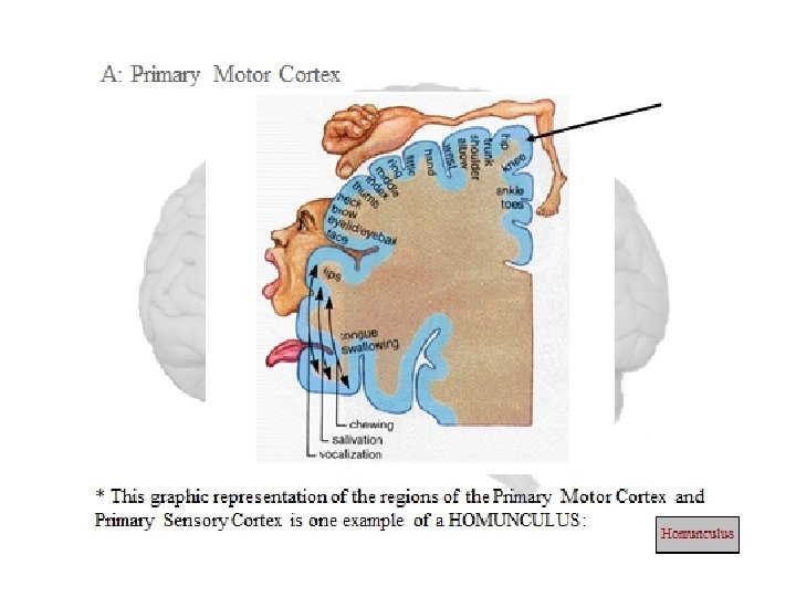
Primary motor area cortex (area 4) § located at the precentral gyrus. § It controls muscle activity in the opposite side but the upper half of the face and the respiratory and abdominal muscles are bilaterally represented. The body is topographically represented in an inverted manner i. e. the feet at the top and the face at bottom, § and the area of representation is proportional to the degree of skilled movements performed by this part. e. g the areas that control the speech muscles and movements of the hands and fingers are much larger than the area that control the trunk muscles.

Function of area 4 • It is the only area that initiates the voluntary fine discrete movements, especially of the hands and fingers • It shares with area 6 in initiating the gross movements • It facilitates the stretch reflex • It is necessary for the occurrence of superficial reflexes.

Premotor area § This lies anterior to the primary motor cortex and it includes mainly area 6, § but areas 8 and 44 are also included. It is less excitable than the primary cortex, but the body representation is roughly the same. § It is the origin of about 30% of the pyramidal fibres and most of the cortical extrapyramidal fibres
 • It shares in planning")
Function of area 6 (premotor or motor association area) • It shares in planning of voluntary movements • It initiates the gross movement (those involving groups of muscles) that help in the performance of specific fine tasks e. g. movements that position the shoulder and the arms at the proper attitude for the hands to perform a fine task. • This is achieved by activating the primary motor cortex both directly by projecting fibres directly to area 4 and indirectly by discharging to the basal ganglia which then discharge to area 4 via the thalamus.

Cont’d • It inhibits the stretch reflex and muscle tone • It shares in controlling autonomic functions • It initiates the subconscious automatic movements e. g. swinging of the arms during walking. • It is necessary for normal flexor response of the plantar reflex. • It contains specialized areas that control specific movements
 : this area lies anterior")
Cont’d • Broca’s area (word formation area, area 44) : this area lies anterior to the lower part of the primary motor cortex and it is essential for normal speech • Eye movement area (frontal eye field area, area 8): this area lies above broca’s area. It directs the eyes voluntarily towards the desired object, and also controls the movements of the eyelids. E. g blinking.

Cont’d • Head rotation area: this area lies above and close to the eye movement area, and it directs the head towards different objects. • Hand skills area: this area lies anterior to the hands and fingers area of the primary motor cortex, and is essential for performance of skilled movements by the hands

Supplementary motor area § This area lies anterior and superior to area 6 and extends on the medial surface of the frontal lobe, so it is sometimes called medial area 6. In it, the body is represented bilaterally in a horizontal plane with the legs posteriorly and the head anteriorly. § It is connected to other motor areas, and is relatively less excitable.
 It supplements area 6 in providing")
Functions of the supplementary motor area • a) It supplements area 6 in providing attitudinal and fixation movements as a background for performance of fine movements. • b) Involvement in planning and programming of complex voluntary movements, with other areas. This is proved by recording a slow surface negative potential over this area about one second before the movement starts (called the readiness potential) as well as an increase in its metabolism and local blood flow.

MAJOR PATHWAY The descending motor tracts: § May be classified into 2 systems known as the pyramidal and extrapyramidal systems. § The neurons of these tracts are called upper motor neurons and they terminate at the cranial and spinal motor neurons. § The latter are called the final common paths and they constitute the lower motor neurons.
 This consists of the corticospinal and corticobulbar tracts")
The pyramidal system (direct activation pathway) This consists of the corticospinal and corticobulbar tracts which arises from the frontal eye field area 8. The corticospinal tract (pyramidal tract) Functions: • Initiation of voluntary movements, especially the fine discrete movements of hands and fingers. • Excitation of the alpha motor neurons, thus facilitating stretch reflex • Facilitation of the superficial reflexes, particularly the flexor plantar response • All these function occurs at opposite side of the body.
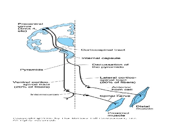

Origin and course § This tract contains about a million nerve fibres. § These are the longest never fibre in the body and originate from area 4 (30%), area 6 and supplementary motor area (30%), and somatic sensory areas (40%). § These fibres descend in the corona radiate, and converge towards the internal capsule where they occupy the genu and anterior two-third of its posterior limb, then they descend in the brain stem as follows:

Cont’d • In the midbrain, they occupy the middle 3 over 5 of the cerebral peduncles. • In the pons, they occupy the basis pontis and are divided into bundles by transverse pontine fibres. • In the upper part of the medulla, the bundles collect in the anterior part forming the pyramid. • However, in the lower medulla, about 80% of the fibres cross to the opposite side (in the motor decussation) and descend in the lateral column of the spinal cord as the lateral corticospinal tract, while the uncrossed fibres descend directly in the anterior column of the spinal cord as the ventral corticospinal tract

Thank you
- Slides: 31