Neuropathology Tumors of the Nervous System Infections of


 • Diffuse – Well")
 • Occur in the optic nerves,")


 • Whorling spindle shaped cells often associated with psammoma body formation")


. • Clinical presentation is with")
 MENINGITIS Neonates - E. coli and Group B Streptococcus Adolescents/young adults –")


 MENINGITIS • Usually a benign illness of children and young adults.")


. • Occurs in immunocompromised hosts. •")

 • Persistent measles virus infection. •")


 • 60% of AIDS patients develop neurologic dysfunction. •")

 Indirect effects Opportunistic Infections Bacteria • Bacterial infections of the CNS in")
 Indirect effects Opportunistic Infections Viruses • Viral infections of the CNS are")
: Indirect effects Opportunistic Infections Fungi and protozoans • Fungal infections of the")


 auto-immune destruction of CNS myelin")


 CAUSES OF DEMENTIA • • Thyroid deficiency B 12 deficiency Drug")


 leads to formation of")


 Cerebral atrophy Superior cerebellar atrophy")


 • Chronic, slowly progressive form of Gullain-Barre • Inflammation")

 • Hereditary Motor and Sensory Neuropathies – HMSM I")
 “Onion bulbs” (layers of Schwann cells from repeated demyelination/remyelination)")
 • Diabetes – Most common cause")


- Slides: 81
Neuropathology: Tumors of the Nervous System Infections of the Nervous System Demyelinating Diseases Degenerative Diseases Metabolic & Toxic Disturbances Peripheral Nerve Disease J. Matthew Velkey matt. velkey@duke. edu 0380 Sealy Mudd/Trent Semans Center
CNS TUMORS • Glial cells (most common “parenchymal” tumor, but still pretty rare: overall incidence ~3 per 100, 000) – Astrocytomas – Oligodendrogliomas – Ependymomas • Neurons (rare in adults, more common in children) – Gangliomas – Medulloblastomas (~20% of childhood CNS tumors) – Neurocytomas • Others – Schwannomas – Meningiomas (also quite common, about as much as astrocytomas)
CNS Tumors tend to follow epidemiologic and anatomic patterns in children and young adults • Childhood locations: – Posterior fossa • Childhood tumors: – Cerebellum – Brainstem – IV Ventricle • Young Adult Locations – Midline or periventricular • Young Adults tumors (low grade) – – Cerebellum Brainstem Hypothalamus Temporal Lobes
Adult CNS Tumors Are More Variable • • • Metastatic tumors Parenchymal Ventricular Dural Spinal Cord – Intramedullary – Extramedullary • Lymphoma Regardless of age, outcome depends on the tumor “grade” World Health Organization (WHO) grading criteria rate neoplasms from I (best) to IV (worst). For CNS tumors: • Grade I - > 10 year survival untreated • Grade II – 5 to 10 year • Grade III – 3 – 5 year • Grade IV - < 3 year
CNS Neoplasia: Pathologic and Clinical Effects • • Mass Effects Focal Neurologic Deficits Seizures Hemorrhage
Astrocytic Tumors • Circumscribed – Pilocytic Astrocytomas (WHO Grade I) • Diffuse – Well Differentiated (Grade II) – Anaplastic Astrocytoma (Grade III) – Glioblastoma (Grade IV)
Pilocytic Astrocytoma • Pilocytic Astrocytomas (WHO Grade I) • Occur in the optic nerves, cerebellum, brain stem, or temporal lobes • Children and young adults • Well circumscribed with little tendency to infiltrate, and frequently cured by surgical excision.
Glioblastoma • • Infiltrative Poorly circumscribed Necrotic Crosses the corpus callosum – “butterfly glioma” • There are two types of glioblastomas recognized – Secondary GBM progress from lower grade tumors – Primary GBM arise de novo with no preceding history • There is mild prognostic benefit to having a secondary glioblastoma
Embryonal Tumors • Tumors composed of cells resembling embryonal cells that occasionally retain an ability for limited divergent differentiation – Medulloblastomas – Atypical Teratoid/Rhabdoid Tumors • Notable features include their high incidence in children and their tendency to seed the neuraxis. • The tumors are rapidly growing and lethal if left untreated, BUT they do respond well to therapy (up to 70% cure rate at some institutions).
Medulloblastoma • Tumor arising in the cerebellum and frequently protrudes into IV ventricle • Most common solid tumor in children • Occurs exclusively in the posterior fossa • The use of radiotherapy, especially when the entire neuraxis is included, has been associated with a cure rate of greater than fifty percent in many institutions. • Chemotherapy has increased this cure rate to approximately 70%.
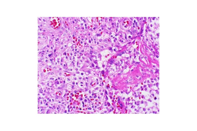
Meningioma • Most common primary tumor of the meninges • One of the most common of all intracranial neoplasms. • The neoplasm is ~3 x more frequent in women • Most remain as discrete masses with a compressive rather than infiltrative relationship to the brain (benign)
Meningioma (Grade I) • Whorling spindle shaped cells often associated with psammoma body formation (“brain sand”)
Acoustic Schwannoma • Any of the cranial nerves (with the obvious exception of CN 1 and 2), are potential sites • Acoustic ‘Neuroma’ CP angle tumor (CN VIII) • Schwannomas are almost always histologically benign, although the size and location of the lesion can present surgical problems.
Neuropathology: Tumors of the Nervous System Infections of the Nervous System Demyelinating Diseases Degenerative Diseases Metabolic & Toxic Disturbances Peripheral Nerve Disease
MENINGITIS • Inflammation of the meninges (arachnoid and pia). • Clinical presentation is with – Headache, vomiting, fever and stiff neck. – Seizures are common in children. – Symptoms are caused by inflammation of the meninges and the subarachnoid space. CSF fills this space. • CSF abnormalities are present which vary with the organism. – Bacteria cause a neutrophilic reaction, ↑protein, ↓glucose – Encapsulated organisms cause a granulomatous reaction, ↑↑protein, normal or ↓glucose. – Viruses cause a lymphocytic reaction, ↑protein, normal glucose – Syphilis cases a plasmacellular reaction.
NEUTROPHILIC (BACTERIAL) MENINGITIS Neonates - E. coli and Group B Streptococcus Adolescents/young adults – Nesseria meningitides Elderly – S. pneumoniae or Listeria monocytogenes
CEREBRITIS
ACUTE FOCAL SUPPURATIVE INFECTIONS • Brain Abscess – Clinical presentation is with focal neurological signs and raised intracranial pressure (which can go on to cause more generalized symptoms). – ↑CSF pressure, WBC and protein. Glucose normal • Subdural empyema • Extradural abscess – Osteomyelitis – Surgical complication
CEREBRAL ABSCESS
CHRONIC BACTERIAL MENINGITIS Mycobacterium tuberculosis • Organisms gain access to the CNS via blood stream. • Caseating granulomas form in the basal meninges. • Parenchymal spread of infection results in a “tuberculoma” which may be mistaken for a tumor. • The infection is indolent but fatal in 4 -6 weeks if it is untreated.
CHRONIC BACTERIAL MENINGITIS Mycobacterium tuberculosis “Tuberculoma”
CHRONIC BACTERIAL MENINGITIS Mycobacterium tuberculosis Caseating granuloma
ACUTE ASEPTIC (VIRAL) MENINGITIS • Usually a benign illness of children and young adults. • There is CSF lymphocytosis, moderate ↑protein • Most common viruses – Coxsackie virus – Echo virus – Nonparalytic polio virus
VIRAL MENINGOENCEPHALITIS • Most commonly caused by Arboviridiae. – Eastern and Western equine, Venezuelan, St. Louis and La Crosse most common in US • Virus is transmitted by mosquitoes and ticks. • Clinical features vary with the virus. • Pathology varies from mild meningitis to severe encephalitis. • Perivascular and parenchymal mononuclear infiltrate and microglial nodules
VIRAL ENCEPHALITIS INCLUSION FORMING VIRUSES • These diseases are generally less common but they are important diagnostic considerations. • Herpes viruses – Herpes simplex – Herpes zoster – Cytomegalovirus • JC virus – Progressive multifocal leucoencephalopathy • Measles virus – Subacute sclerosing panencephalitis
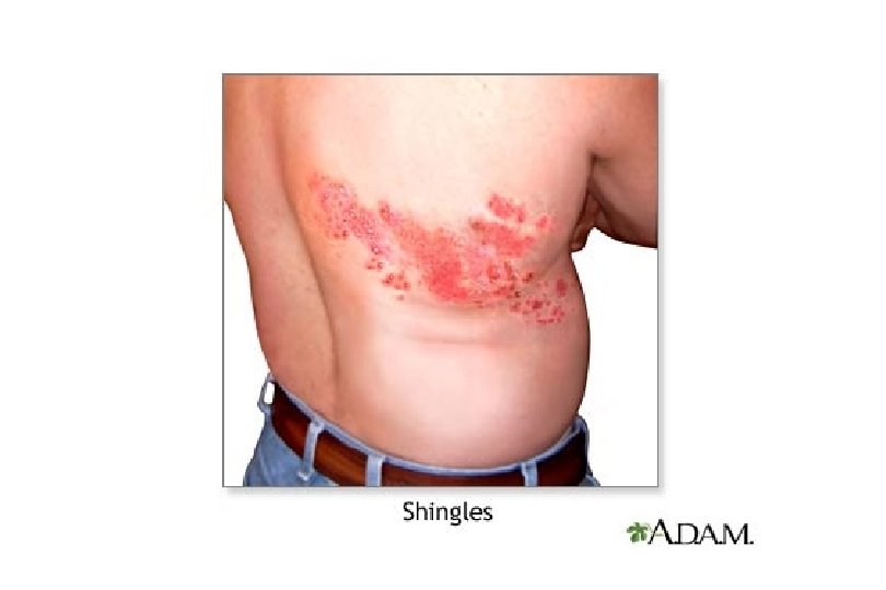
VIRAL ENCEPHALITIS Herpes Viruses • HSV-1 causes “cold sores”. – Virus resides latent in the trigeminal ganglion. – Reactivation may cause Herpes encephalitis which is necrotizing and localized to the temporal lobes. • HSV-2 infects infants via birth canal. – It also causes a necrotizing encephalitis. • Herpes zoster (Shingles) affects older adults – Reactivation of chickenpox (Varicella) infection. – Causes a radiculopathy. • Cytomegalovirus – Causes encephalitis in fetuses infected in utero and in immunocompromised adults, especially AIDS patients.
VIRAL ENCEPHALITIS Herpes simplex
PROGRESSIVE MULTIFOCAL LEUCOENCEPHALOPATHY • Polyoma virus (JC virus). • Occurs in immunocompromised hosts. • Clinical presentation is with dementia, weakness and ataxia. • Death ensues within 6 months. • Virus infects oligodendroglia and causes demyelination.
PROGRESSIVE MULTIFOCAL LEUCOENCEPHALOPATHY Progressive multifocal leukoencephalopathy. Section stained for myelin showing irregular, poorly defined areas of demyelination, which become confluent in places. Inset, Enlarged oligodendrocyte nucleus represents the effect of viral infection by Polyoma virus
VIRAL ENCEPHALITIS Measles Virus Subacute Sclerosing Panencephalitis (SSPE) • Persistent measles virus infection. • The disease has largely disappeared due to vaccination programs. • However, it is a rare complication in live vaccine recipients. • Elevated measles virus antibody titer is found in the CSF.
SUBACUTE SCLEROSING PANENCEPHALITIS Measles
POLIOMYELITIS Poliovirus • Disease is caused by a ss. RNA virus which is a member of the picorna group of enteroviruses. • Virus is spread by the fecal-oral route. – Causes a mild gastroenteritis in most people. – In a few people, it invades the CNS. • Viral binding site is present on lower motor neurons. • Clinical presentation is with fever, malaise, headache, meningitis and subsequent paralysis. • Death results from respiratory failure with a mortality of 5 -25%. • Lower motor neurons show chromatolysis and neuronophagia. • Vaccines have largely eliminated the disease.
Motor neurons infected by Polio virus
POLIOMYELITIS Spinal cord anterior horn cells infected by virus
HUMAN IMMUNDEFICIENCY VIRUS 1 (HIV-1) • 60% of AIDS patients develop neurologic dysfunction. • Neuropathology is seen in 80 -90% of AIDS patients. – The neuropathologic changes include direct effects of HIV-1 infection and indirect effects - opportunistic infection and CNS lymphoma. • Direct effect of HIV-1 infection – HIV-1 Meningoencephalitis. – Vacuolar myelopathy. – AIDS-associated myopathy and peripheral neuropathy.
HIV-1 Meningoencephalitis • Manifests clinically with dementia referred to as “AIDS-related cognitve-motor complex”. • Neuropathologic findings include: – Multinucleated giant cells – Microglial nodules – Myelin pallor
HIV (AIDS) Indirect effects Opportunistic Infections Bacteria • Bacterial infections of the CNS in AIDS are uncommon but do occur. • Caustive organisms include: – Mycobacterium avium intracellulare – Mycobacterium tuberculosis – Treponema pallidum • rare
HIV (AIDS) Indirect effects Opportunistic Infections Viruses • Viral infections of the CNS are very common – Cytomegalovirus – Polyoma virus • Causes Progressive multifocal leucoencephalopathy (PML) – Herpes simplex – Herpes zoster • Causes radiculopathy – Epstein Barr virus • Causes B cell lymphoma
HIV (AIDS): Indirect effects Opportunistic Infections Fungi and protozoans • Fungal infections of the CNS are common – Cryptococcus • very common – Aspergillus • very common – Coccidiodes • residents of the Southwest – Histoplasma • residents of the Mississippi valley – Candida sp. (yeast) • Toxoplama gondii – Very common, often treated empirically. – The brain shows necrotizing focal infection with abscesses. • Acanthomoeba – Rare
FUNGAL MENINGOENCEPHALITIS Cryptococcus neoformans • • Cryptococcus organisms are encapsulated spheres 5 - 15 µ diameter. Usually in immunocompromised host; can be indolent or rapidly progressing There may be minimal tissue reaction or extensive reaction. India ink examination of the CSF is used for diagnosis
CEREBRAL TOXOPLASMOSIS • Very common in HIV infected patients • Symptoms develop over 1 -2 weeks and may be focal or diffuse • CT and MRI show multiple ring enhancing lesion which must be distinguished from CNS lymphoma, TB and fungal infection
Neuropathology: Tumors of the Nervous System Infections of the Nervous System Demyelinating Diseases Degenerative Diseases Metabolic & Toxic Disturbances Peripheral Nerve Disease
MULTIPLE SCLEROSIS • T-cell mediated (aka type IV hypersensitivity) auto-immune destruction of CNS myelin • The onset is acute, at age 20 - 40 years, more common in women • Symptoms are separated in space and time: – Optic neuritis – Cranial nerve signs / opthalmoplegia – Motor / sensory loss, spasticity, bladder incontinence • Clinical course is quite variable with relapses and remissions • Risk factors – European descent – HLA haplotype A 2, B 7, DW 2. • Prevalence is 1 per 1000 in US and Europe. CONTRAST with acute disseminated encephalomyelitis and acute necrotizing hemorrhagic encephalomyelitis which are post-infective demyelinating diseases (due to cross reactivity with pathogen-associated antigens) usually in young adults and children and present with more global symptoms (headache, lethargy, coma)
MULTIPLE SCLEROSIS White matter plaques
CENTRAL PONTINE MYELINOLYSIS • Caused by rapid correction of hyponatremia • Demyelination of corticospinal tracts resulting in rapidly evolving paralysis • Susceptible populations – Alcoholics – Debilitated patients
Neuropathology: Tumors of the Nervous System Infections of the Nervous System Demyelinating Diseases Degenerative Diseases Metabolic & Toxic Disturbances Peripheral Nerve Disease
ALZHEIMER DISEASE • Most common cause of dementia in the elderly. • Affects over 5 million Americans with an estimated annual cost of $172 billion. • 2: 1 Female predominance. • Duration 5 - 20 years.
OTHER (and treatable) CAUSES OF DEMENTIA • • Thyroid deficiency B 12 deficiency Drug reaction Depression Central nervous system neoplasm Subdural hematoma Cardiovascular disease
RISK FACTORS FOR AD • • • Family history Head trauma Hematologic malignancies Down’s syndrome Genes Gene Chr PS-2 1 PS-1 14 APP 21 Apo. E 4 yrs inheritance onset auto dom 40 -70 yrs auto dom 30 -60 yrs auto dom 45 -65 yrs 19 susceptibility > 60
Gross appearance of the brain NORMAL ALZHEIMER DISEASE
ALZHEIMER DISEASE NEUROPATHOLOGY Inappropriate cleavage of Amyloid Precursor Protein (APP) leads to formation of insoluble amyloid fibers that induces neuritic reaction and eventual death. • • • Cortical atrophy and synapse loss Neuritic plaques Neurofibrillary tangles Amyloid angiopathy Granulovacuolar degeneration “plaques” “tangles”
PARKINSON DISEASE • Age of onset is generally after 60. – Early onset cases occur, especially in families. • More common in males • Affects 0. 5 million Americans with an estimated annual cost of $5. 6 billion. • Extrapyramidal motor symptoms. • 20% of patients develop dementia. • Duration 5 - 15 years.
PARKINSON DISEASE • • Defect is due to loss of dopaminergic neurons in the substantia nigra and brainstem. 75% of cases have Lewy bodies histopathologically. Postencephalitic Parkinsonism is characterized by neurofibrillary tangles. Rarely PD is caused by the neurotoxin MPTP (methyl phenyl tretrahydro pyridine) Synthetic opioid contaminant 1976 • Treatment is with L-Dopa and similar drugs. Parkinson Normal
HUNTINGTON DISEASE • • Age of onset is 35 -45 years; duration is approximately 15 years. Loss of basal ganglia, particularly caudate and putamen with secondary loss of globus pallidus Chorea is most noticeable symptom, but there can be personality changes and dementia. Inherited in an autosomal dominant fashion. – CAG repeats in “huntingtin” gene on chr 4
AMYTROPHIC LATERAL SCLEROSIS • • Age of onset is in mid to late life. Male predominance. Duration 3 - 5 years Symptoms are caused by degeneration of corticospinal tract. • Familial cases may be due to superoxide dismutase gene mutation on chr 21.
AMYOTROPHIC LATERAL SCLEROSIS normal spinal cord Motor cortex - ALS spinal cord
Neuropathology: Tumors of the Nervous System Infections of the Nervous System Demyelinating Diseases Degenerative Diseases Metabolic & Toxic Disturbances Peripheral Nerve Disease
VITAMIN DEFICIENCIES AFFECTING THE CNS • Thiamine deficiency caused by alcohol abuse or chemotherapy. – Wernicke encephalopathy – psychotic symptoms and opthalmoplegia – Korsakoff syndrome – memory disturbance and confabulation • Hemorrhage and necrosis in the mamilary bodies and periventricular regions
WERNICKE’S ENCEPHALOPATHY
VITAMIN DEFICIENCIES AFFECTING THE CNS • Vitamin B 12 deficiency - gastric resection, pernicious anemia. – Ataxia, numbness and tingling in the lower extremities • Subacute combined degeneration of the spinal cord
ETHANOL TOXICITY (chronic abuse) Cerebral atrophy Superior cerebellar atrophy
Neuropathology: Tumors of the Nervous System Infections of the Nervous System Demyelinating Diseases Degenerative Diseases Metabolic & Toxic Disturbances Peripheral Nerve Disease
PERIPHERAL NEUROPATHY • The causes of peripheral neuropathy are diverse. – Inflammatory/ Infectious – Hereditary – Nutritional and Metabolic – Toxic – Traumatic
Peripheral Nerve Injury and Repair • Segmental Demyelination –Schwann cell injury • Remyelination – repair of injury to Schwann cells • • • Axonal Degeneration – injury to nerve cell body or axon results in muscle fiber atrophy. Reinnervation – recovery from injury to axon (sprouts). Myopathy – disease of muscle
Clinical Presentation • • • Weakness Numbness Paresthesias Loss of sensation Symptoms correspond to dermatome
PERIPHERAL NEUROPATHY Inflammatory/ Infectious Causes • Guillain-Barré syndrome, acute • Chronic inflammatory demyelinating polyradiculoneuropathy (CIDP) • Leprosy • Diphtheria • Varicella-Zoster (Shingles)
Guillain-Barrè • Rapid, life threatening ascending paralysis – 1 -3 cases per 100, 000 persons per year in US • Inflammation and demyelination of spinal and peripheral nerves. • Preceded by an acute flu like illness. • Idiosyncratic T cell mediated immune response to peripheral nerve myelin. • Usually resolves with support of respiratory function.
Chronic inflammatory demyelinating polyradiculoneuropathy (CIDP) • Chronic, slowly progressive form of Gullain-Barre • Inflammation with demyelination and remyelination of peripheral nerves. • Idiosyncratic T cell mediated immune response to peripheral nerve myelin.
Infectious Polyneuropathy • Lepromatous leprosy – Schwann cells invaded by M. leprae • Tuberculoid leprosy – inflammation associated with M. leprae injures the nerves • Diphtheria - exotoxin injures the sensory ganglia • Varicella-Zoster (shingles)- reactivated chicken pox virus leads to painful vesicles along the dermatome
PERIPHERAL NEUROPATHY Hereditary Causes (rare) • Hereditary Motor and Sensory Neuropathies – HMSM I -Charcot-Marie-Tooth (hypertrophic neuropathy)– auto dom – HMSN II -autosomal recessive – HMSN III Dejerine-Sottas disease -auto rec • Hereditary Sensory and Autonomic Neuropathies • Neuropathy associated with inherited metabolic disease – – Adrenoleucodystrohy Refsum’s disease Porphyria Familial amyloid polyneuropathy
HEREDITARY NEUROPATHY Charcot-Marie-Tooth (HMSN I) “Onion bulbs” (layers of Schwann cells from repeated demyelination/remyelination)
PERIPHERAL NEUROPATHY Nutritional and Metabolic Causes (more common) • Diabetes – Most common cause • Renal failure • Thiamine (B 1) deficiency – Consequence of alcohol abuse – Chemotherapeutic agents • Other vitamin deficiencies – Cobalamin (B 12) – Pyridoxine (B 6) – α-tocopherol (E) • Ethanol
Diabetic Neuropathy • 50% of diabetics will develop peripheral neuropathy after 25 years of disease • Distal symmetric sensory or sensorimoter neuropathy – Decreased sensation in the lower extremities (“stocking and glove” distribution) • Autonomic neuropathy
Diabetic Neuropathy
PERIPHERAL NEUROPATHY Toxic Causes • Lead • Arsenic • Chemotherapeutic agents – Cisplatin – Vincristine • Organic solvents – Glue sniffing – Industrial exposure
TOXIC NEUROPATHY Wallerian degeneration of individual axons
PERIPHERAL NEUROPATHY Traumatic • Lacerations Wallerian degeneration • Avulsion Traumatic neuroma, proliferation of nerve twigs • Compression neuropathy Carpal tunnel syndrome Morton neuroma
Traumatic neuroma Proliferation of small nerves encased in perineurium Patho. Pic image 7846
Carpal tunnel syndrome • Occupational hazard • Office workers • Can be treated with physical therapy and antiinflammatory agents • Severe cases progress to neurological deficits which must be corrected surgically