Neuropathology of Cytochrome b Deficiency Boleslaw Lach Mark

Neuropathology of Cytochrome b Deficiency • Boleslaw Lach, Mark Tarnopolsky, Janet , Simons, Samantha Marin, Lauren Mac. Neil and Steve Sommer Mc. Master University

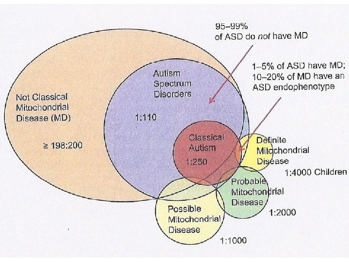
CYTOCHROME B DEFICIENCY ØFive complexes Ø> 80 proteins Øm. DNA – encodes 13 polypeptides , ØEncoded by m. DNA ØOne out of 11 polypeptides of complex III ØLocated in the inner membrane ØMostly sporadic, very uncommon, probably less than 7% of mitochondrial disorders ØRarely transmitted to offspring ØSome restricted to muscle ØFunctionally associated with complex III ØMutations: Stop codon , deletions, frame shift, termination and missense

Cytochrome B deficiency with cardiac and neurological manifestations Mutation Amino Acid Reduced Complex III Activity Histopathology (muscle) Exercise Intolerance Fixed Weakness Elevated Resting Lactate Clinical Manifestations Reference m. 14787_14790 del frameshift Yes Mild RRF, variable COX staining + - + Juvenile parkinsonism, MELAS, WPW, LV hypertrophy, epilepsy De Coo(13) m. 14792 C > G H 16 D NR, complex I def. Normal - - + Dystonia, seizures, external ophthalmoplegia, episodic central apnea Ronchi (18) Schuelke (11) Yes Normal + + - Fasting rhabdomyolysis, cognitive and motor delay, ataxia, LV hypertrophy, CRI, Septo-optic dysplasia G 166 X Yes Many RRFs, all COX+ + MELAS, menorrhagia Keightley (9) m. 15243 G>A G 166 E Yes Heart – Macrovesicular lipid storage - - + Hypertrophic cardiomyopathy, cardiac failure Valnot (7) m. 15498 G>A G 251 D Yes Heart – Increased lipid deposits - - - Histiocystoid cardiomyopathy, hepatic steatosis, ATN Andreu (8) Wibrand (12) Blakely (10) m. 14849 T>C S 35 P m. 15242 G>A m. 15579 A>G Y 278 C m. 15699 G>C R 318 P , RRF (17%), all COX+ + Bilateral deafness, cognitive dysfunction, GH deficiency, cataracts, retinitis pigmentosa, epilepsy Yes, and Mild RRF 1%), complex I def. normal COX staining + + + Bilateral deafness, MELAS, fertility problems Yes

CLINICAL HISTORY – CASE #1 Ø Born at normal gestational age with subtle dysmorphic features (epicanthal fold and high arch palate) Ø Global developmental delay of unknown origin with attention deficit hyperactivity disorder (ADHD) Ø Corrective surgery for strabismus at the age of 3 Ø At 6 years of age –, episode of acute creatinine kinase elevation and myoglobinuria after episode of gastrointestinal innocent fasting; frequent myalgias, nausea and occasional vomiting accompanying exercise or minute trauma; no visual or hearing loss Ø Progressive muscle weakness and scoliosis requiring treatment with bracing at the age of 15 Ø MR spectroscopy revealed elevated lactate peak in the basal ganglia and thalami; EKG – Wolfe-Parkinson-White syndrome; echocardiogram – mild left ventricular progressive dysfunction with decreased ejection fraction Ø Elevated serum lactate 6 -8 mmol/L (normal = 2. 2 mmol/L), normal CSF lactate Ø Biopsies at the age of 6 and 14 Ø Family history: mother with mild proximal muscle weakness and normal muscle biopsy Ø He died in his sleep at the age of 21.

6 yo. biopsy NADH ,

14 yo. ,

Post-mortem: quadriceps Ragged red fibers , NADH COX ORO

Diaphragm , Myocardium ORO

Parietal White Matter S? tau? Calretinin , Tau Neurofilaments

Occipital White Matter Subependymal era Synaptophysin , Tau Synaptophysin Calretinin

Neurofilament OCCIPITAL CORTEX NF , GFAP

Basilar Artery ,

Case #1 • • Mild cortical dysplasia White matter heterotopia- multifocal Cardiomyopathy , Mitochondrial myopathy

CLINICAL HISTORY – CASE #2 Ø Prematurely born at 27 weeks and 5 days gestational age (placental abruption) a) required brief respiratory support b) dysmorphic features (microcephaly, epicanthal fold, high arched palate, limited visual acuity to bright light, rapidly progressing to complete blindness) Ø At 7 months – beginning of seizures, hypotonia , Ø At 3 years – rigidity, spasticity, decortication, lower extremity clonus, kyphoscoliosis and progressive respiratory failure Ø Died at the age of 5 years and 4 months Ø Family history: mother with history of migraines, strabismus, myalgias and depression; brother and two maternal uncles with myalgias and intermittent exercise intolerance; maternal grandmother ? ? Ø Laboratory findings: normal lactate in CSF and blood; CT – cerebral atrophy; MRI – delayed myelination and increased lactate; EEG – high voltage waves in the left temporal region; EKG – normal Ø Muscle biopsies: 5 months – not remarkable; at 5 years – end stage muscle biopsies. mother, maternal grandmother and maternal great aunt – normal; brother – mitochondrial microangiopathy

Occipital lobe– neuronal loss& WM atrophy Temporal lobe – meningeal heterotopia HE HETERTOPIA NF GFAP ,

TEMPORAL LOBE , NF

MBP CALR SYN , SYN CALR NF

SMA , SMA CD 34 SMA

GFAP , NF Olivary nucleus

Case #2 • • Cerebral cortical dysplasia Grey matter heterotopia in white matter Grey matter heterotopia in meninges , Severe vasculopathy Cerebellar degeneration Brain atrophy Severe myopathy (end stage)
 - Substitution of serine")
Patient 1 Patient 2 - Novel sequence (m 15161 T>C) - Substitution of serine with proline at amino acid position 139 - Heteroplasmy in muscle 20%, not detected in blood - 50% reduction in complex I and III in muscle biopsy - No mutation in mother – de novo , mutation - Novel sequence (m 15152 G>A) - Substitution of glycine with serine at amino acid position 139 - Heteroplasmy in lymphocytes 58% - Heteroplasmy in many brain areas: 5068% - Same variant in mother’s muscle sample with 18% heteroplasmy – maternal inheritance Same enzyme deficiency due to different pattern of inheritance. Severity of pathological changes and clinical manifestations related to degree and presence of heteroplasmy Shared developmental brain abnormalities, especially neuronal migration.

DEVELOPMENTAL CNS ABNORMALITIES IN MITOCHONDRIAL DISORDERS Ø Agenesis of corpus callosum (Shevell et al, 1994) Ø Massive leptomeningeal heterotopia (complex I + IV) Ø Agenesis of corpus callosum with Leigh’s syndrome and microencephaly (Samson et al, 1994) Ø Cerebellar , hypoplasia (Lincke et al, 1996) Ø Partial agenesis of corpus callosum (PDH) and microencephaly (Lissen et al, 1999) and complex I + IV von Kleist-Retzow (2003) Ø Agenesis of corpus callosum, olivary dysplasia, absence of pyramidal system Ø Infantile cerebral lactic acidosis (Chow et al, 1987, Brown et al, 1998) Ø Dandy-Walker malformation (complex II, von Kleist-Retzow, 2003)

Thank you ,
- Slides: 24