
NEUROMUSCULAR SYSTEM basic principles P Kuera FBMI Kladno
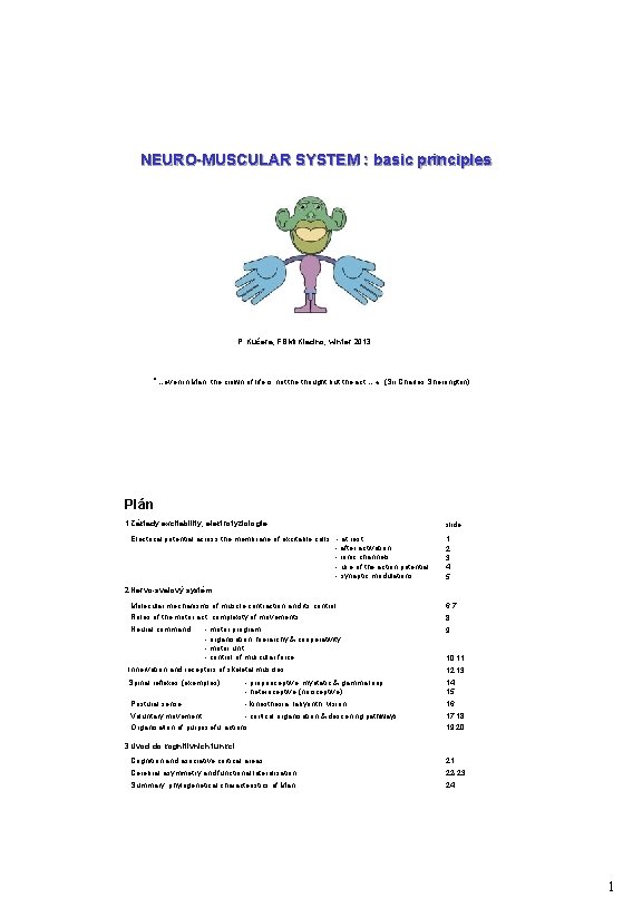
NEURO-MUSCULAR SYSTEM : basic principles P. Kučera, FBMI Kladno, winter 2013 "…even in Man, the crown of life is not the thought but the act… « (Sir Charles Sherrington) Plán 1 Základy excitability, elektrofyziologie Electrical potential across the membrane of excitable cells: - at rest - after activation - ionic channels - use of the action potential - synaptic modulations slide: 1 2 3 4 5 2 Nervo-svalový systém Molecular mechanisms of muscle contraction and its control 6, 7 Roles of the motor act, complexity of movements 8 Neural command: 9 - motor program - organisation: hierarchy & cooperativity - motor unit - control of muscular force 10, 11 Innervation and receptors of skeletal muscles 12, 13 Spinal reflexes (exemples) - proprioceptive: myotatic & gamma loop - heteroceptive (nociceptive) 14 15 Postural sense - kinesthesia, labyrinth, vision 16 Voluntary movement - cortical organisation & descening pathways 17, 18 Organisation of purposeful actions 19, 20 3 Úvod do kognitivních funkcí Cognition and asociative cortical areas 21 Cerebral asymmetry and functional lateralisation 22 -23 Summary: phylogenetical characteristics of Man 24 1
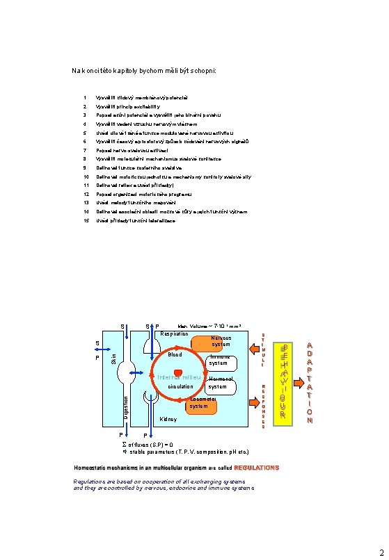
Na konci této kapitoly bychom měli být schopni: 1. Vysvětlit klidový membránový potenciál 2. Vysvětlit princip excitability 3. Popsat akční potenciál a vysvětlit jeho binární povahu 4. Vysvětlit vedení vzruchu nervovým vláknem 5. Uvést cílové tkáně a funkce modulované nervovou aktivitou 6. Vysvětlit časový a prostorový způsob kódování nervových signálů 7. Popsat nervo-svalovou aktivaci 8. Vysvětlit molekulární mechanismus svalové kontrakce 9. Definovat funkce kosterního svalstva 10. Definovat motorickou jednotku a mechanismy kontroly svalové síly 11. Definovat reflex a uvést příklad(y) 12. Popsat organizaci motorického programu 13. Uvést metody funkčního mapování 14. Definovat asociační oblasti mozkové kůry a jejich funkční význam 15. Uvést příklady funkční lateralizace S Man: Volume ~ 7. 10 7 mm S P Respiration Nervous system S Blood Skin P Immune system internal milieu circulation Hormonal system Locomotor system Digestion P 3 Kidney S T I M U L I R E S P O N S E S B E H A V I O U R A D A P T A T I O N P of fluxes (S, P) = 0 stable parameters (T, P, V, composition, p. H etc. ) Homeostatic mechanisms in an multicellular organism are called REGULATIONS Regulations are based on cooperation of all exchanging systems and they are controlled by nervous, endocrine and immune systems 2
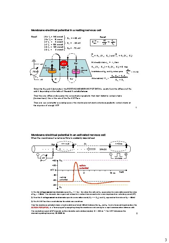
Membrane electrical potential in a resting nervous cell [ Na+ ]e = 145 mmol/l [ Na+ ]i = 15 mmol [ K+ ]e = 4 mmol/l [ K+ ]e = 136 mmol/l [ Cl- ]e = 120 mmol/l [ Cl- ]i = 8 mmol/l Recall : ENa = + 60 m. V E i-e = ion EK = - 93 m. V - RT ln Ci Ce z. F ECl = - 72 m. V INa = GNa (Vm - ENa) and IK = GK (Vm - EK) K+ K channel Vm ase Na channel GK -75 m. V GNa Na+ K+ int At steady state INa = - IK then: Na+ ATP ext GNa (Vm - ENa) + GK (Vm - EK) = 0 thus: GK Substitution of Eion and Vm values gives: ~ 11 -13 GNa ENa + GK EK Alternatively: Vm ~ GNa + GK Given the Na and K distribution, the RESTING MEMBRANE POTENTIAL results from the diffusion of Na and K depending on the ratio of Na and K conductances. Thus the ionic diffusion dissipates the concentration gradients that must however remain stable (homeostasis): this is the role of the Na, K, ATPase Thus ions are constantly circulating across the membrane but electrochemical gradients remain stable at the expense of energy: ATP 1 Membrane electrical potential in an activated nervous cell When the membrane of a nervous fibre is suddenly depolarised: Na+ - - - t = t t L K+ + - - - - t = 0 ms SPACE m. V +60 ENa +30 action potential m. V 0 -30 -60 -90 + stimulus 5 10 ms TIME resting potential EK 1) The Na voltage sesnsitive channels open (GNa >>> GK) , Na enters the cell and Vm approaches for some milliseconds the value of ENa ~ + 60 m. V. The channels close again and remain for a certain time insensitive to a new depolarisation: refractory period (RP). 2) Then the K voltage sensitive channels open for some milliseconds (GK >>> GNa) and Vm approaches the value of EK ~ -90 m. V. 3) The A, K, ATPase then re-establishes the initial ionic conditions. Thus the membrane potential shows a brief oscillation of about 100 m. V between the ENa and EK. Such a transient depolarisation, the ACTION POTENTIAL, is a "binary signal" propagating along the membrane and serving for a rapid communication between cells. The conduction speed of AP depends on fiber diameter and isolation (myelin): 0. 1 -100 ms-1. The 1/RP determines the maximal signalling frequency: 20 -2000 Hz 2 3
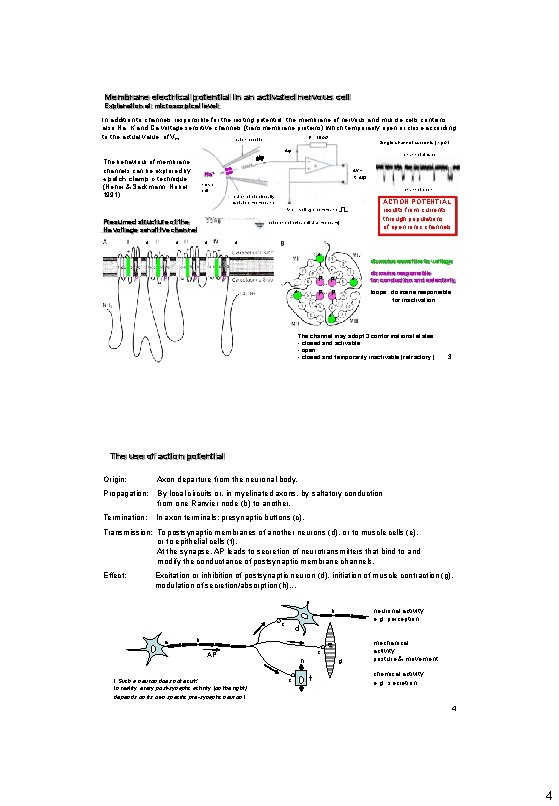
Membrane electrical potential in an activated nervous cell Explanation at microscopical level: In addition to channels responsible for the resting potential, the membrane of nervous and muscle cells contains also Na, K and Ca voltage sensitive channels (transmembrane proteins) which temporarily open or close according to the actual value of Vm. R = 10 G patch pipette Single channel currents (~ p. A) ip The behaviour of membrane channels can be explored by « patch clamp » technique (Neher & Sackmann, Nobel 1991) channel closed V = R. ip Na+ nerve cell channel open patch of electrically isolated membrane Presumed structure of the Na voltage sensitive channel 4 4 ACTION POTENTIAL results from currents through populations of open ionic channels voltage command Vc reference (extracellular medium) 4 4 4 domains sensitive to voltage P P P P 4 domains responsible for conduction and selectivity loops domains responsible for inactivation 4 The channel may adopt 3 conformational states: - closed and activable - open - closed and temporarily inactivable (refractory) 3 The use of action potential Origin: Axon departure from the neuronal body. Propagation: By local circuits or, in myelinated axons, by saltatory conduction from one Ranvier node (b) to another. Termination: In axon terminals: presynaptic buttons (c). Transmission: To postsynaptic membranes of another neurons (d), or to muscle cells (e), or to epithelial cells (f). At the synapse, AP leads to secretion of neurotransmitters that bind to and modify the conductance of postsynaptic membrane channels. Effect: Excitation or inhibition of postsynaptic neuron (d), initiation of muscle contraction (g), modulation of secretion/absorption (h). . . b c a d b c AP ! Such a neuron does not exist! In reality, every post-synaptic activity (on the right) depends on its own specific pre-synaptic neuron ! neuronal activity: e. g. perception h c e g f mechanical activity: posture & movement chemical activity: e. g. , secretion 4 4
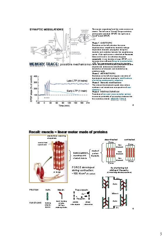
SYNAPTIC MODULATIONS Two spines originating from the same neuron are shown. The left one is “strong” (large excitatory postsynaptic potential, EPSP), the right one is “weak” (small EPSP). MEMORY TRACE: possible mechanisms Phase 1: IONOTROPIC Excitation on the left activates the spine: depolarisation, amplified by dendritic voltage dependent channels propagates along the dendrite and irradiates towards the neighbouring spines. If the right spine is activated at the same time and if such a co-activation happens repeatedly, it can develop a larger EPSP, i. e. it becomes more efficient (long term potentiation LTP). The communication among the dendritic spines depend on electrotonic propagation (spine impedance), dimensions (cytoskeleton) distribution of channels and modulation by inhibitory inputs. Phase 2: METABOTROPIC Excitation on the left also triggers cascades of biochemical reactions leading to modifications of existing postsynaptic receptors. Phase 3: Genomic modifications The induced biochemical signals may induce synthesis and membrane incorporation of new receptors. Phase 4: RESTRUCTURATION Formation of new and more complex spines: increased probability of association between the two excitatory stimuli: MEMORY TRACE 5 Recall: muscle = linear motor made of proteins muscle fibers consisting of myofibrils decontracted Z-line nerves from spinal cord thin filaments SARCOMERES repeating units: striated muscle made of protein filaments m. biceps Z-line thick filaments thin filaments Z-line FORCE developed during contraction: ~100 N/cm 2 of section contracted Z-line No shortening but sliding of filaments: L=working stroke (cca 9 nm) L Actin PROTEIN: Actin Myosin Tropomyosin Ca++ Myosin + Troponin FUNCTIONS: binding sites for myosin head: binding of actin ATPase working stroke inhibits acto-myosin allows interaction Actin L 6 5
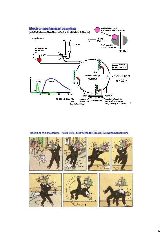
Electro-mechanical coupling acetylcholine from presynaptic nerve terminals (excitation-contraction events in striated muscle) sarcolemma AP T- tubule nicotinic ACh receptor-channel sarcoplasmic reticulum + Na + Ca++ ATP Ca++ contraction relaxation AM cross bridge cycling delay stroke: work + heat ~ 25 % Force AP 10 20 40 50 ms ATP RESERVES for 20 s creatine 60 s lactic acid long effort CO 2 energy metabolism ADP+P creatine phosphate glucose(glycogene) fatty acids/glucose + O 2 7 Roles of the muscles: POSTURE, MOVEMENT, HEAT, COMMUNICATION 8 6
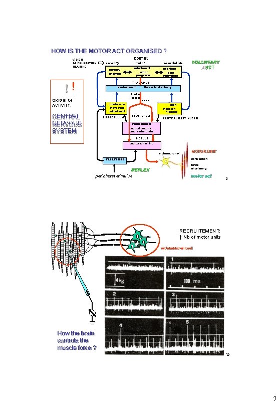
HOW IS THE MOTOR ACT ORGANISED ? VISION ACCELERATION HEARING ! CORTEX motor sensory selection of motor programs sensory analysis associative intention plan motivation VOLUNTARY ACT THAL AMUS moduation of tractus cortico ORIGIN OF ACTIVITY: the cortical activity spinal posture vs movement adjustment CENTRAL NERVOUS SYSTEM CEREBELLUM plan initiation filtering BRAINSTEM CENTRAL GREY NUCLEI modulation of spinal circuits and motor units MOELLE activation of MU motoneuron MOTOR UNIT contraction: RECEPTORS REFLEX peripheral stimulus force shortening motor act 9 RECRUITEMENT: ↑ Nb of motor units motoneuronal pool How the brain controls the muscle force ? 10 7
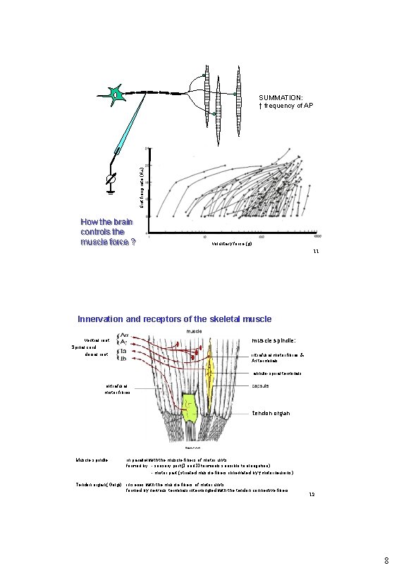
Unit firing rate (Hz) SUMMATION: ↑ frequency of AP How the brain controls the muscle force ? voluntary force (g) 11 Innervation and receptors of the skeletal muscle spindle: ventral root Spinal cord dorsal root intrafusal motor fibres & A terminals annulo-spiral terminals extrafusal motor fibres tendon organ Muscle spindle: in parallel with the muscle fibers of motor units formed by - sensory part (I and II terminals sensible to elongation) - motor part (striated muscle fibers innervated by motorneurons) Tendon organ (Golgi) in series with the muscle fibers of motor units formed by nervous terminals intermingled with the tendon connective fibers 12 8
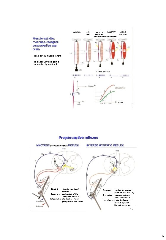
2 passive elongation Muscle spindle: mechano-receptor controlled by the brain 1 initial length 3 4 extrafusal Intrafusal contraction (A stimulation) 5 extra- & intrafusal contraction - records the muscle length - its sensitivity and gain is controlled by the CNS Ia fibre activity muscle length 13 Proprioceptive reflexes MYOTATIC (STRETCH/JERK) REFLEX Stimulus: muscle elongation (gravity!) Response: contraction of the elongated muscle Importance: maintains postural (antigravitational tone) INVERSE MYOTATIC REFLEX Stimulus: tendon elongation (muscle contraction!) Response: relaxation of the contracted muscle Importance: limits the force, defends against the muscle lesion 14 9
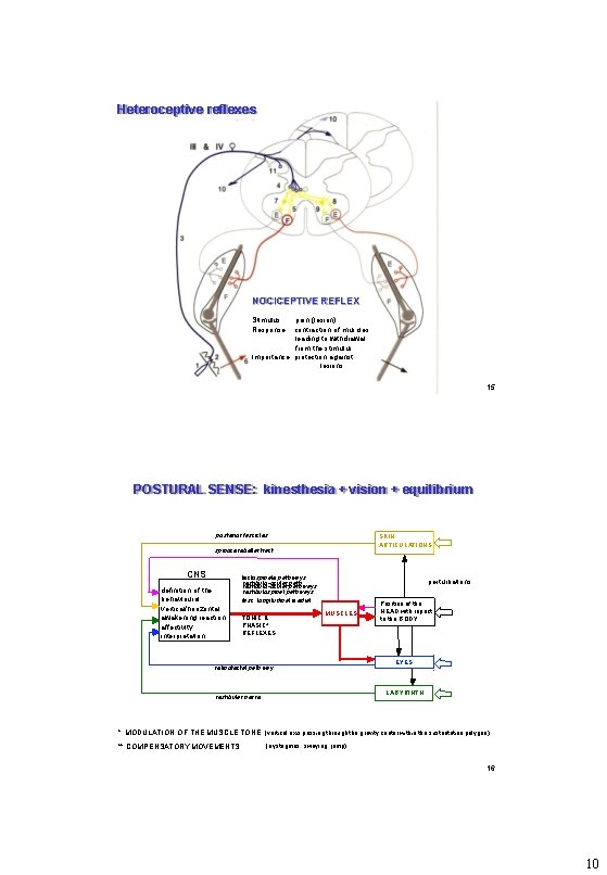
Heteroceptive reflexes NOCICEPTIVE REFLEX Stimulus: pain (lesion) Response: contraction of muscles leading to withdrawal from the stimulus Importance: protection against lesions 15 POSTURAL SENSE: kinesthesia + vision + equilibrium posterior fascicles SKIN , ARTICULATIONS spinocerebellar tract CNS definition of the behavioural vertical/horizontal, awakening reaction, affectivity, interpretation tectospinale pathways vestibulo-ocular pathways vestibulospinal pathways fasc. longitudinal medial TONIC & PHASIC* REFLEXES perturbations MUSCLES retinotectal pathway Position of the HEAD with repect to the BODY EYES LABYRINTH vestibular nerve * MODULATION OF THE MUSCLE TONE (vertical axis passing through the gravity center within the sustentation polygon) ** COMPENSATORY MOVEMENTS (nystagmus, swaying, jump) 16 10
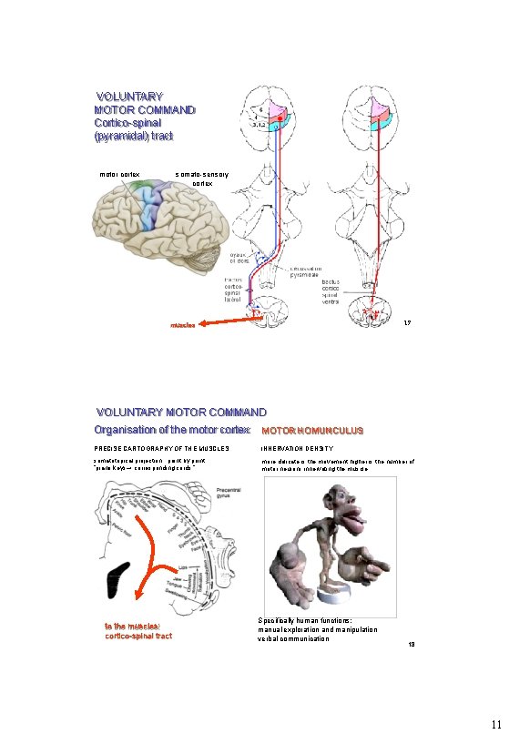
VOLUNTARY MOTOR COMMAND Cortico-spinal (pyramidal) tract motor cortex somato-sensory cortex 17 muscles VOLUNTARY MOTOR COMMAND Organisation of the motor cortex MOTOR HOMUNCULUS PRECISE CARTOGRAPHY OF THE MUSCLES: INNERVATION DENSITY : somatotopical projection : point by point “piano keys→ corresponding cords ” more delicate is the movement higher is the number of motor neurons innervating the muscle to the muscles: cortico-spinal tract Specifically human functions: manual exploration and manipulation verbal communication 18 11
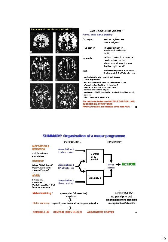
Increase of the blood perfusion But where is the pianist? Functional cartography Principle: active regions are more irrigated Realisation: measurement of the blood perfusion MRIf Example: which cerebral structures are involved in the discrimination of formes by the right hand? Task : successively explore 2 objects, then decide if they are identical - understanding and recall of instructions - tactile exploration - extraction from the sensory information of the characteristical features of the object - mental reconstruction of the object - memorisation of the object - comparaison with the mental image of the other object - decision - motor command: response The task is distributed over MULTIPLE CORTICAL AND SUBCORTICAL STRUCTURES. All these structures are indicated on the slide Nb 8. 19 SUMMARY: Organisation of a motor programme PREPARATION MOTIVATION & INTENTION Association & Limbic cortex I will (must) write my signature CONTEXT Central Grey Nuclei Motor cortex Association & (Pre)motor cx Where? Size? Speed? Paper? Blackboard? Standing? Sitting? STATE Eyes open? Equilibrium? Position: shoulder->wrist Force vs resistance … Motor learning : EXECUTION Association & Sens. -mot. cx Cerebellum APRAXIA: aperceptive (observation) cognitive Motor memory: implicit (non-declarative), « procedural » CEREBELLUM CENTRAL GREY NUCLEI ACTION no paralysis but impossibility to execute complex movements ASSOCIATIVE CORTEX 20 12
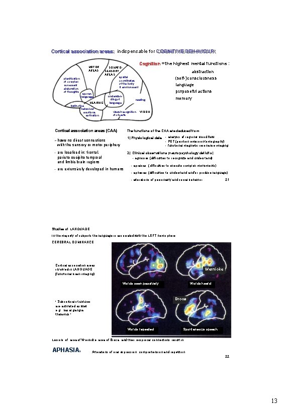
Cortical association areas: indispensable for COGNITIVE BEHAVIOUR MOTOR AREAS planification of complex movement elaboration of thoughts Cognition = the highest mental functions : SOMATOSENSORY AREAS abstraction (self-)consciousness language purposeful actions spatial coordinates of the body & environment spoken language HEARING instinctive behaviour emotions motivation understanding of language reading visual recognition of objects memory VISION Cortical association areas (CAA) The functions of the CAA are deduced from: - have no direct connections with the sensory or motor periphery 1) Physiological data: - analysis of regional blood flow - PET (positron emission tomography) - functional magnetic resonance imaging - are localised in: frontal, parieto-occipito-temporal and limbic brain regions 2) Clinical observations (neuropsychology deficits): - agnosias (difficulties to recognize and understand) - are extensively developed in humans - apraxias (difficulties to execute complex movements) - aphasias (difficulties to understand and/or produce language) 21 - alterations of personality and social behavior Studies of LANGUAGE In the majority of subjects the language is associated with the LEFT hemisphere: CEREBRAL DOMINANCE Cortical association areas involved in LANGUAGE (functional brain imaging) Wernicke Words seen passively Words heard Broca ! Subcortical structures are activated as well: e. g. , basal ganglia, thalamus ! Words repeated Spontaneous speech Lesions of area of Wernicke, area of Broca, and their reciprocal connections result in APHASIAs Alterations of oral expression, comprehension and repetition 22 13
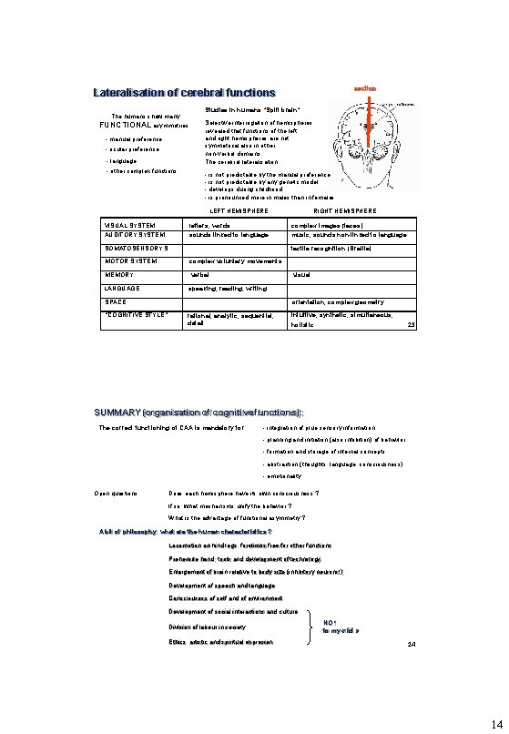
section Lateralisation of cerebral functions The humans show many FUNCTIONAL asymmetries: - manual preference - ocular preference - language - other complex functions Studies in humans “Split brain” Selective interrogation of hemispheres revealed that functions of the left and right hemispheres are not symmetrical also in other, non-verbal domains The cerebral lateralisation : - is not predictable by the manual preference - is not predictable by any genetic model - develops during childhood - is pronounced more in males than in females LEFT HEMISPHERE VISUAL SYSTEM AUDITORY SYSTEM Ietters, words sounds linked to language SOMATOSENSORY S. MOTOR SYSTEM MEMORY LANGUAGE RIGHT HEMISPHERE complex images (faces) music, sounds non-linked to language tactile recognition (Braille) complex voluntary movements verbal visual speaking, reading, writing SPACE “COGNITIVE STYLE” orientation, complex geometry rational, analytic, sequential, detail intuitive, synthetic, simultaneous, holistic 23 SUMMARY (organisation of cognitive functions): The correct functioning of CAA is mandatory for - integration of pluri-sensory information - planning and initiation (also inhibition) of behavior - formation and storage of internal concepts - abstraction (thoughts, language, consciousness) - emotionality Open questions : Does each hemisphere have its own consciousness ? If so, what mechanisms unify the behavior ? What is the advantage of functional asymmetry ? A bit of philosophy: what are the human characteristics ? Locomotion on hind legs: forelimbs free for other functions Prehensile hand: tools and development of technology Enlargement of brain relative to body size (inhibitory neurons!) Development of speech and language Consciousess of self and of environment Development of social interactions and culture Division of labour in society Ethics, artistic and spiritual expresion NO ! to my « id » 24 14
- Slides: 14